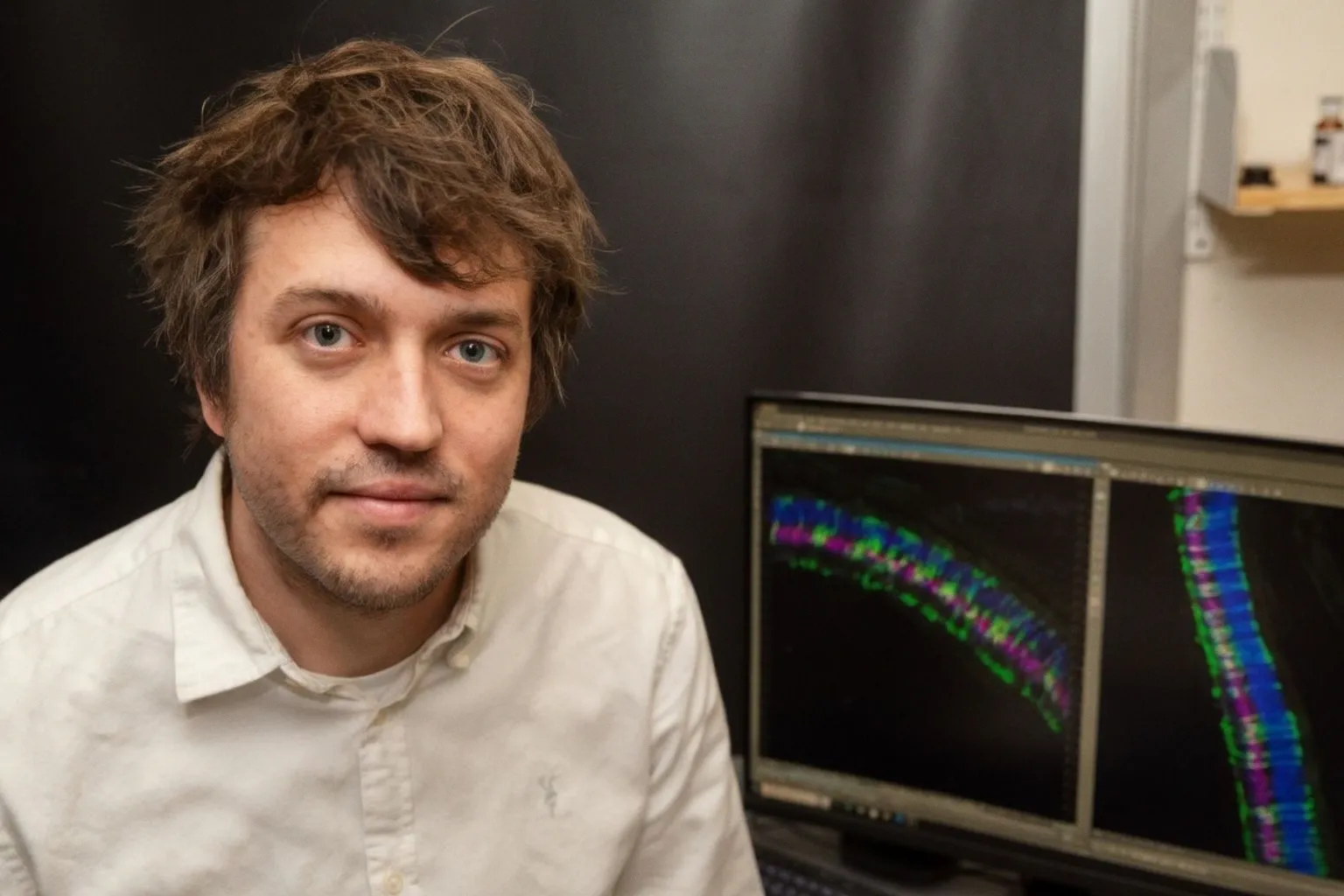
Researchers at University of Rochester Medicine have put a 3D retinal organoid model to work on a stubborn question in juvenile Batten disease: why do photoreceptors continue to die even when the underlying lysosomal defect is understood? According to Batten Disease News, the team's organoid work points squarely at retinal pigment epithelium (RPE) dysfunction as a driver of secondary photoreceptor degeneration in CLN3 disease. The finding reframes CLN3 as not just a neuronal storage problem, but a tissue-level failure where the support layer stops doing its job.
What the organoid actually shows
Organoid systems matter here because they let researchers isolate RPE from photoreceptors and watch the cascade unfold in three dimensions rather than in flat culture. The Rochester group reports that RPE dysfunction directly precedes photoreceptor loss in their model, separating cause from correlation. In the same preclinical system, restoring expression of acid ceramidase — the enzyme deficient in CLN3 — mitigated cellular damage. That is a practical signal for anyone designing a therapeutic pipeline: if the RPE is the upstream failure point, intervention timelines may need to move earlier than photoreceptor rescue alone would suggest.
Why this lands on the biobanking side
For ocular biobanking and research biologics, juvenile Batten disease has long been a procurement challenge. Affected donor tissue is rare, age-matched controls are scarce, and the standard histopathology snapshot rarely captures the RPE-photoreceptor relationship in living tissue. A reproducible 3D organoid model built from patient-derived cells changes that calculus. It offers a renewable, quality-controlled platform for screening acid ceramidase-restoration strategies without requiring fresh juvenile donor eyes every cycle. Eye banks coordinating CLN3 families and research consortia should expect growing demand for fibroblasts, iPSCs, and carefully consented retinal tissue as downstream groups try to replicate or extend the finding.
The wider retinal regeneration context
The Rochester study arrives alongside two other preclinical signals worth tracking. A SUNY Upstate Medical University team, publishing in PNAS, found that aging impairs the ability of retinal glial cells to be reprogrammed into replacement neurons, with anti-inflammatory steroids only partially restoring that response. Separately, Scripps Research and collaborators identified erucamide as an endogenous protective molecule whose levels fall during photoreceptor loss, with preclinical restoration slowing degeneration. Medical College of Georgia researchers, supported by the National Eye Institute, also linked cholesterol ester accumulation to vascular leakage and neuroretinal damage in diabetic retinopathy. Taken together, the September cluster points toward a field increasingly focused on the support environment — RPE, glia, lipid handling, inflammation — rather than photoreceptors alone.
The honest read from the bench: CLN3 therapy is moving from "fix the gene" toward "fix the tissue," and that shift will quietly reshape what ocular biobanks are asked to collect, preserve, and ship.