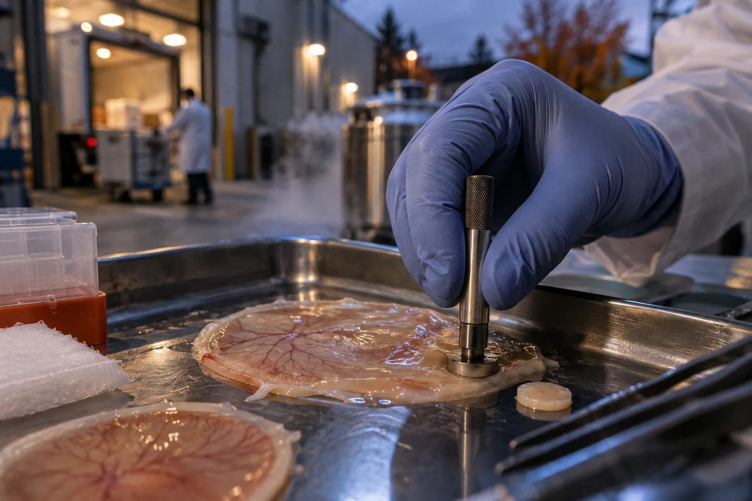
In perfused post-mortem donor retina, ON-bipolar cells can lose light sensitivity within minutes, while photoreceptors retain more than half of their light-evoked response for at least 30 minutes. The tissue is already moving toward collapse, but its layers do not cross that threshold together.
That temporal mismatch is the central problem in photoreceptor degeneration models in donor retina. A retinal explant can preserve laminated neural architecture for up to a month, allowing drug screening and viral-vector optimization in human tissue. A donor eye, by contrast, contains the original macular geometry, the native RPE interface, age-associated extracellular deposits, vascular injury, and the molecular residue of disease. One model offers control. The other offers biological history.
Neither is a complete substitute for the other.
Preserving native architecture: what an explant keeps intact
A dissociated culture can isolate photoreceptors, Müller glia, RPE cells, or retinal vascular components with considerable experimental precision. That precision is also its weakness. Once the tissue is reduced to a cell population, the spatial relationships that regulate survival begin to disappear: the photoreceptor outer segment no longer faces an intact RPE apical surface, synaptic contacts become partial or absent, and the diffusion geometry of oxygen, glucose, retinoids, and trophic factors is simplified into a culture-medium problem.
Organotypic retinal explants preserve more of the original arrangement. The outer nuclear layer remains aligned with the outer plexiform layer; photoreceptor terminals remain embedded in a partially preserved synaptic environment; Müller cell processes continue to span the retinal thickness; and the RPE, when deliberately retained, can still function as a boundary between neural retina and the underlying choroidal compartment.
This is not equivalent to preserving a living eye. The choroidal circulation is gone, the immune environment is altered, and the tissue is exposed to an artificial oxygen and nutrient gradient. Yet the explant retains a form of spatial causality that cell lines and many organoid systems cannot reproduce. When a photoreceptor dies, the surrounding tissue is still capable of responding through neighboring Müller cells, microglia, bipolar neurons, and damaged synaptic compartments.
For ex vivo retinal degeneration assessment, that distinction determines the question the model can answer.
An explant is particularly useful when the experiment asks:
- whether a viral vector reaches a defined retinal layer;
- how a candidate compound diffuses through laminated tissue;
- whether photoreceptors remain structurally connected to their post-synaptic partners;
- how quickly degeneration propagates after a controlled insult;
- whether a rescue strategy preserves tissue architecture rather than merely increasing cell survival markers.
The culture window can extend up to a month in human post-mortem retinal explants. That duration is long enough to observe progressive structural changes, compare treatment schedules, and follow delayed responses that would be invisible in an acute slice. But the longer the explant remains outside the donor eye, the more carefully its measurements must be interpreted. Culture itself imposes transcriptional and metabolic stress, and those changes can become entangled with the disease mechanism under investigation.
The explant is therefore not a frozen fragment of the donor retina. It is a living preparation with a clock.
An explant preserves retinal geography, but it does not preserve the original physiological timeline.
Post-mortem kinetics: viability is not a single variable
Donor tissue photoreceptor viability is often treated as though it were a yes-or-no property. The retina is either alive enough for an assay, or it is not. That binary view obscures the sequence of failures occurring across the tissue.
Electrical responsiveness, membrane integrity, mitochondrial activity, outer-segment morphology, synaptic preservation, and transcriptional stability do not decline at the same rate. A donor retina may still produce a measurable response while already carrying irreversible injury in one cellular compartment. Conversely, a weak electrical response does not necessarily mean that every photoreceptor has lost structural integrity. The assay captures one layer of the pathology.
Ex vivo perfusion studies make this separation visible. Under post-mortem hypoxia, ON-bipolar cells can lose light sensitivity within minutes, whereas photoreceptors retain more than 50% of their response for at least 30 minutes. The implication is not that photoreceptors are resistant to death. It is that the neural retina enters dysfunction through a staggered cascade rather than a synchronized collapse.
That cascade matters when comparing a controlled degeneration model with donor tissue. If an experiment begins too late after procurement, a researcher may interpret hypoxia-induced synaptic failure as primary photoreceptor degeneration. If an explant is assessed only by outer nuclear layer thickness, early functional loss may be missed. If viability is inferred from one metabolic stain, cells with preserved membrane integrity but compromised phototransduction may be counted as successfully maintained.
A useful comparison must therefore separate three clocks:
1. The procurement clock — the interval between donor death, enucleation, transport, dissection, and experimental stabilization.
2. The physiological clock — the order in which photoreceptors, bipolar cells, RPE, Müller glia, and microglia lose or alter function.
3. The culture clock — the period during which the explant adapts to artificial media, oxygenation, temperature, and mechanical isolation.
These clocks interact, but they are not interchangeable. A tissue collected rapidly may still contain disease-associated mitochondrial dysfunction or RPE senescence. A slower procurement interval may amplify hypoxic damage without erasing the donor’s pre-existing pathology. A long culture period may reveal degeneration kinetics while simultaneously generating stress pathways that were not present in the donor eye.
For this reason, post-mortem photoreceptor structural integrity should be documented alongside functional assays, not used as a proxy for them. Outer-segment length, nuclear-layer organization, synaptic ribbon preservation, and the condition of the external limiting membrane provide a morphological timeline. Electrophysiology adds another. Neither is sufficient alone.
The RPE-dependent pathway: when removing support accelerates the crime
The retinal pigment epithelium is not a passive substrate beneath the photoreceptors. It participates in outer-segment phagocytosis, retinoid handling, metabolic exchange, barrier formation, and the regulation of local inflammatory signaling. Remove it, and the photoreceptor compartment does not simply lose one neighboring cell type. It loses an integrated support interface.
In an in vitro human retinal detachment model, removal of the RPE accelerated photoreceptor death. The effect peaked at day three, when AIF-positive cells increased tenfold compared with control retinas. AIF, or apoptosis-inducing factor, is not merely a generic marker of tissue deterioration; its increase indicates that the mode and timing of cell death are being reshaped by the loss of RPE support.
This is one of the clearest reasons donor retina and engineered degeneration models should not be treated as equivalent platforms. A controlled model can remove the RPE deliberately and establish a reproducible injury trajectory. That is experimentally valuable. It allows researchers to ask which pathways become active after detachment, which interventions delay photoreceptor loss, and whether a candidate therapy acts directly on the photoreceptor or indirectly through the tissue environment.
But an RPE-depleted preparation does not recreate every form of retinal disease. In age-related macular degeneration, the RPE may be senescent, overloaded by lipofuscin, altered by complement activity, or functionally compromised while remaining physically present. In diabetic retinal disease, the surrounding microvasculature, pericytes, inflammatory mediators, and metabolic conditions contribute to injury. In geographic atrophy, the border between surviving and atrophic tissue contains a spatially organized pathology that cannot be reduced to an acute RPE removal event.
The difference is mechanistic:
| Experimental platform | What it preserves | What it exposes most clearly | Principal limitation |
|---|---|---|---|
| Human organotypic retinal explant with RPE | Laminated neural retina and, when retained, the RPE–photoreceptor interface | Tissue-level degeneration, vector distribution, synaptic and cellular remodeling | No intact circulation, systemic immune input, or full choroidal exchange |
| RPE-depleted retinal explant | Neural retinal architecture without the principal outer support layer | Acute photoreceptor stress and RPE-dependent survival pathways | Models a defined injury rather than the full chronology of chronic disease |
| Donor retina from diseased eye | Native pathology, age-related architecture, macular geometry, and disease-associated cellular states | Human degeneration pathways as they existed at death | Post-mortem interval and pre-analytical history can obscure the original disease trajectory |
| iPSC-derived retinal organoid or photoreceptor preparation | Controlled developmental stage and scalable experimental material | Cell-intrinsic maturation, transplantation behavior, and genetic manipulation | Does not fully reproduce native macular organization or post-mortem human pathology |
The key question is not whether RPE removal is realistic. It is whether the removal corresponds to the biological transition being studied. For acute detachment-associated degeneration, it may be an appropriate perturbation. For chronic macular degeneration, it risks compressing years of cellular adaptation into a short, highly artificial injury sequence.
Inner retinal remodeling reveals the duration of disease
Photoreceptor degeneration is often measured from the outside inward: outer-segment shortening, loss of nuclei in the outer nuclear layer, disruption of the ellipsoid zone, and eventual collapse of the photoreceptor mosaic. Human donor tissue shows why that sequence is incomplete.
When the outer nuclear layer degenerates, the inner retina does not remain an indifferent scaffold. ON-bipolar cells retract dendrites, and Kv1.3 potassium channels become upregulated. These are not secondary details. They indicate that the loss of photoreceptor input changes the electrical and structural state of neurons deeper in the retinal circuit.
A controlled explant can capture some of this remodeling, particularly when tissue architecture is preserved long enough for dendritic retraction and glial responses to emerge. Yet donor tissue provides a different kind of evidence: it records the accumulated consequences of degeneration after the retina has had time to reorganize. The remaining inner retinal cells have been exposed to prolonged synaptic deprivation, altered extracellular potassium, inflammatory signaling, and changes in metabolic demand.
This is where donor retina becomes indispensable for validating a model. An explant may reproduce a sequence of photoreceptor death, but does it reproduce the downstream remodeling observed in human disease? Does the timing of bipolar-cell dendrite retraction match the donor pattern? Does Kv1.3 upregulation occur in the same cellular compartments? Are Müller glia and microglia positioned as they are in a diseased human macula, or are they responding primarily to dissection and culture stress?
The answers determine whether the model has captured a disease pathway or only a general injury response.
Donor tissue also preserves the geometry of pathology. Macular cones, parafoveal rods, the RPE monolayer, Bruch’s membrane, and the choroidal interface are arranged according to a human spatial map that organoids do not fully reproduce. Even when an organoid displays laminated retinal features, its developmental pattern is not identical to the mature human macula. It may provide excellent access to cell-intrinsic mechanisms while remaining less informative about the spatial distribution of drusen biogenesis, RPE senescence, vascular proximity, or geographic atrophy borders.
That does not diminish organoids. It clarifies their position. They are powerful instruments for controlling developmental age, genotype, and treatment exposure. They are not replacements for human donor tissue when the research question depends on native macular architecture.
Donor tissue quality begins before the microscope
The pathology visible in a donor retina is inseparable from how the tissue reached the laboratory. Procurement is not administrative prelude; it is the first experimental manipulation.
The relevant variables include the interval from death to enucleation, storage temperature, transport duration, dissection method, whether the retina remains attached to the RPE, and whether the tissue is fixed, cryopreserved, perfused, or placed directly into culture. Mechanical separation can damage the outer segments and leave the RPE surface irregular. Delayed stabilization can exaggerate hypoxic injury. Inconsistent orientation can turn a spatial transcriptomic comparison into a mapping problem.
For researchers building a donor-tissue series, the metadata should be treated as part of the specimen rather than as an accessory record. A retina with excellent lamination but an uncertain post-mortem interval may be less useful for electrophysiological interpretation than a more modest specimen with a well-documented procurement history.
The practical requirements are specific:
- Record procurement and stabilization intervals in a consistent format rather than reducing them to a general statement about rapid collection.
- Separate central, parafoveal, and peripheral regions when the disease mechanism has a spatial component.
- Preserve matched regions for morphology, molecular profiling, and functional assays whenever tissue quantity permits.
- Document whether the RPE remained attached, was intentionally removed, or was lost during dissection.
- Interpret negative findings cautiously when tissue quality limits detection of low-abundance transcripts or fragile cellular structures.
- Use multiple readouts so that structural preservation is not mistaken for preserved phototransduction.
This is also where the comparison between explants and donor tissue becomes operational. An explant study can standardize dissection, medium, oxygenation, and treatment exposure across specimens. A donor-tissue study carries more uncontrolled variation but gains access to biological states that cannot be generated quickly in culture.
The trade-off is not simply control versus realism. It is reproducibility versus historical depth.
iPSC-derived photoreceptors meet the degenerating retina
Stem cell-derived photoreceptors occupy a third position in the comparison. They are not donor tissue, and they are not merely simplified explants. Their value lies in the possibility of controlling developmental age, genetic background, cell dose, and preparation quality before introducing them into a degenerating environment.
In degeneration models, transplanted human iPSC-derived photoreceptors have survived for up to 24 weeks and have shown structural and functional integration. The outcome, however, depends strongly on the developmental age of the donor cells and the severity of degeneration in the recipient. A photoreceptor preparation that remains immature may possess greater plasticity but less immediate functional competence. A more mature preparation may offer a more appropriate cellular phenotype while losing some capacity to integrate into a hostile retinal environment.
The recipient tissue is not a neutral landing site. In advanced degeneration, the outer nuclear layer may be reduced, the outer limiting membrane altered, Müller glia reactive, and synaptic targets displaced. In a less severe host, there may be more residual circuitry but also greater competition for space and connectivity. The same donor cell product can therefore produce different outcomes depending on the pathology it encounters.
This is where human donor retina can refine transplantation studies. Diseased donor tissue can reveal the physical and molecular terrain that a transplanted photoreceptor must navigate: the state of the RPE, the integrity of the interphotoreceptor matrix, the organization of surviving photoreceptors, and the extent of inner retinal remodeling. Without that context, integration may be judged by cell survival alone.
Preclinical studies have tested transplanted cell doses in the range of 150,000 to 500,000 cells, but dose is only one variable in the transplantation equation. Cell identity, developmental stage, delivery geometry, recipient degeneration severity, and the preservation of host synaptic architecture all influence whether structural proximity develops into functional connectivity.
A model that records only the persistence of transplanted cells risks confusing occupation with integration. The more demanding question is whether the cells establish the correct outer-retinal relationships and participate in light-evoked signaling without creating a new source of disorganized activity.
Choosing the model around the pathological event
The most defensible comparison begins with the event under study, not with a preference for a particular platform.
If the question concerns acute loss of RPE support, a controlled explant with and without RPE may provide the cleanest contrast. If the question concerns the progression of age-associated macular pathology, donor tissue is needed to anchor the experiment in native human architecture and chronic cellular history. If the question concerns vector penetration, tissue-level distribution, or treatment timing, an organotypic explant offers a practical middle ground. If the question concerns developmental maturation or the behavior of a defined cell product, iPSC-derived photoreceptors provide the necessary control before the cells are challenged by degenerative tissue.
A robust study will often use these platforms sequentially rather than forcing one to answer every question:
1. Define the cell-intrinsic mechanism in a controlled photoreceptor or organoid preparation.
2. Test spatial behavior in a human organotypic explant with preserved retinal layers.
3. Compare the resulting pathology with donor retina from relevant disease states.
4. Assess transplantation or rescue in a recipient environment whose degeneration severity is documented rather than assumed.
5. Return to donor tissue to determine whether the experimental pathway resembles human remodeling.
This sequence creates a chain of evidence. It also exposes where the model breaks. A compound may preserve photoreceptor nuclei in an explant but fail to restore synaptic organization. A vector may reach the outer nuclear layer while producing little functional rescue. A transplanted cell may survive for 24 weeks yet remain poorly integrated. Each apparent success requires a second measurement at a different biological level.
The strongest model is not the one that looks most human under the microscope; it is the one whose failure can be localized.
The unresolved interval between death and disease
Ex vivo models and donor tissue answer different temporal questions. The explant can reveal how degeneration unfolds after a defined perturbation. The donor retina shows what remains after disease, death, procurement, hypoxia, and tissue handling have already acted on the same architecture.
That distinction leaves a difficult interval between the two. How much of the donor retina’s molecular state reflects chronic disease, and how much reflects the final hours of oxygen loss? When does post-mortem remodeling begin to obscure the pathway that researchers are trying to reconstruct? Can perfusion restore a meaningful light-evoked response without restoring the metabolic conditions required for long-term phototransduction? And how long can donor photoreceptors retain functional capacity under ex vivo perfusion before apparently recoverable activity becomes irreversible injury?
There is no single answer because the retina does not fail as one object. Photoreceptors, bipolar cells, RPE, Müller glia, microglia, and vascular compartments enter different states on different schedules. The task of retinal pathobiology is to keep those schedules separate long enough to see the causal sequence.
For photoreceptor degeneration models in donor retina, the most reliable strategy is therefore comparative rather than exclusive: preserve the native tissue when disease architecture is the question, use organotypic explants when spatial control is required, and bring in iPSC-derived cells when developmental or transplantation variables must be isolated. Each platform illuminates a different section of the pathological cascade.
The remaining uncertainty is precise, not philosophical: the field still lacks a definitive boundary between reversible post-mortem functional decline and the point at which human photoreceptor metabolism has crossed into irreversible damage. That boundary will determine how donor tissue is interpreted, how explants are calibrated, and whether an apparent rescue represents restored retinal function—or only the final measurable signal before the tissue goes dark.