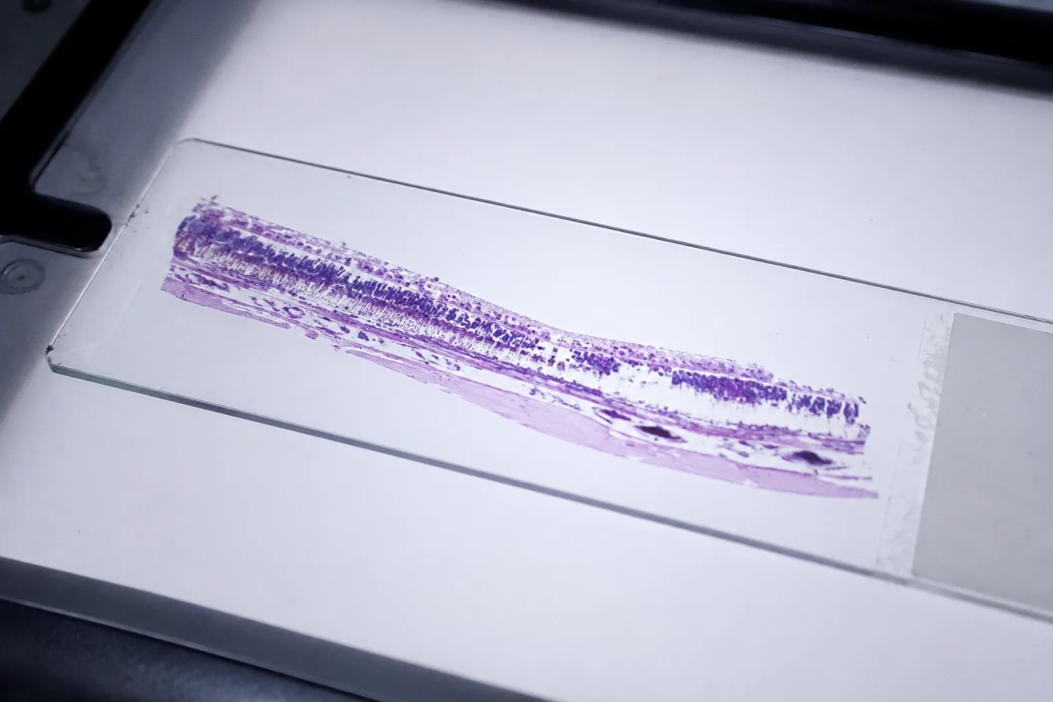
In a histopathology series of 37 donor eyes assembled through the MidAmerica, Iowa, and Utah eye banks, a quietly contradictory picture emerges at the margins of expanding geographic atrophy lesions: rod photoreceptors are dying faster than cones, and they are doing so in territory where the retinal pigment epithelium itself has not yet disappeared. The lesion, as the clinic sees it on fundus autofluorescence, is an island of RPE loss — a dark patch where the orange-red fundus glow has been extinguished. The post-mortem tissue sees something else: a halo of outer segment disintegrity extending outward from that dark patch, where photoreceptor architecture has begun to fray from the distal tips inward, even while the underlying RPE layer still appears, by routine stains, largely intact.
This is the territory where donor tissue earns its value. The clinic captures function, geography, and rate of progression in the living patient; the biobanked eye, properly fixed and sectioned, captures the cellular taxonomy of a lesion in the act of expanding — the dying cells, the misplaced cells, the activated complement caught mid-storm on Bruch's membrane, the choroidal capillaries slipping silently out of perfusion, the microglia gathering at the margin. What follows is a close reading of that taxonomy, traced through the cascade of biological events that converts a patch of stressed RPE into a steadily widening atrophic scar.
Mapping RPE Phenotypic Diversity in AMD Donor Eyes
The first surprise of a well-stained AMD donor eye is that the RPE is not a uniform monolayer collapsing into nothing. It is a cast of characters. In the systematic expansion by Zanzottera and colleagues, sixteen distinct RPE phenotypes have now been catalogued across AMD donor tissue, and that catalogue has continued to grow as additional morphologies — sloughed, bilaminar, dissociated, intraretinal, subducted, and several others — have been described in donor series from collaborating eye banks. Each phenotype represents a different way an RPE cell can fail to remain where it belongs.
The sloughed RPE cell has detached from Bruch's membrane and lies free within the subretinal space, often rounding up as its basal integrin contacts give way. The bilaminar cell has, in effect, duplicated itself into a transient double layer before either cell disappears. The dissociated phenotype describes RPE cells that have lost their tight-junctional neighbors and now sit isolated, their apical processes no longer interlocking with photoreceptor outer segments. Intraretinal RPE refers to cells that have migrated inward, past the outer limiting membrane, into the neural retina — a translocation that is rare in healthy tissue but documented repeatedly in AMD donor series, particularly adjacent to regions of photoreceptor dropout. Subducted RPE cells appear to have slipped downward through a fenestrated or compromised Bruch's membrane into the choriocapillaris.
Donor RPE does not simply vanish — it migrates, duplicates, detaches, and buries itself, and the sixteen phenotypes now on the books are the cartography of those exits.
At the leading edge of an expanding lesion, one phenotype tends to dominate: hypertrophic RPE. In donor tissue examined under multi-channel fluorescence microscopy, these cells contain cytoplasm packed with granules that fluoresce in both far-red and green-red channels — the spectral signature of lipofuscin-derived material combined with internalized outer segment debris that has not been cleared. They are large, multinucleated-appearing, and visibly stressed, exhibiting hallmarks of RPE senescence: enlarged morphology, SA-β-galactosidase activity, and a senescence-associated secretory phenotype that releases cytokines into the local microenvironment. Their position, immediately peripheral to complete RPE loss, makes them the prime suspect in any sequence that asks which RPE cell dies next, and through which door.
Photoreceptor Degeneration Patterns Beyond the Atrophic Margin
The second surprise is geographic: the photoreceptors are not, as a first approximation, simply following the RPE downhill. In clinicopathologic correlations where post-mortem donor eyes have been mapped back to in-life imaging, the area of reduced photoreceptor segment integrity (rPSi) frequently exceeds the area of complete RPE loss — sometimes substantially so. Rod outer segments shorten, fragment, and lose their orderly stacking, while the underlying RPE, in the same eye and at the same eccentricity, still stains positive for cytokeratin and still sits on an apparently continuous Bruch's membrane.
Rod loss is, in nearly every published donor series, greater than cone loss distal from the lesion edge. The reasons are partly structural — rods dominate the parafoveal and perifoveal regions where GA typically begins, and their outer segments are longer and more lipid-rich, placing heavier metabolic demand on the underlying RPE — but the asymmetry persists even where rods and cones coexist. In one carefully quantified clinicopathologic case, the mean distance between the external limiting membrane and the RPE basal lamina at the outer junctional zone was reduced by approximately 50% in affected retina compared with age-matched controls, a compression that reflects the inward collapse of outer segments as the apical RPE-photoreceptor interface fails. Microglial activation, with Iba1-positive cells clustering in the outer plexiform and outer nuclear layers at the lesion margin, sits alongside the photoreceptor story as a parallel effector arm — clearing debris, but also releasing cytokines that may accelerate segment loss.
What the tissue shows, in other words, is photoreceptor degeneration proceeding in two registers. There is the frank loss that follows complete RPE atrophy, and there is a slower, segment-first disintegration that can begin while the RPE is still present. Both registers almost certainly matter for vision, since the second register may explain the paracentral scotomas that patients describe in regions where fundus imaging still appears unremarkable — a clinical observation that, until the donor tissue provided the histologic counterpart, sat awkwardly with any model that treated GA as a clean RPE-first disease.
Complement Activation and Bruch's Membrane Lipid Dynamics
Bruch's membrane, in the geographic atrophy donor eye, is a layered crime scene. Between the RPE basal lamina above and the choriocapillaris endothelium below, the membrane thickens with age and accumulates lipids, advanced glycation end-products, and apolipoprotein B–containing particles that together constitute the precursor matrix of drusen biogenesis. The hypertrophic RPE cells at the lesion margin are not innocent bystanders to this accumulation; their cytoplasm, fluorescent in far-red and green-red channels, indicates ongoing failed outer segment clearance, and their swollen apical surface is itself a source of membranous debris shed basally onto Bruch's membrane.
Alongside the lipid story, the donor tissue captures a parallel immune story. Immunostaining of the leading edge consistently reveals increased deposition of membrane attack complex (MAC) on RPE cells — the terminal complement complex C5b-9 punching channels through the RPE plasma membrane, with the morphology of pore-forming lesions visible at the ultrastructural level. Complement factor H (CFH), the principal soluble regulator of the alternative pathway, is also detected within Bruch's membrane in these regions, its presence a marker that the regulatory system has been engaged, though apparently not effectively enough to halt lesion expansion. Together, these findings support a model in which local complement activation, dysregulated at the RPE–Bruch's interface, contributes directly to RPE cell injury at the lesion margin, while lipid accumulation sets the stage on which that injury is amplified.
The expanding GA margin is not a line of starvation alone — it is a line of immune injury, written into Bruch's membrane as MAC deposition and unresolved CFH engagement.
The choroidal side of the membrane tells a related story. Loss of choriocapillaris fenestrations, endothelial attenuation, and luminal narrowing are documented in donor tissue peripheral to GA lesions, and they raise a question the field has not yet fully resolved: whether choriocapillaris loss strictly precedes, strictly follows, or proceeds in lockstep with RPE loss across clinical phenotypes. The honest answer at present is that the chronology appears to be lesion-dependent, and that no single donor series has yet settled the question — including the choroidal neovascularization counterpart, where vessel invasion, rather than vessel loss, defines the alternative trajectory.
Mechanisms of Cell Death: Apoptosis versus Regulated Necrosis
How, mechanistically, an RPE cell at the lesion margin actually dies is the question the tissue alone cannot fully answer — apoptosis and necrosis leave different footprints, and both footprints have been found. Dunaief and colleagues, using TUNEL labeling on AMD donor tissue, documented a statistically significant increase in TUNEL-positive cells not only in the RPE but also in the inner choroid, the outer nuclear layer, and the inner nuclear layer of donor eyes with both geographic atrophy and exudative AMD. Apoptosis, in other words, is present, and it is not confined to the RPE monolayer.
Yet apoptosis is unlikely to be the whole story. Experimental models of oxidative-stress-induced RPE death, particularly those employing 4-hydroxynonenal (4-HNE) or sodium iodate (NaIO3), demonstrate hallmarks of regulated necrosis — specifically necroptosis — with activation of the receptor-interacting serine/threonine kinases RIPK1 and RIPK3 and the downstream executioner MLKL. These models do not reproduce every feature of human GA, and they should be read as mechanistic probes rather than as direct portraits of the donor lesion, but they have shifted the consensus view: human RPE in geographic atrophy probably dies through more than one pathway, and the mix may differ from patient to patient, from lesion edge to lesion core, and from one AMD subphenotype to another.
| Death pathway | Trigger signature in RPE | Key molecular markers | Evidence in human GA donor tissue |
|---|---|---|---|
| Apoptosis | TUNEL-positive nuclei, chromatin condensation | Caspase-3 cleavage, PARP fragmentation | Increased TUNEL-positive cells in RPE, inner choroid, ONL, INL (Dunaief et al.) |
| Necroptosis | Oxidative membrane injury, organelle swelling | Phospho-RIPK1, phospho-RIPK3, phospho-MLKL | Inferred from 4-HNE and NaIO3 model work; direct donor-tissue evidence still being assembled |
| Ferroptosis | Lipid peroxidation under iron dysregulation | GPX4 suppression, lipid ROS accumulation | Mechanistically plausible given Bruch's lipid load and RPE iron handling; human donor evidence emerging |
Whether ferroptosis is a major contributor in the human post-mortem GA eye remains, at the time of writing, an open question. The complete set of molecular triggers that determines which death pathway an individual RPE cell enters in human donor GA tissue has not yet been enumerated, and donor tissue — paired with spatial transcriptomics and targeted proteomics on serial sections — is the substrate on which that enumeration will be built.
Clinicopathologic Correlation of Lesion Expansion Rates
The single most cited quantitative observation from clinicopathologic GA correlation is also the one most often misread: an RPE loss rate of approximately 2.432 mm² per year, recorded in a single long-term tracked case in which in-life imaging was later matched against post-mortem histology. The number is real, and the case is well documented, but it is a single case. Population-level fundus autofluorescence studies routinely report median GA expansion rates that vary by lesion size, by fellow-eye status, by the presence of subretinal drusenoid deposits, and by whether the lesion is unifocal or multifocal, and these medians should not be replaced in clinical reasoning by any single post-mortem-derived figure.
What the donor tissue does give the clinic, and what no imaging modality can, is the structural counterpart of that expansion. In the same eye that produced the 2.432 mm²/year figure, the histologic section revealed not just a patch of absent RPE but a graded transition through the lesion phenotypes catalogued above — hypertrophic RPE at the margin, dissociated cells trailing inward, sloughed cells and intraretinal cells marking the older core. The rate of expansion, in other words, is the macroscopic footprint of a microscopic sequence, and the sequence is what donor tissue is uniquely positioned to record. Donor retinal spatial transcriptomics, layered onto the histopathology, is beginning to add molecular resolution to that sequence: cell-type-resolved gene expression at the lesion margin, in the peri-lesion halo, and in the apparently uninvolved peripheral retina, all from the same eye.
Expansion rate is the visible echo of a cellular sequence — sixteen RPE phenotypes, a halo of disintegrating outer segments, MAC deposition on a stressed monolayer — written into the chorioretinal landscape one cell at a time.
A Position from the Bench
The temptation, when sixteen RPE phenotypes sit on a slide, is to treat the taxonomy as the answer. It is not. The taxonomy is the vocabulary; the question remains whether the cells of any one donor eye entered those phenotypes through complement injury, oxidative stress, lipid overload, or some combination specific to that individual's lesions and that individual's lifetime of exposures. Donor tissue, precisely sectioned, properly fixed, and matched against the donor's clinical record, can narrow that question for each case — and the field's job, over the next several years, is to bind the histopathology more tightly to the molecular layer, so that the cellular sequence at the lesion margin becomes not just visible but readable in its full biochemical detail.
For now, the donor eye offers something rarer than a number: a sequence. RPE stressed, hypertrophic, fluorescent with undegraded outer segment debris and bearing the molecular signatures of RPE senescence. MAC assembling on its apical surface while CFH gathers within Bruch's membrane without fully regulating. Photoreceptor outer segments shortening beyond the lesion edge while the RPE beneath them still sits, for the moment, in place. Microglia clustering at the outer retina, clearing debris and amplifying signals. The choriocapillaris thinning below, the outer nuclear layer thinning above. The optic nerve head, notably, untouched — GA is a chorioretinal disease, and the donor tissue reminds us that the pathology keeps to its territory. Read together, that sequence is the closest approximation the field has to a real-time portrait of geographic atrophy in the act of expanding, and the biobanked eye, section by section, is the camera that has been recording it.