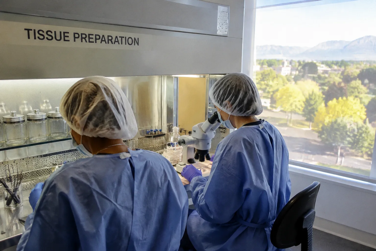
A standard assessment may record dead or compromised cells, while the clinical graft later achieves the functional result expected of preserved tissue.
The distinction matters because the number on a viability report is not a pure readout of donor quality. It is the result of several overlapping histories: the condition of the tissue at procurement, the time spent in preservation medium, the response of the endothelium to cold stress, and the method and timing of assessment.
Across mixed cohorts of donor corneas held in cold-storage media, a mean baseline dead endothelial cell rate of approximately 4.9% has been documented. That figure should not be treated automatically as evidence of donor pathology or as a direct forecast of graft failure. It is a measurement made after a preservation process that changes the tissue at molecular and cellular levels.
The right interpretation is not that the 4.9% is meaningless. It is that the number needs a biological context.
Molecular Signatures of Hypothermic Stress in Endothelial Tissue
The endothelial monolayer on the posterior surface of the cornea is a particularly sensitive part of the graft. These cells maintain corneal deturgescence and are expected to remain functional after procurement, storage, transport, preparation, and implantation. Once the tissue is removed from the donor eye and placed in a preservation medium at approximately 2–8 °C, the endothelium is no longer operating under its native conditions.
Cold storage suppresses many metabolic processes, but it does not turn cellular biology off. The cells continue to respond to their environment. Their energy use, membrane behavior, protein handling, adhesion, and stress responses are altered by the temperature and by the absence of the normal aqueous and vascular context.
A paired-comparison study of donor corneal endothelium after 10 days of cold storage found 1,264 upregulated genes and 2,058 downregulated genes when the stored tissue was compared with immediately preserved controls. The scale of that difference is important. It shows that hypothermic storage is not a neutral pause between procurement and transplantation. It produces a distinct biological state.
The affected pathways were also coherent rather than random. Transcripts associated with inflammation changed. Components of the unfolded protein response changed, consistent with cellular stress and altered protein-folding conditions. Transcripts connected with cell-cell junctions also shifted. Those junctions are central to the organization of the endothelial monolayer and to its barrier function.
This evidence supports a careful conclusion: cold-stored donor endothelium carries a molecular signature of hypothermic stress. It does not, by itself, establish that every transcript change represents permanent structural injury, nor does it establish that the changes resolve completely after rewarming. The study documents the state of the tissue after storage; it does not provide a complete time course of molecular recovery.
Cold storage does not freeze endothelial biology in place. It leaves a measurable stress signature on the tissue.
That qualification is especially important for research use. A laboratory using cold-stored corneas to study native endothelial biology may mistake a preservation response for a disease-associated or pharmacologically induced signal. The problem is not that the transcriptomic data are unreliable. The problem is that they answer a narrower question than researchers sometimes assume.
They describe endothelium that has experienced procurement and hypothermic storage. That can be exactly the material needed for a study of preservation injury, post-storage recovery, or graft preparation. It is a less clean model of untreated, in-situ corneal endothelium.
For work involving wound healing, cell junctions, inflammatory signaling, or drug response, the storage history therefore belongs in the experimental interpretation. Two tissue samples can be matched by donor and anatomical region yet differ in how strongly the preservation process has shaped their molecular profile. The storage medium, duration, handling sequence, and interval before assessment are not administrative details; they are part of the biological model.
Quantifying the 4.9% Baseline: Histological Reality of Dead Cell Rates
The molecular results explain why a stored cornea should not be treated as biologically unchanged. Histology shows how that alteration appears at the cellular level.
Histological analysis of 28 donor corneas preserved in cold-storage media reported a mean dead endothelial cell rate of 4.9%, with a standard deviation of 3.3% and a range from 0.6% to 10.5%. The distribution is more informative than the mean alone. Some corneas emerged with very little detectable cell death, while others showed a substantially higher fraction of dead or visibly compromised endothelial cells.
That spread is a reminder that preservation does not affect every donor cornea identically. Tissue condition at procurement, donor-related variables, handling, and the specific storage history can all contribute to the result. A single percentage on a report is therefore a snapshot of one tissue after one preservation trajectory.
Viability stains such as trypan blue are commonly used to identify cells with compromised membrane integrity. In a histological or laboratory assessment, stained cells may be counted as nonviable according to the assay’s criteria. But the result still depends on the assay conditions and on when the tissue is examined. A stain records what is detectable at that moment; it does not necessarily predict the later physical distribution of those cells during every stage of graft preparation.
This is where the phrase “baseline dead cell rate” can become misleading. Baseline does not mean native. It means the reference condition established by the particular preservation and assessment protocol. The approximately 4.9% value belongs to a cold-storage cohort and should not be transported uncritically to fresh, unstored tissue or to every preservation medium.
The standard deviation is also not a footnote. At 3.3 percentage points, it is large relative to the mean. The reported range shows that the cohort contains materially different tissue responses. A cornea at the lower end of the range and one near the upper end may share the same storage category while presenting very different histological appearances.
That does not make the measurement useless. It makes it contextual.
A laboratory or eye bank can use the rate to monitor a preservation process, compare protocols, or identify tissue that requires additional review. What it should not do is treat the number as a standalone diagnosis of donor quality. The question is whether the measured cell loss is compatible with the intended clinical use, the rest of the endothelial assessment, and the evidence supporting the storage protocol.
What the number can and cannot tell you
A cold-storage viability result can support several reasonable observations:
- It can show that a measurable fraction of cells has lost membrane integrity by the time of assessment.
- It can help distinguish tissue with a relatively low or high burden of detected cell death within the same protocol.
- It can reveal variability between donor corneas processed under nominally similar conditions.
- It can identify that the tissue has been biologically affected by storage, even when the graft remains suitable for its intended use.
The same result cannot, on its own, establish:
- that all detected dead cells will remain attached to the graft;
- that the measured rate represents irreversible failure of the surviving monolayer;
- that the tissue will produce a poor clinical outcome;
- or that a molecular stress signal has fully resolved after warming.
Those limits are not an argument against viability testing. They are an argument for reading the test as one layer of evidence rather than as the entire graft assessment.
Reconciling Laboratory Artifacts with Clinical Graft Success
The difference between laboratory appearance and clinical performance becomes most useful when it is stated plainly. Bench measurements and graft outcomes are not interchangeable endpoints.
The National Eye Institute–funded Cornea Preservation Time Study examined whether longer cold preservation, within the studied clinical framework, translated into worse keratoplasty performance. Its reported finding was that donor corneas stored in cold media for up to 11 days did not show a negative effect on keratoplasty graft success rates.
That is a clinically meaningful result, but it needs to be kept within its evidentiary boundaries. The CPTS finding supports a conclusion about graft success rates. It does not separately establish that cold storage has no effect on every component of postoperative clarity, endothelial density, rejection, or other individual outcomes. Nor does it erase the molecular and histological changes documented in laboratory studies.
The more defensible reading is narrower: cold storage can produce measurable cellular and transcriptomic stress without that stress necessarily translating into a reduction in the studied graft-success outcome within the validated storage window.
The two datasets are not contradicting each other. They are looking at different points in the biological chain.
- Transcriptomic analysis asks how the cells respond at the level of gene expression.
- Histology asks how many cells appear dead or structurally compromised under the selected assessment conditions.
- Clinical follow-up asks whether the transplanted graft succeeds as a surgical treatment.
A graft can carry evidence of storage stress and still retain enough functional endothelium to support the outcome measured in a clinical study. That possibility is not a claim that every stressed cell recovers, or that every donor cornea behaves the same way. It is a reminder that endothelial reserve and clinical success cannot be inferred from one laboratory variable in isolation.
The same caution applies to tight-junction transcripts. Their movement during cold storage indicates that junction-related biology has changed. It does not prove that the change is permanent, and it does not prove that it completely resolves on rewarming. The available evidence supports describing a stress-associated alteration, not assigning it a guaranteed recovery pathway.
For tissue programs, this distinction changes how an apparently unfavorable number should be handled. A result around 4–5% detected dead cells may warrant review, especially when it is accompanied by other concerns. But it is not automatically equivalent to an unacceptable graft. The relevant interpretation depends on the full endothelial assessment, the storage duration, the protocol used, and whether the tissue remains within the conditions supported by clinical evidence.
The CPTS result provides operational confidence about graft success within its studied context. It should not be expanded into a blanket claim that every laboratory consequence of cold storage is clinically irrelevant.
Comparative Performance of Modern Preservation Media at 14 Days
The clinically studied 11-day window and laboratory investigations of 14-day storage answer different questions. The former is tied to graft-success evidence. The latter examines how donor tissue behaves under an extended preservation interval and how different media compare at the end of that period.
A comparison of donor corneas preserved for 14 days reported the following endothelial measurements:
| Parameter at day 14 | Kerasave | Optisol-GS |
|---|---|---|
| Endothelial cell density | 2,312 ± 98 cells/mm² | 2,335 ± 128 cells/mm² |
| Central endothelial cell mortality | 0.54 ± 0.40% | 0.14 ± 0.14% |
| Variation in cell density | Narrower spread | Wider spread |
The two media produced similar mean endothelial cell densities in that comparison. Optisol-GS showed the lower reported central mortality, while Kerasave showed the narrower spread in cell-density measurements. Those are useful comparative findings, but they should not be turned into a universal ranking of the media or into a clinical superiority claim.
A 14-day bench result is not the same as a 14-day clinical outcome. The study demonstrates how the tissues measured in that experiment looked at the end of storage. It does not establish that the same differences will produce different graft-success rates, nor does it show that one formulation is responsible for every favorable or unfavorable feature of the result.
The low mortality values at day 14 also should not be directly compared with the approximately 4.9% baseline rate as if the difference had one proven explanation. The cohorts, protocols, assessment methods, tissue handling, and definitions may differ. A lower reported mortality percentage may reflect the characteristics of the study material and method as much as the formulation itself. The data do not establish that a particular medium actively caused the entire difference through membrane stabilization or metabolic recovery.
That does not make the comparison unhelpful. It makes the comparison more precise. At day 14, the reported measurements suggest that both media were capable of preserving the examined donor corneas with relatively low central mortality and maintained cell density under the study conditions. Kerasave’s tighter density distribution and Optisol-GS’s lower central mortality are separate observations; one should not be used to imply an overall winner without a defined endpoint.
For eye banks considering a change in medium or evaluating extended storage, several practical questions follow:
1. Are the assessment methods comparable? A mortality percentage obtained by one staining or counting method may not map directly onto a percentage generated by another.
2. Is the endpoint laboratory or clinical? Endothelial density and central mortality at day 14 provide bench information. They do not substitute for prospective graft-outcome data.
3. How wide is the specimen-to-specimen variation? A mean can conceal a broad distribution, particularly when the sample is small or the tissue has heterogeneous starting conditions.
4. Does the protocol match the intended use? A research result under extended storage should not automatically redefine routine clinical acceptance criteria.
5. What is being compared: the medium or the entire workflow? Temperature control, transport, handling, preparation, and timing of assessment can influence the final measurement alongside the preservation formulation.
The clinically supported 11-day window therefore remains distinct from the 14-day comparison. The latter can inform preservation research and protocol development. It does not, by itself, expand the clinical evidence base.
A low day-14 mortality percentage is a useful laboratory observation, not a substitute for outcome evidence.
The Mechanism of Post-Storage Endothelial Cell Drop-off
One of the more revealing observations in this area comes from laboratory incubation. When cold-stored donor corneas with detectable dead cells are transferred into culture medium at warmer incubation temperatures, some of the dead cells detach from the tissue.
That observation helps explain why a histological count and a later view of the endothelial surface may not be identical. It also provides a plausible mechanism for change between assessment stages. But the setting matters: the documented observation concerns incubation in culture medium. It does not establish that the same process occurs at the same rate during clinical rewarming, graft preparation, or implantation.
This distinction is essential. It is tempting to describe the dead cells as being washed away before the graft reaches aqueous humor, but the available evidence does not support that clinical sequence. The laboratory result shows detachment during incubation. It does not demonstrate that rewarming and rehydration in the clinical workflow remove a defined fraction of cells before exposure to the recipient’s anterior chamber.
The observation also should not be attributed automatically to trypan-blue-positive cells as a specific clinical population. Trypan blue can identify compromised membrane integrity under assay conditions, but the evidence described here does not establish that every cell classified in that way is the same cell later observed to detach, or that the staining result predicts detachment in a recipient eye.
What can be said with confidence is more limited and more useful: post-storage incubation can alter the visible or measurable cellular burden, and timing can therefore influence the result of an endothelial assessment.
That has implications for interpretation of specular microscopy donor cornea viability data. An image taken immediately after removal from cold storage may capture tissue before the laboratory detachment process has occurred. A later assessment after incubation may show a different surface population. The difference does not necessarily mean that one measurement is wrong. It may mean that the tissue has changed between measurements.
The practical problem is that clinical workflows and research workflows do not always reproduce one another. A research protocol may specify a defined warming period, culture medium, incubation temperature, and imaging interval. A tissue bank or surgical team may assess the cornea at another point in the handling sequence. Unless those conditions are recorded, two apparently similar viability percentages can describe different biological states.
The mechanism behind the detachment remains incompletely characterized in the evidence considered here. Disruption of cell-cell adhesion is a plausible area for further study, particularly because junction-related transcripts also change during hypothermic storage. But a shared association is not proof of a causal chain. The transcriptomic finding does not demonstrate that it causes cell drop-off, and the culture observation does not demonstrate that the junctional changes are the sole or primary trigger.
Other explanations may also need to be considered in future work: altered membrane integrity, mechanical sensitivity during handling, changes in the extracellular environment, and differences in how dead cells remain attached to the Descemet membrane. The important point for present interpretation is not to choose one mechanism prematurely. It is to acknowledge that the measured cell burden can change after storage and that the timing of measurement is part of the result.
What This Means for Donor Corneal Assessment
For eye banks and tissue coordinators, a reported dead-cell percentage should be treated as a process-sensitive measurement. The first question is not simply whether the number is above or below an idealized threshold. It is how the number was generated and what else the assessment shows.
A result around the reported 4.9% mean may be compatible with cold-storage artifact, particularly when interpreted alongside the documented distribution and the broader endothelial examination. That does not make a higher value irrelevant. It means that the value should not be isolated from storage duration, donor tissue condition, cell density, morphology, and the intended clinical application.
A useful assessment record should preserve the context that allows the result to be interpreted later:
- the storage medium and temperature range;
- the duration of hypothermic storage;
- the time between removal from storage and imaging or staining;
- the viability assay and its criteria;
- the method used to estimate cell density and mortality;
- and any handling or incubation step performed before the final assessment.
These details become particularly important when a cornea is near the edge of an acceptance range or when different laboratories are comparing results. Without them, an apparent difference in mortality may reflect timing or methodology rather than a true difference in tissue condition.
For surgeons, the clinical evidence provides a counterweight to an overly literal reading of the bench report. The CPTS finding supports the conclusion that cold storage for up to 11 days, within the studied conditions, did not negatively affect keratoplasty graft success rates. That is a reason to avoid treating every storage-associated abnormality as a direct prediction of failure.
It is not a reason to ignore the endothelial report. Clinical success data describe a population and an outcome. They do not guarantee that every individual graft with the same reported percentage will behave identically. The report remains part of the decision; it is simply not the whole decision.
For researchers, the central caveat is even sharper. Cold-stored tissue is not native tissue. It is tissue that has passed through a defined environmental perturbation and carries measurable molecular and histological consequences of that passage. If the research question concerns hypothermic stress, preservation injury, or post-storage behavior, that history is the subject. If the question concerns untreated corneal biology, the history is a confounder that must be acknowledged.
The same applies to experiments involving tight junctions, inflammatory pathways, or cellular metabolism. A shifted transcript may be biologically important without being a marker of permanent damage. A stained cell may be nonviable under the assay without being evidence that the entire endothelial monolayer is failing. A cell that detaches during culture may help explain a changing count without proving what happens during clinical implantation.
The Limits of the Current Interpretation
The evidence supports a clear but bounded position.
Hypothermic storage changes donor corneal endothelium. The transcriptome shifts after prolonged cold storage, including pathways associated with stress, inflammation, protein handling, and cell-cell junctions. Histological assessment of cold-stored tissue has documented a mean dead cell rate of approximately 4.9%, with substantial variation between donor corneas. In culture, some dead cells detach during warmer incubation. In clinical data, storage in cold media for up to 11 days did not negatively affect the studied keratoplasty graft-success rates. Separate 14-day comparisons have reported preserved cell density and low central mortality under particular laboratory conditions.
Those findings can coexist without requiring a stronger claim than the data support.
They do not prove that every transcriptomic change reverses after rewarming. They do not prove that clinical rewarming removes a predictable fraction of dead cells before aqueous-humor exposure. They do not show that trypan-blue-positive cells are the specific population responsible for any clinical shedding process. They do not establish that the CPTS finding covers every postoperative endpoint separately. And they do not establish why mortality values in one day-14 comparison were lower than the baseline dead-cell rate reported across mixed cold-storage cohorts.
The remaining unknowns are therefore concrete. The molecular trigger of post-storage cell detachment is not fully mapped. The relationship between storage duration, donor characteristics, medium composition, and post-storage behavior requires further study. The extent to which different assessment protocols measure the same biological state is also not settled.
That uncertainty should not be mistaken for failure of the preservation system. It is a reason to use more disciplined language around the data. A cold-stored cornea can show real cellular injury and still produce a successful graft. A viability stain can identify a real laboratory abnormality without functioning as a complete clinical forecast. The number is not false; it is incomplete.
The most defensible position for procurement programs is to keep both sides of that statement in view. Donor corneal endothelial cell preservation artifacts are genuine and measurable. They affect molecular readouts, histology, and the interpretation of specular microscopy donor cornea viability. At the same time, the documented culture findings and graft-success data do not justify treating every storage-associated cell-loss signal as evidence of clinical failure.
The report is a snapshot of tissue after preservation. The graft outcome is the result of what remains functional across the entire surgical process. Confusing those two measurements is how a cold-storage artifact becomes a false positive for graft risk.