Regenerative Therapeutics
Synthetic corneal hydrogels: cellular integration dynamics
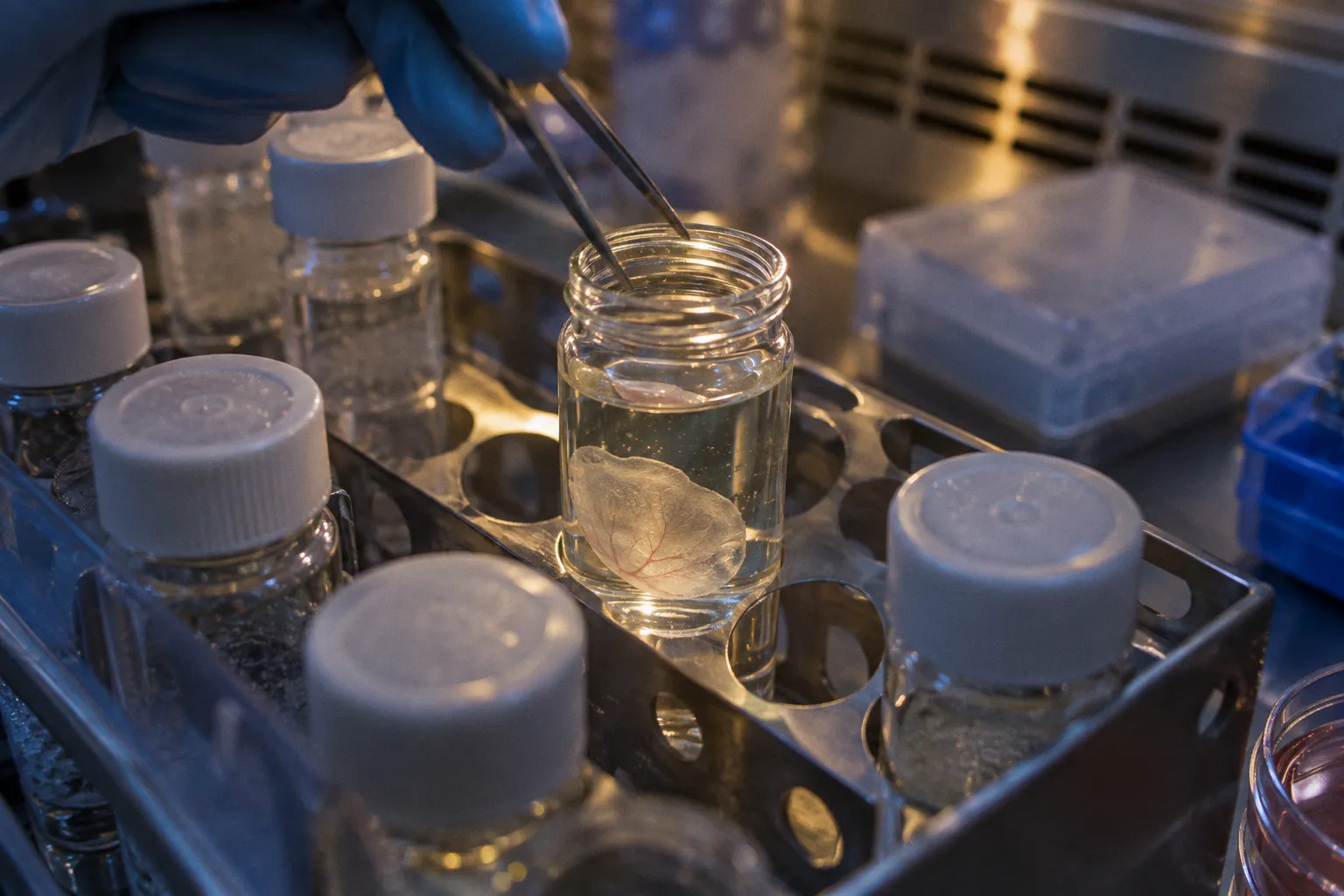
A synthetic corneal hydrogel is only useful if it can do more than fill a defect. The implant must remain optically clear, stay…
Bridging ocular biobanking logistics and vision research.
A synthetic corneal hydrogel is only useful if it can do more than fill a defect. The implant must remain optically clear, stay…
Ocular single-cell RNA-seq donor variability is not a single nuisance variable. It is a stacked variance problem. Donor genetics…
Every corneal surgeon has a quiet superstition. Mine used to be about enucleation time — the faster you get the tissue out of the…
A donor-derived retinal pigment epithelium culture can look structurally intact while already failing at one of its most…
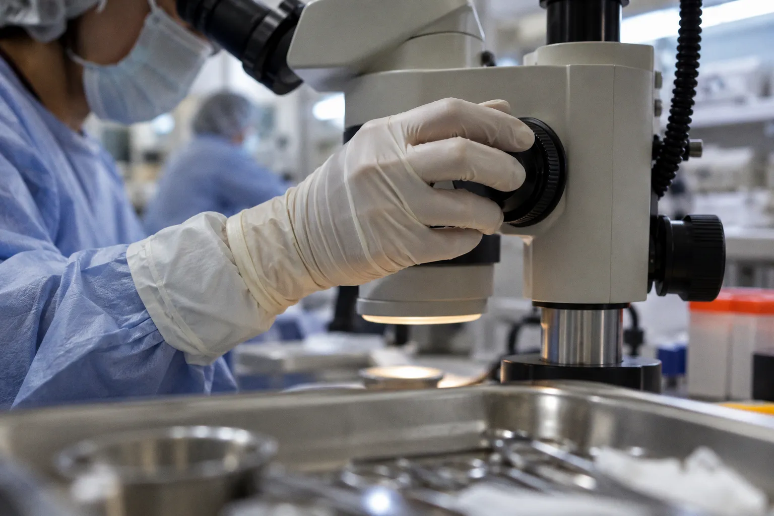
Donor eye procurement operates under two different performance models. Clinical transplantation prioritizes tissue that can meet…
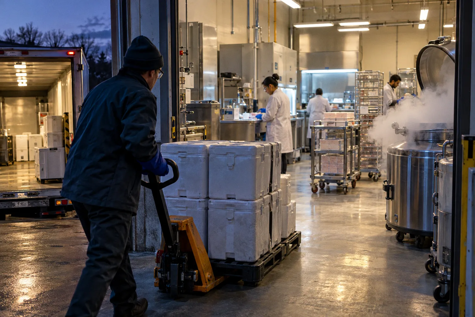
The numbers are humbling. In translational ophthalmology, the scaffold often gives way before the cells have had time to…
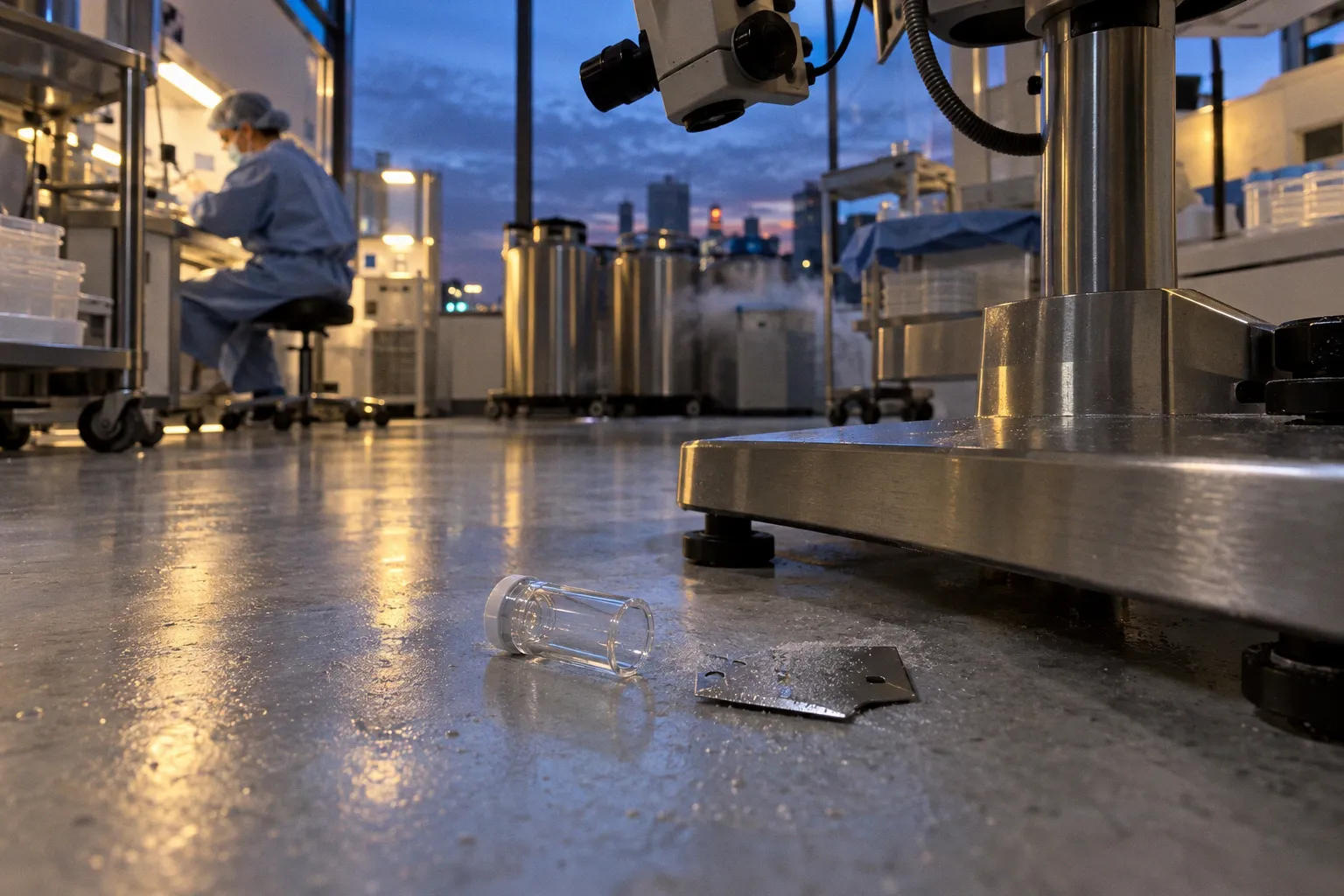
Mitochondrial DNA variants in donor retinal tissue are not a secondary annotation layer. They are part of the tissue-quality…
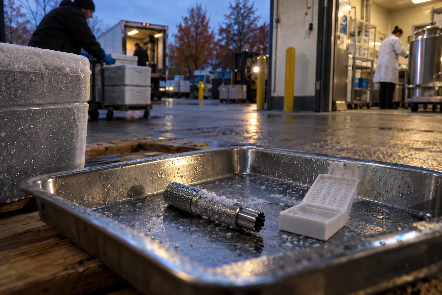
The accepted clinical shorthand is familiar: glaucoma raises intraocular pressure because aqueous humor cannot leave the eye…
In human donor retina, the first casualty of ischemia is not the photoreceptor but the nerve fiber layer, where vacuolation…
Scaffold-free retinal organoid transplantation removes one problem and exposes another. Without a synthetic matrix, there is no…
The 52,705 differentially methylated sites identified in age-related cataract anterior lens capsules did not arrive through…
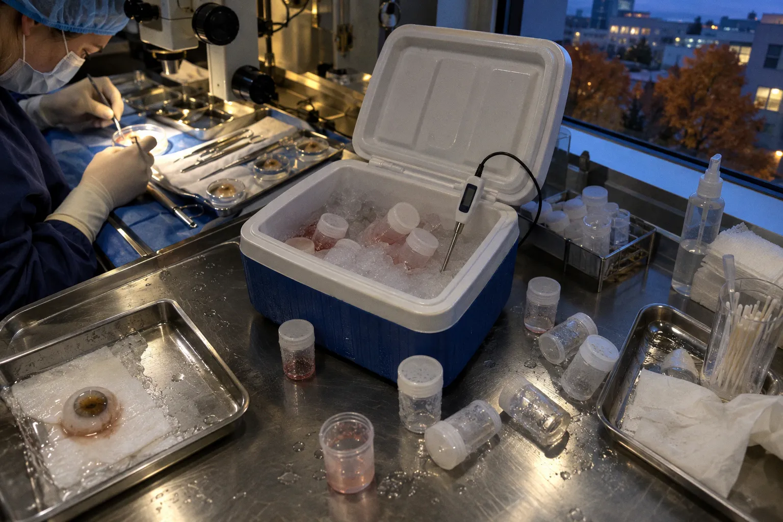
By convention, donor corneal endothelium is treated as tissue that tolerates hypothermic transport with the calm of a specimen in…
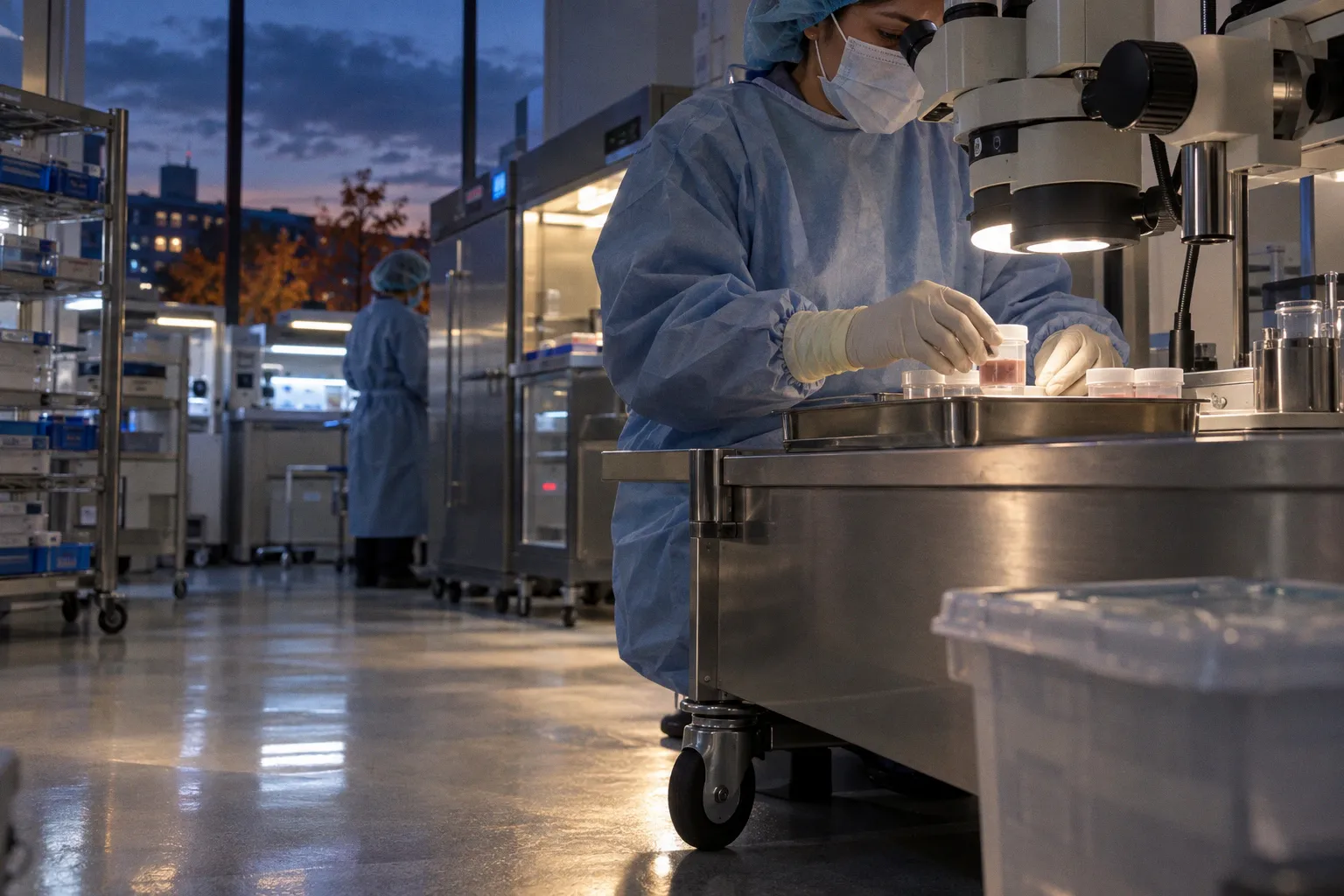
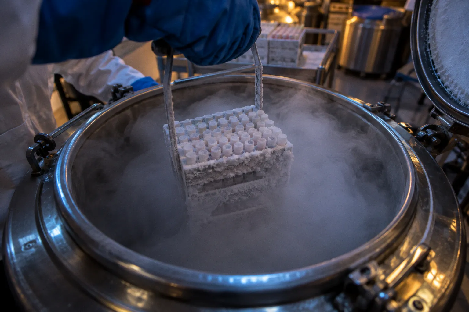
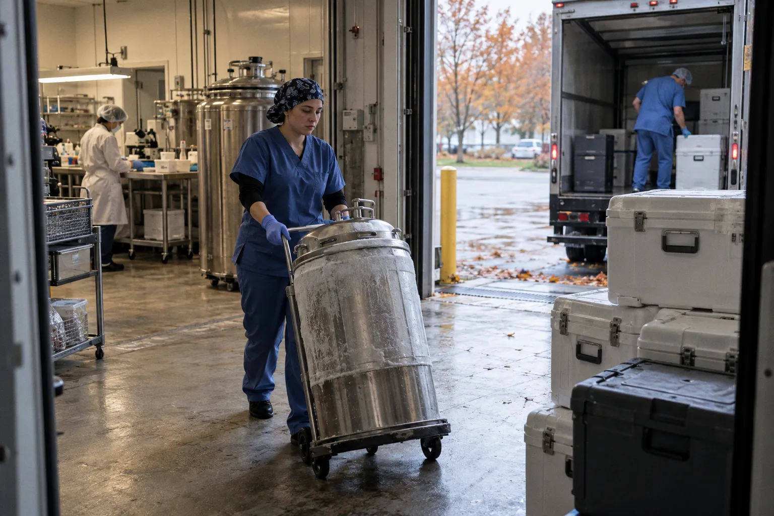
The donor eye tissue cold chain is governed by a narrow operational window, not by a single storage temperature. Hypothermic…
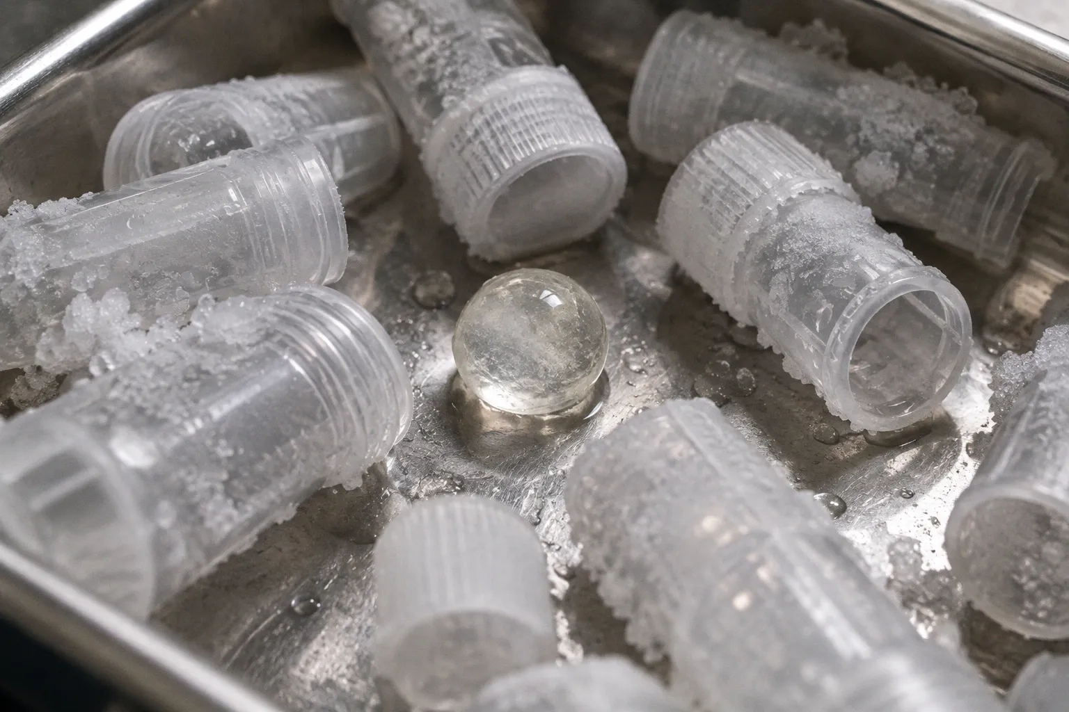
A corneal surgeon waiting on a vesicle batch and a process development engineer staring at a single T-175 flask have more in…
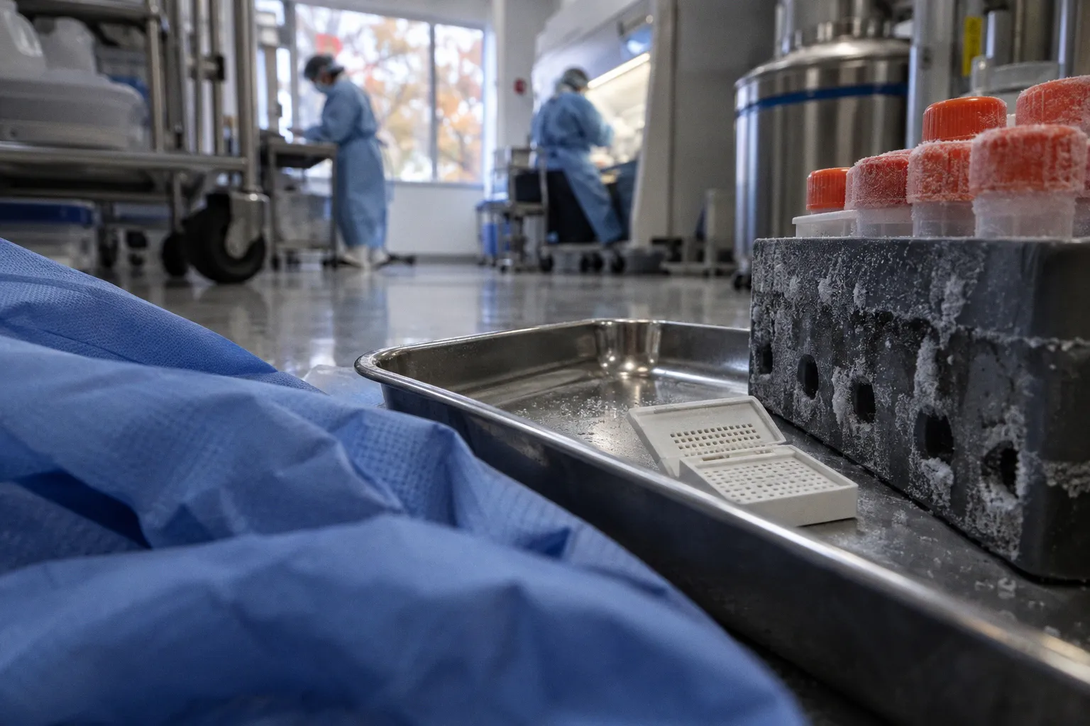
Post-mortem retinal protein degradation rates are not governed by elapsed time alone. The controlling variable is the interaction…
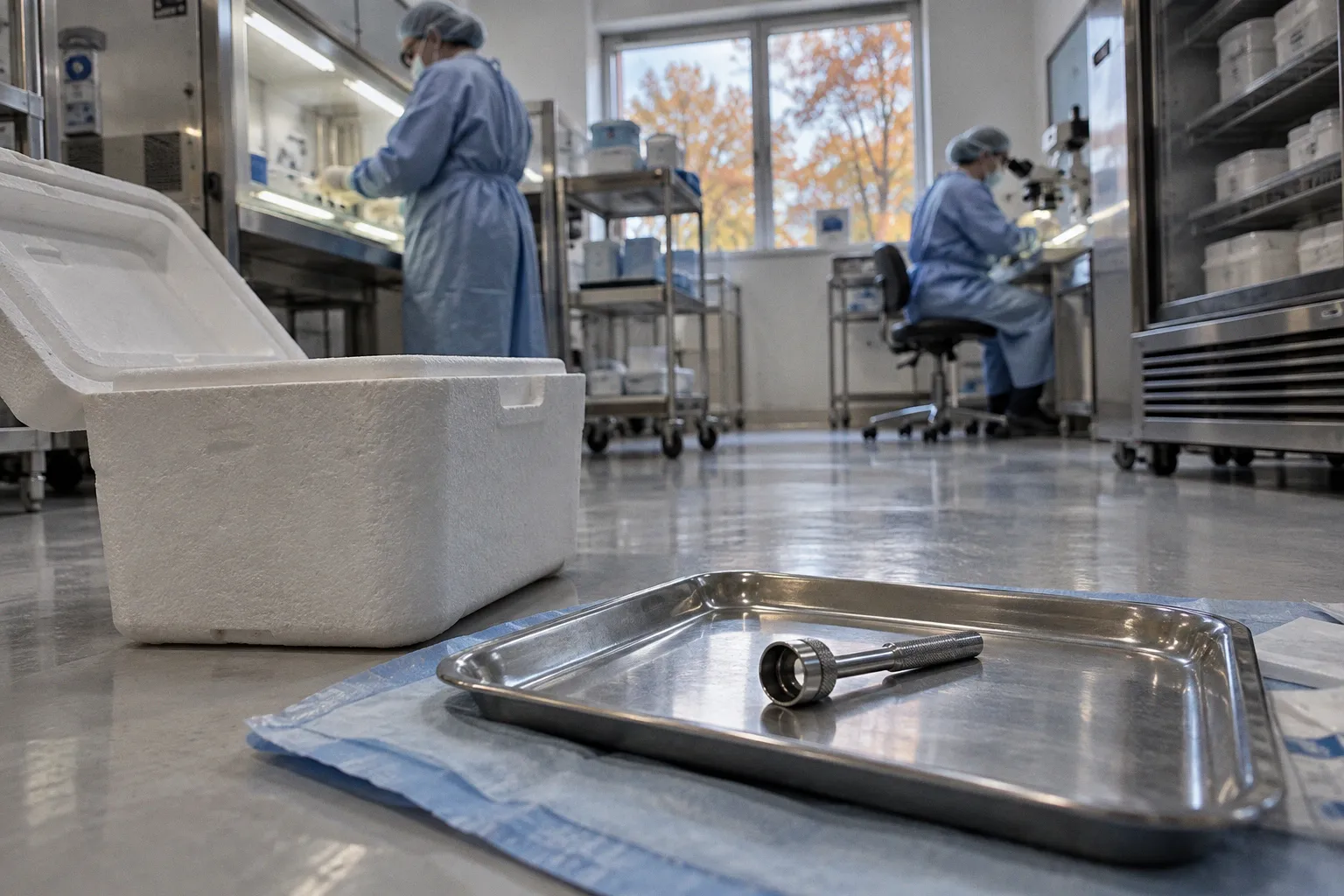
The accepted clinical shorthand is deceptively tidy: cold storage is simple and organ culture is elaborate. Therefore, the first…
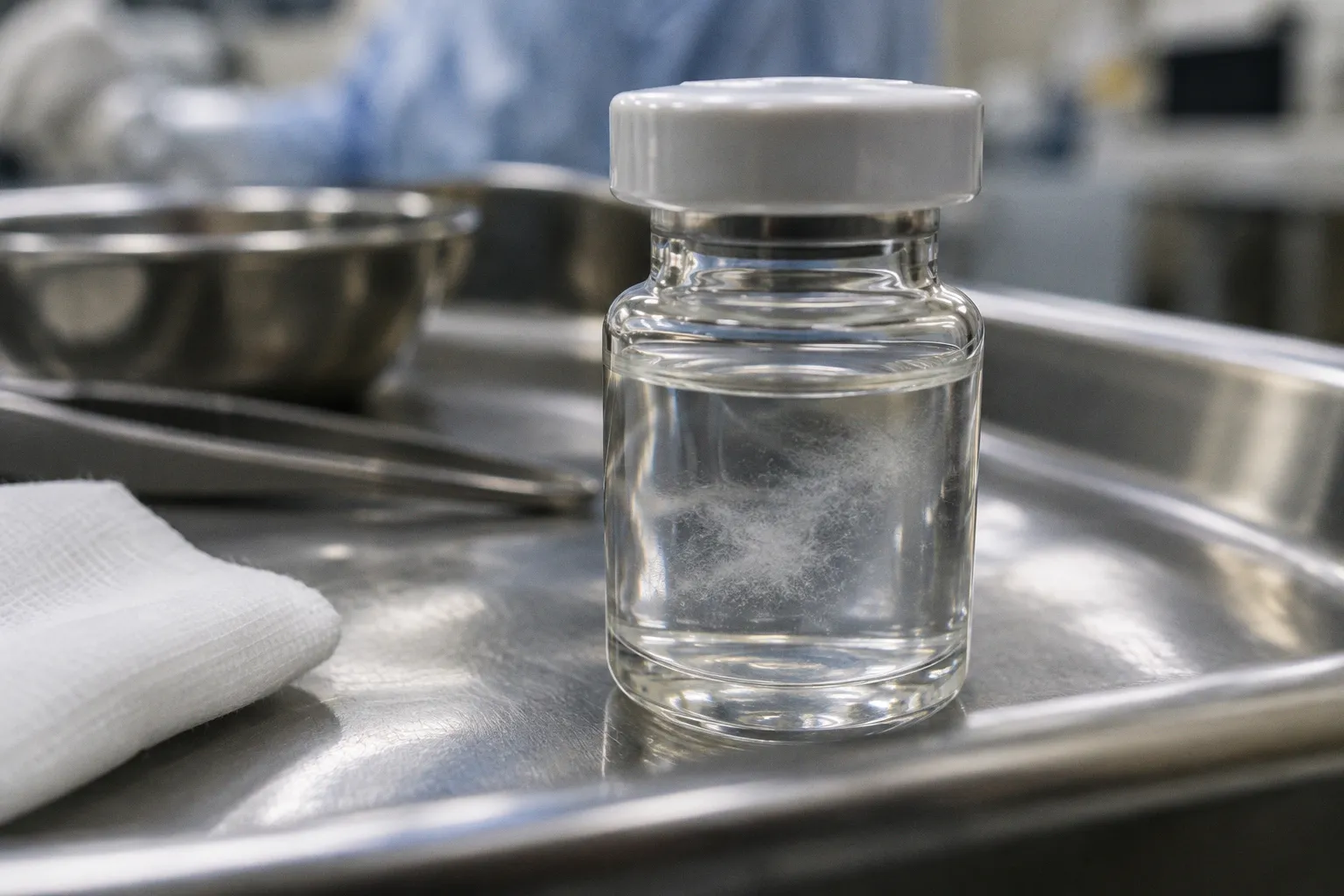
More than one in ten cell cultures worldwide have been reported to carry Mycoplasma contamination that no inverted microscope can…
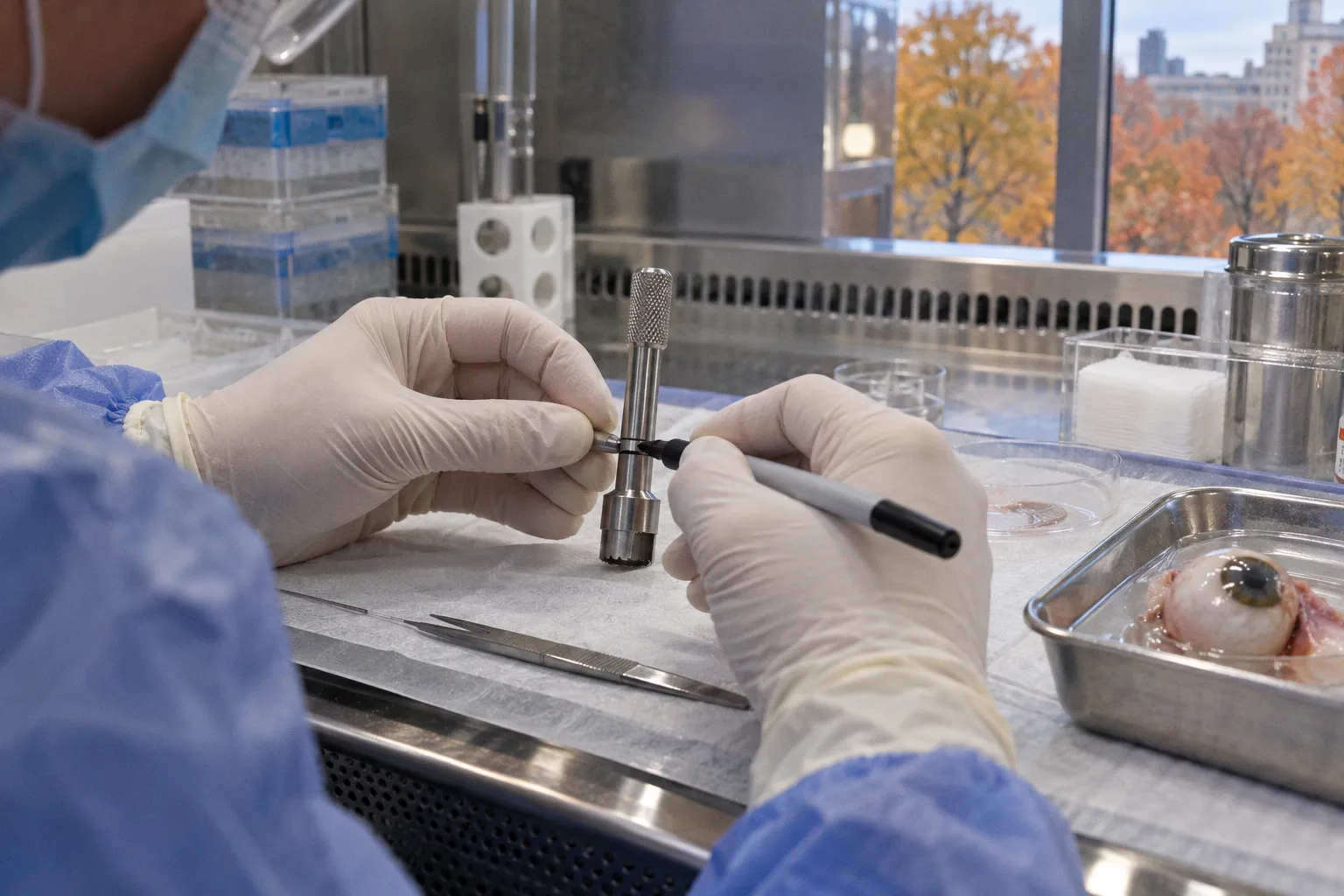
For ocular tissue chromatin accessibility post-mortem stability is not an abstract quality attribute. It is a time-dependent…
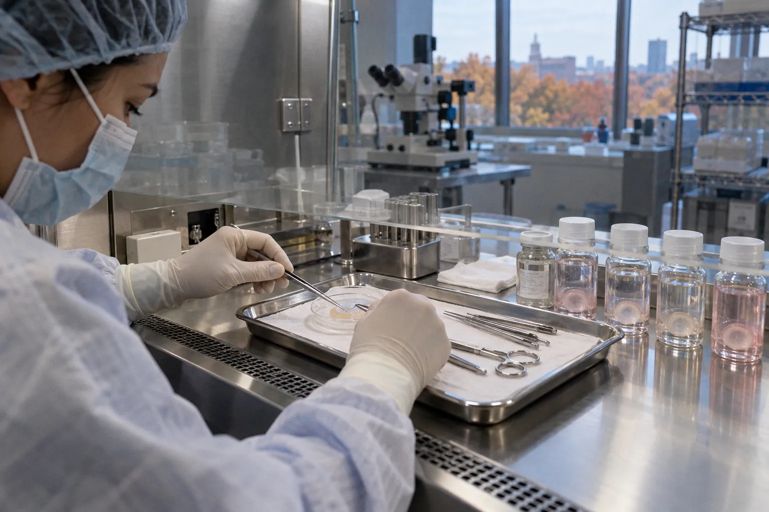
The textbook narrative is reassuring: donor corneas are harvested, cooled, and shipped within a tightly regulated window, and the…
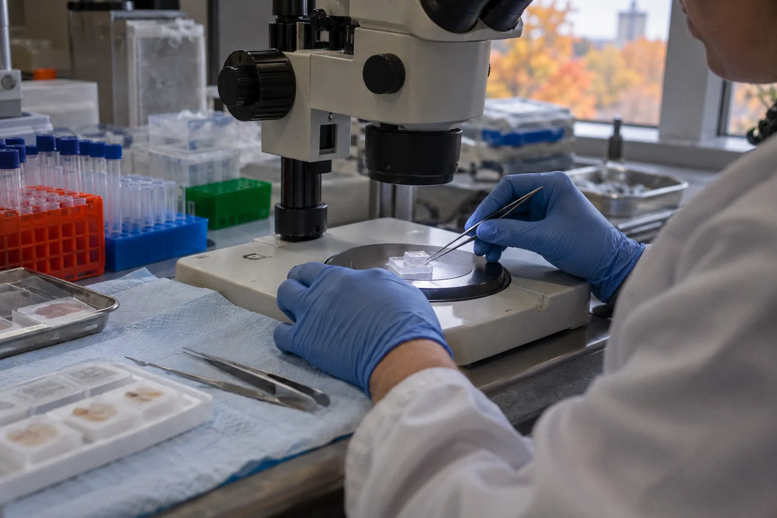
A geographic atrophy lesion is not molecularly uniform, even when it appears sharply outlined in a donor macula section. The…
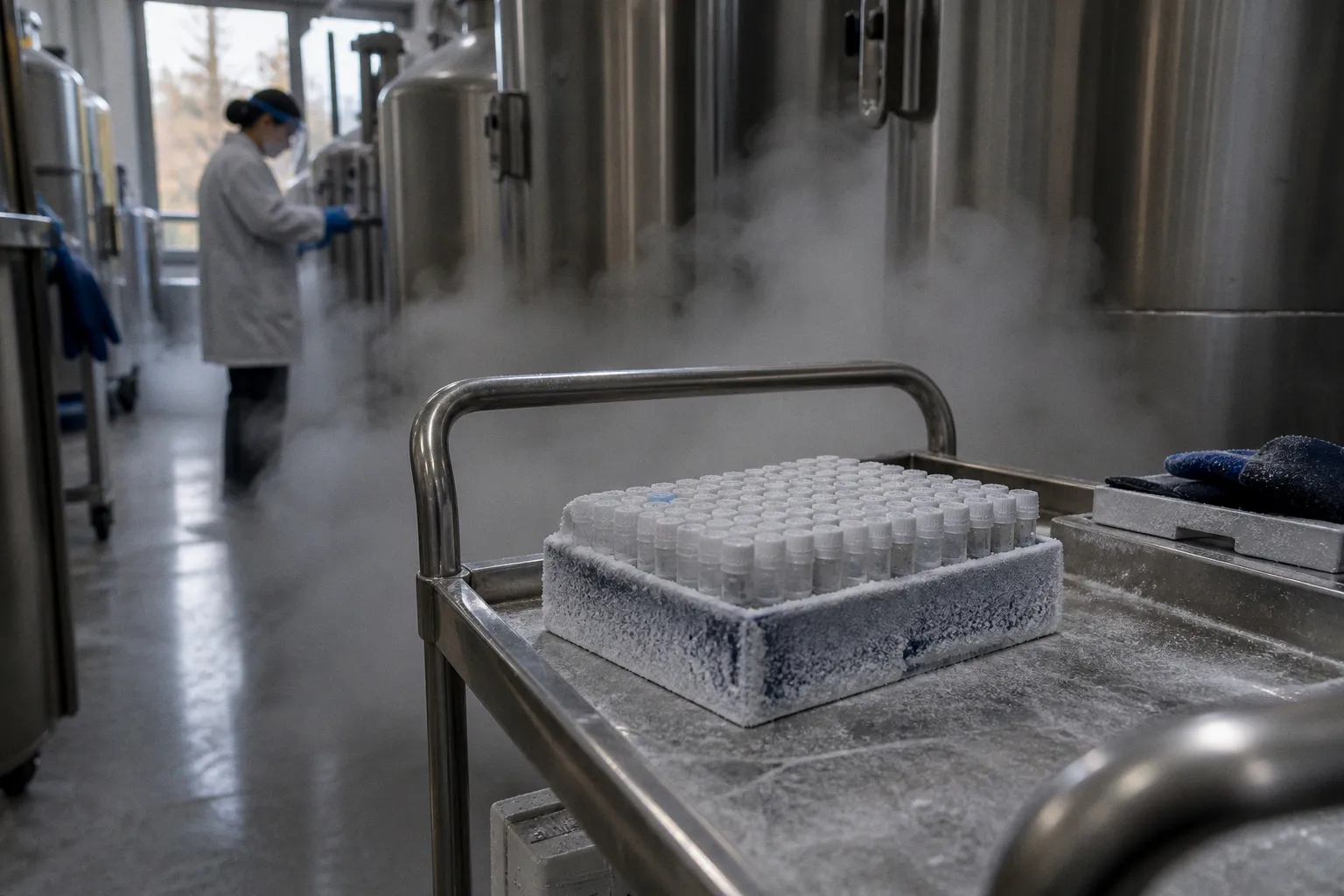
Donor variability is not the same thing as a batch effect. Conflating the two is one of the central failure modes in retinal…
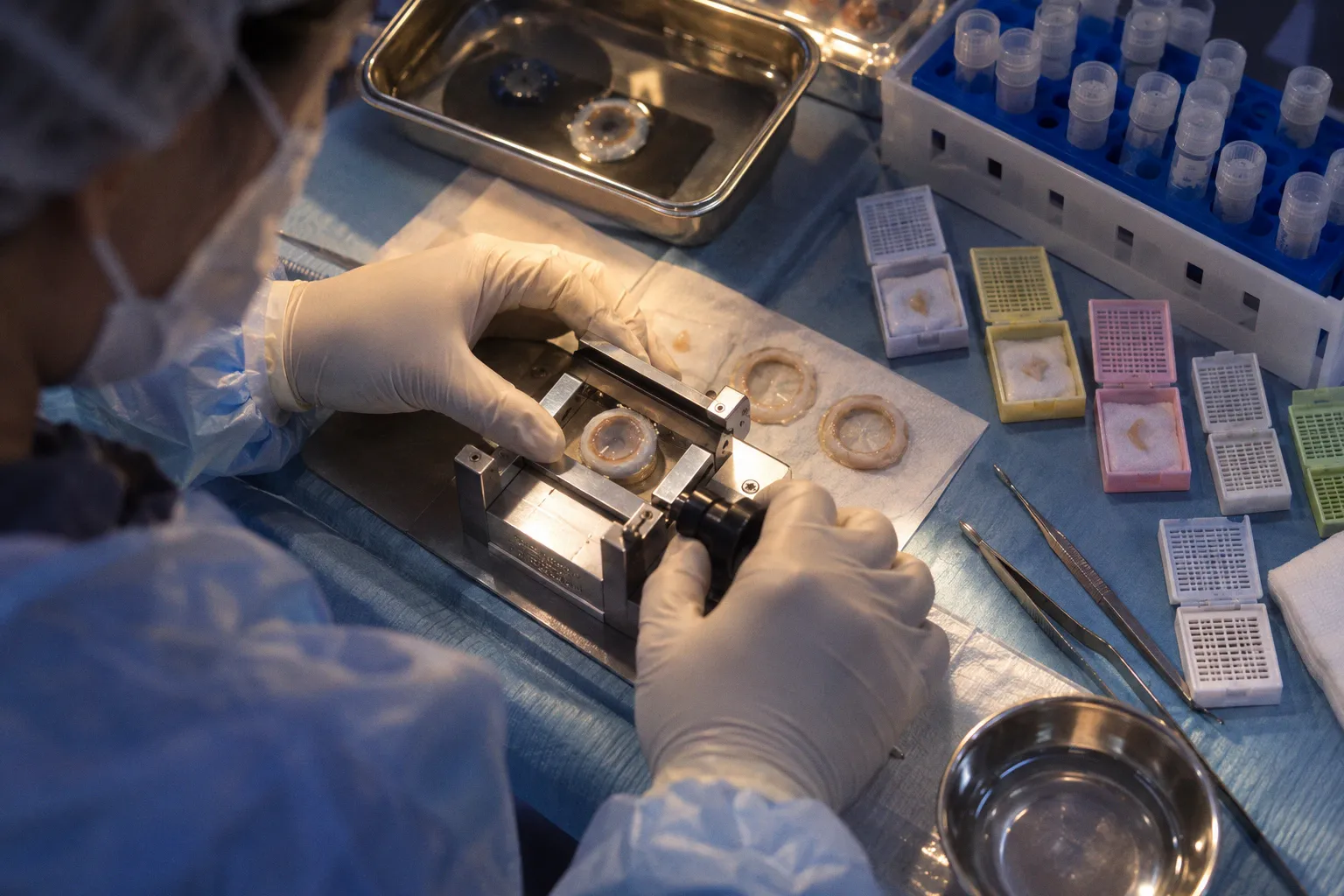
The accepted clinical shorthand is simple: limbal epithelial stem cells reside at the corneal periphery, and damaged limbus means…

AAV ocular gene delivery can look convincing in a rodent retina and still lose its clinical relevance when the vector reaches…
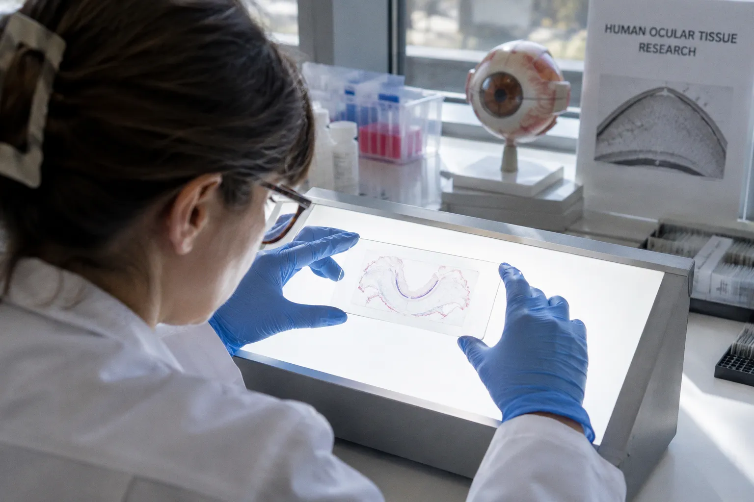
Bulk RNA-seq and single-cell RNA-seq do not measure the same biological object. Bulk sequencing produces an averaged expression…
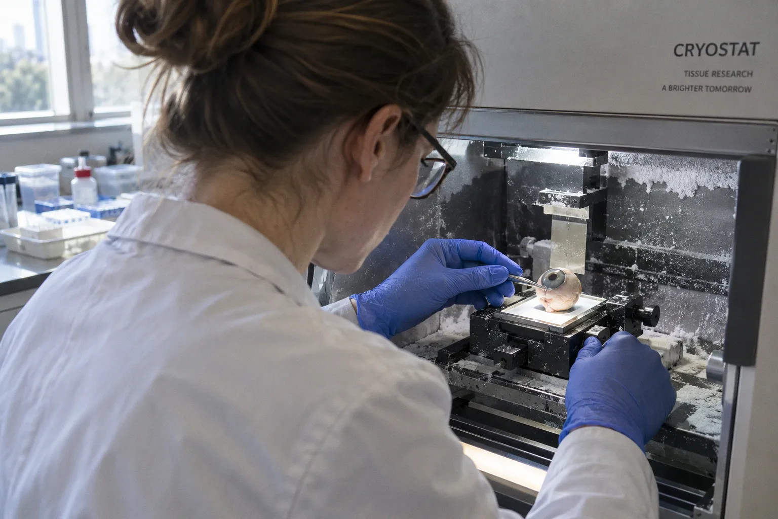
The first structural failure in photoreceptor outer segment shedding is not necessarily photoreceptor death. It is a failure of…
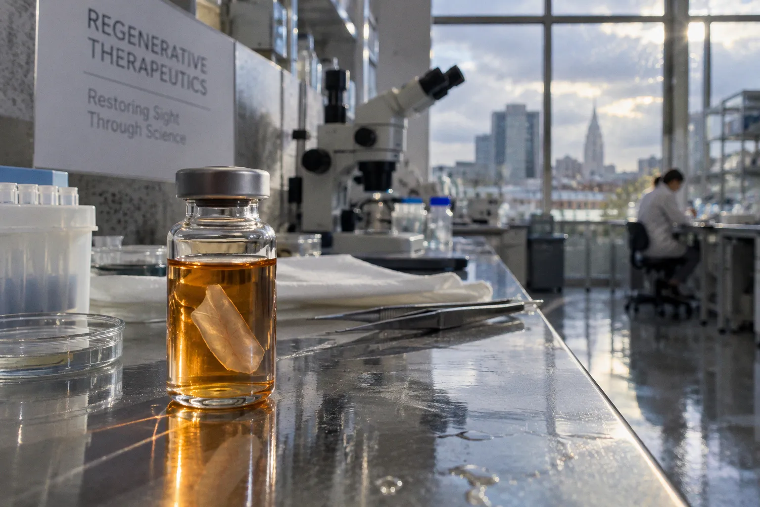
Photoreceptor cell survival in retinal progenitor grafts is often lost in the first days after surgery, before the graft has a…
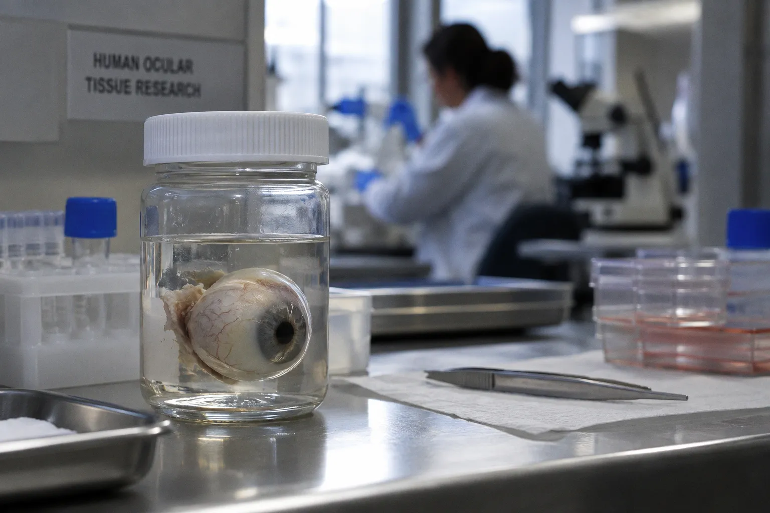
The proteomic catalog of healthy human retrobulbar optic nerve donor tissue — 2,711 non-redundant proteins resolved from 8,729…
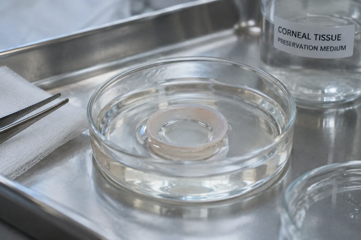
The accepted assumption is simple: if a donor corneal rim looks intact, it should provide a reasonable starting material for…
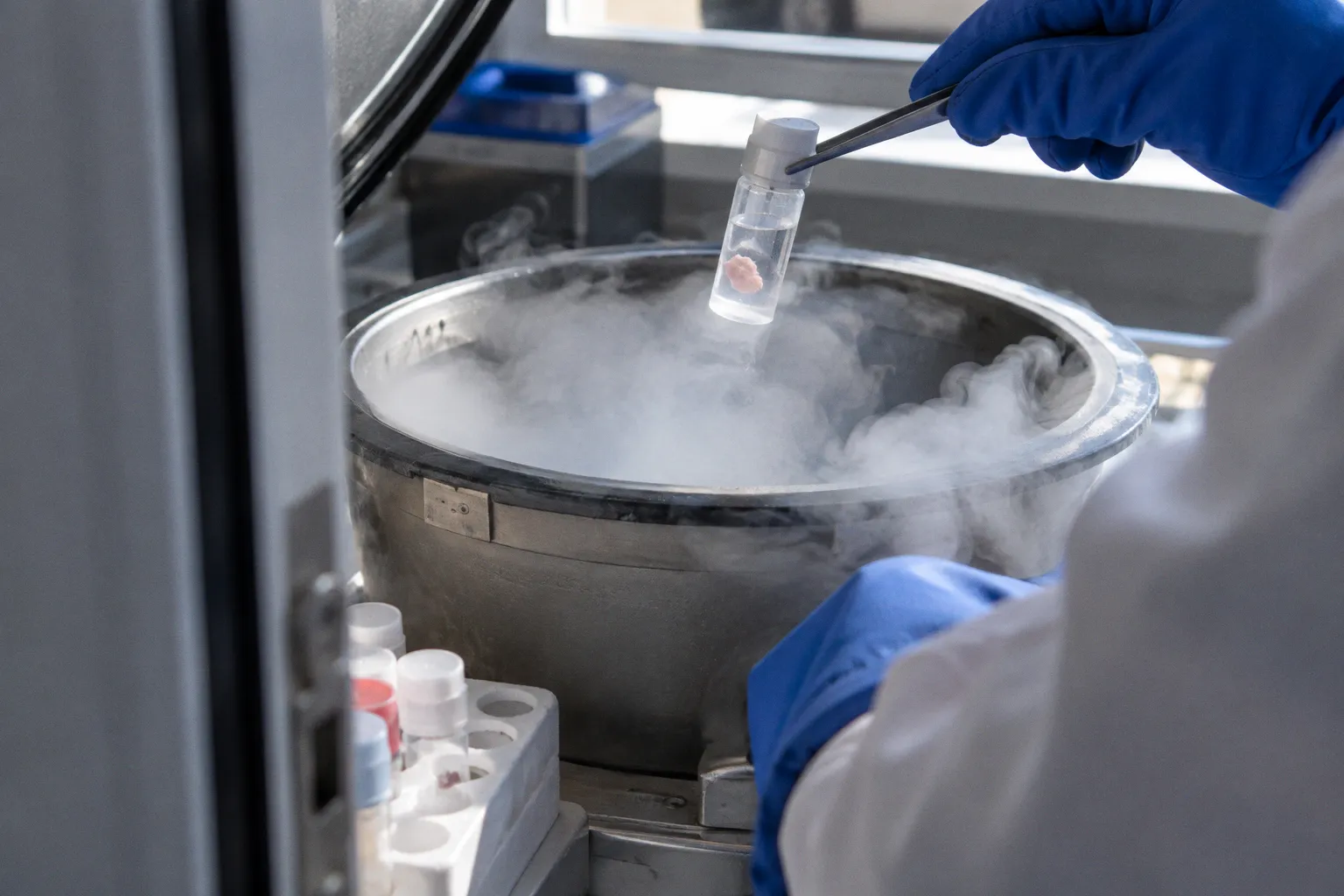
When a donor retina comes out of cryogenic storage, the first thing a tissue coordinator often checks is not whether the cells…

Mitochondrial protein expression in donor retinal tissue is not a single readout. It is a layered measurement shaped by disease…

The accepted premise is that DMEK graft preparation is primarily a problem of handling an extremely thin tissue. The harder fact…

The central problem in ocular cell therapy is often not whether the cells are biologically capable of helping damaged tissue. It…

A human retina is not a single molecular specimen. It is a layered cellular system in which abundant neuronal populations coexist…

The accepted clinical assumption is simple: a donor cornea that looks clear and meets an initial endothelial cell-density…

In the postmortem human retina, a single radial cell type remains responsible for much of the tissue’s biochemical housekeeping …
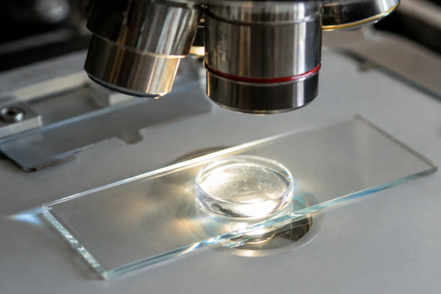
Every corneal surgeon who has transplanted tissue knows the arithmetic of failure. Approximately 90% of total corneal volume is…
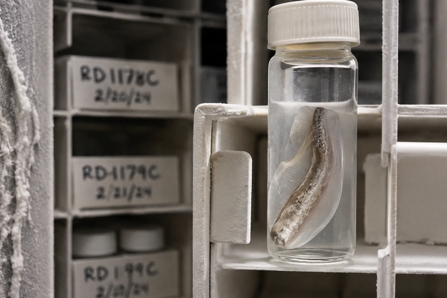
In a 2018 LC-MS/MS survey of healthy human donor retinas, researchers catalogued an average of 1,974 detectable proteins in the…
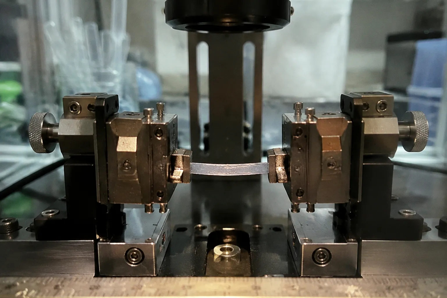
The standard assumption is familiar: cut the cornea into strips, pull them until the tissue yields, and the resulting…
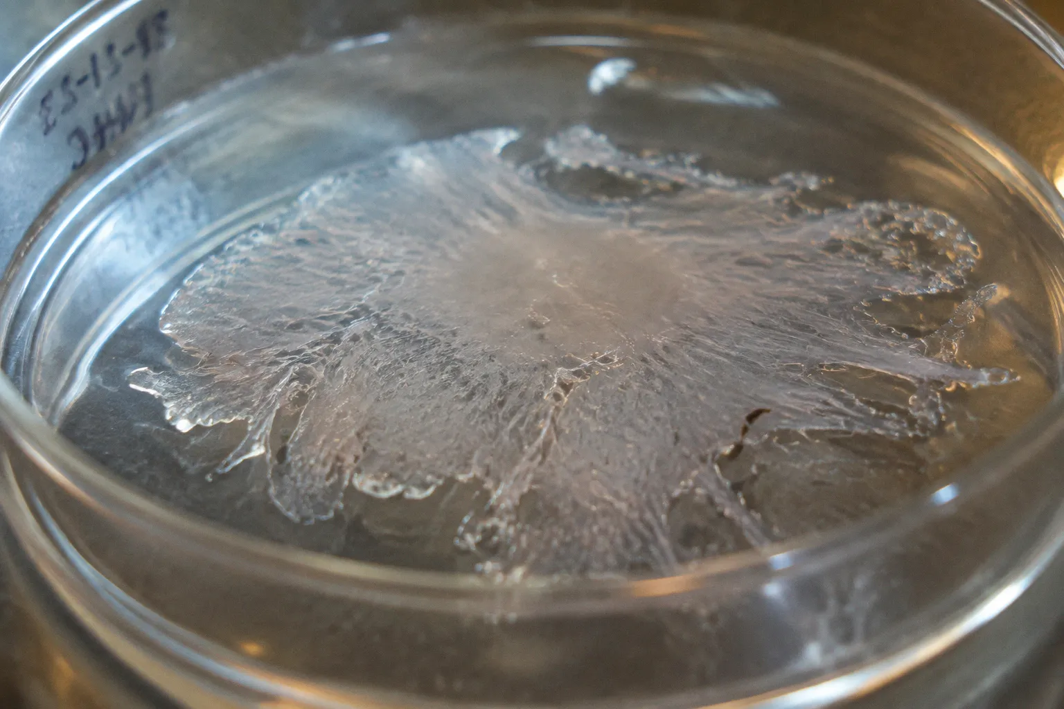
In tissue procurement suites, the moment the protease bath ends is often the moment the entire downstream experiment is decided…
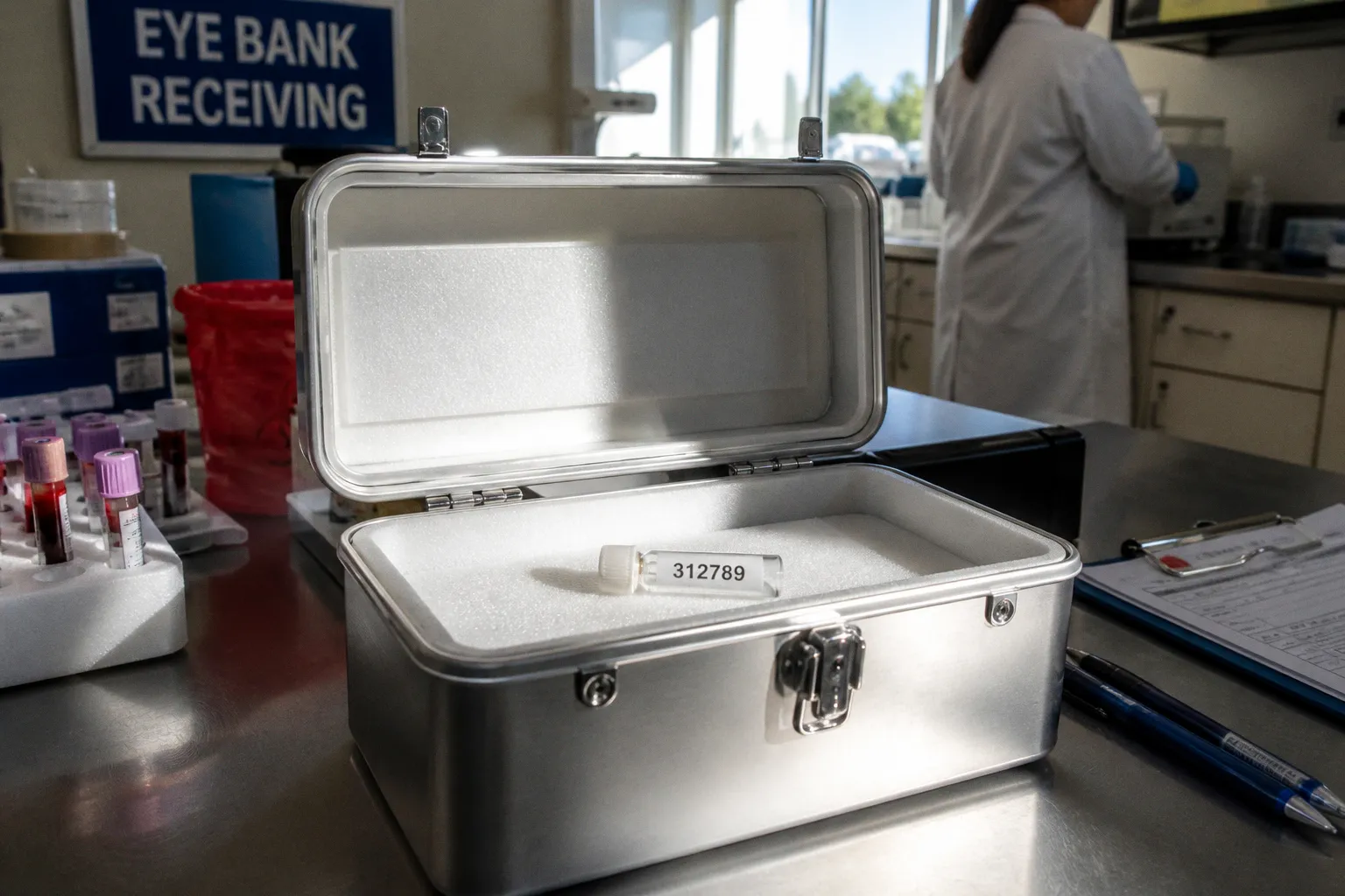
The 24-hour post-mortem clock is not a bureaucratic window but a cellular one. Within hours of circulatory arrest, corneal…
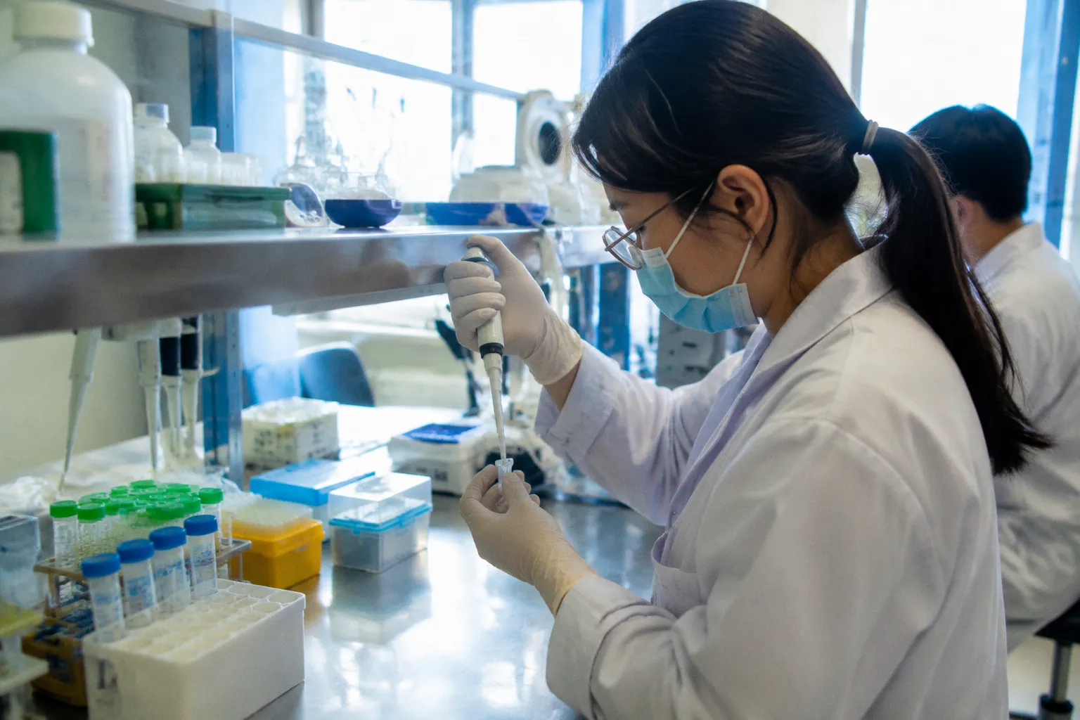
Extracellular vesicle therapy for retinal repair is not a cell replacement system. It is a biologic delivery architecture. The…
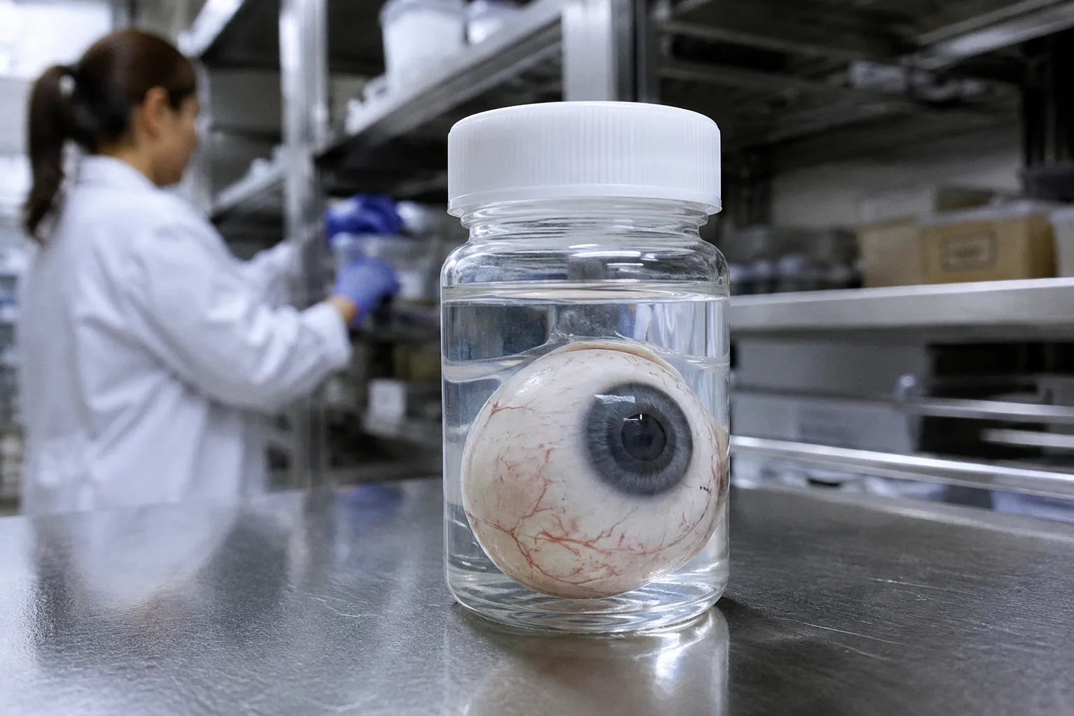
The accepted assumption is simple: once the donor dies, the epigenome begins to betray the experiment. In ocular research, that…
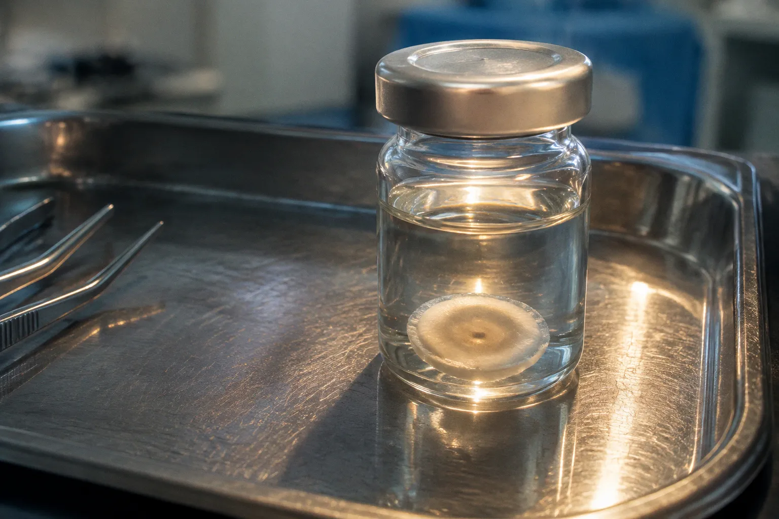
A Fuchs dystrophy donor cornea can look acceptable at the eye-bank bench and still carry changes that matter for research, graft…
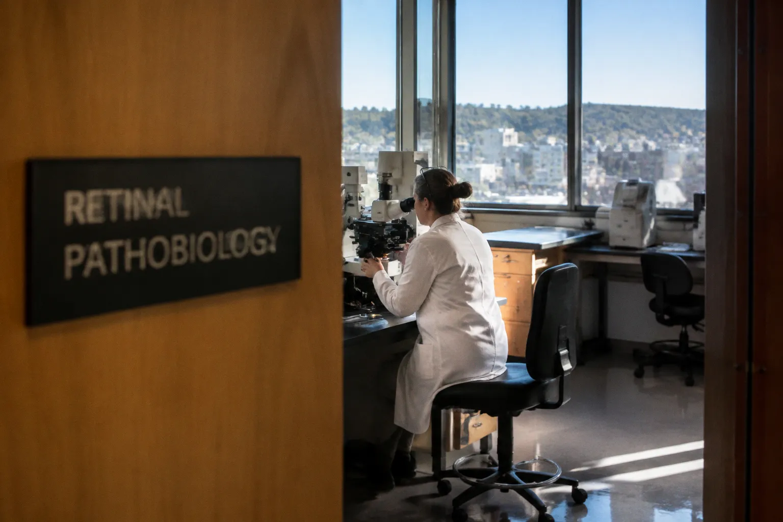
The central conflict in dry age-related macular degeneration is not whether the retinal pigment epithelium or the…
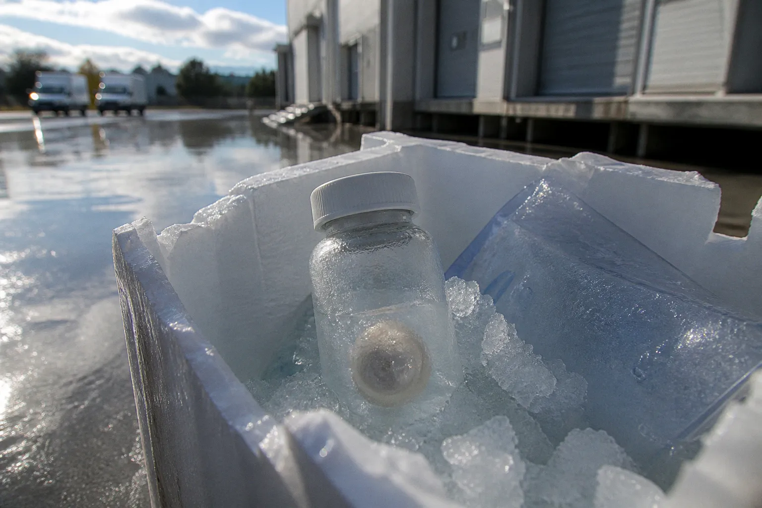
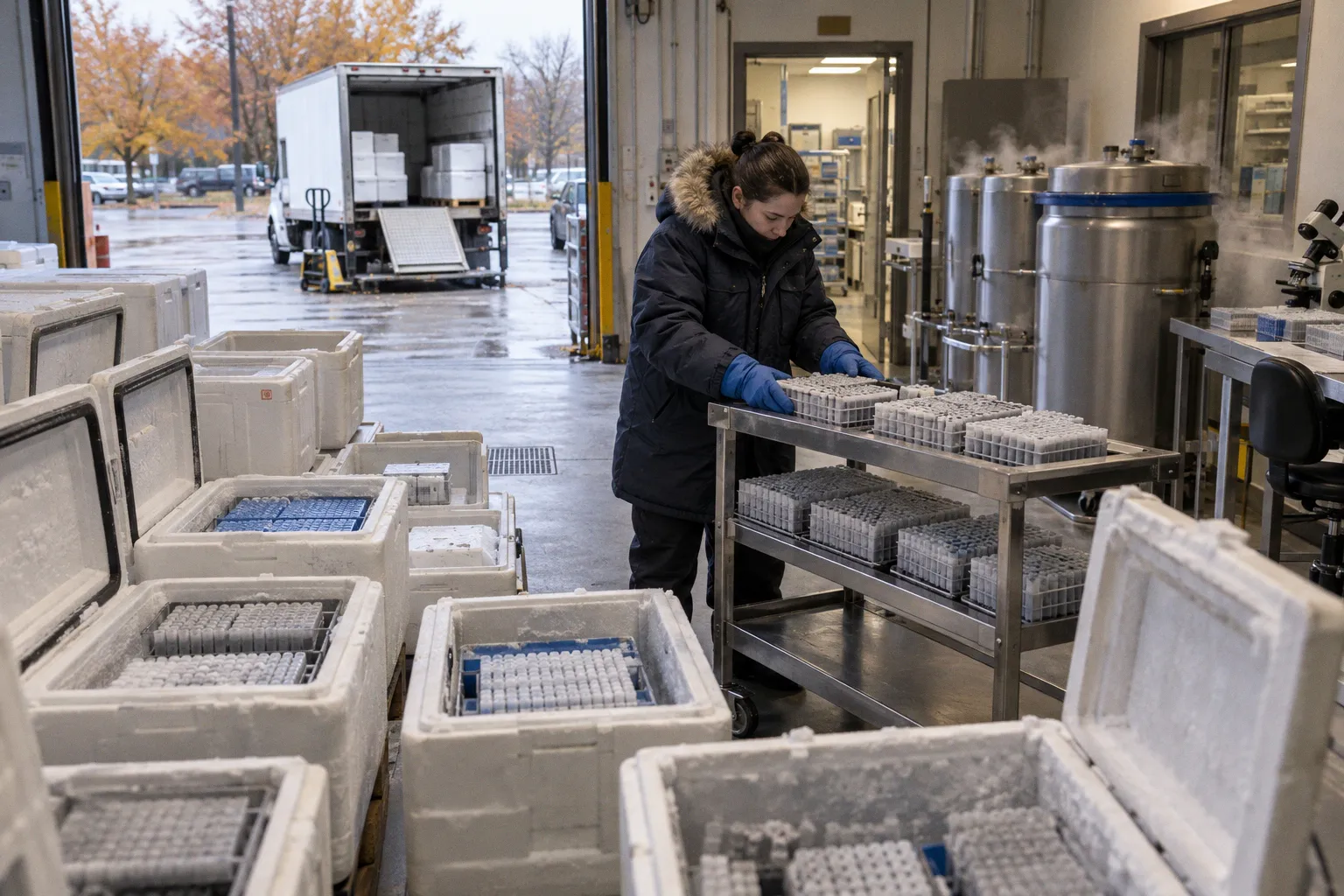
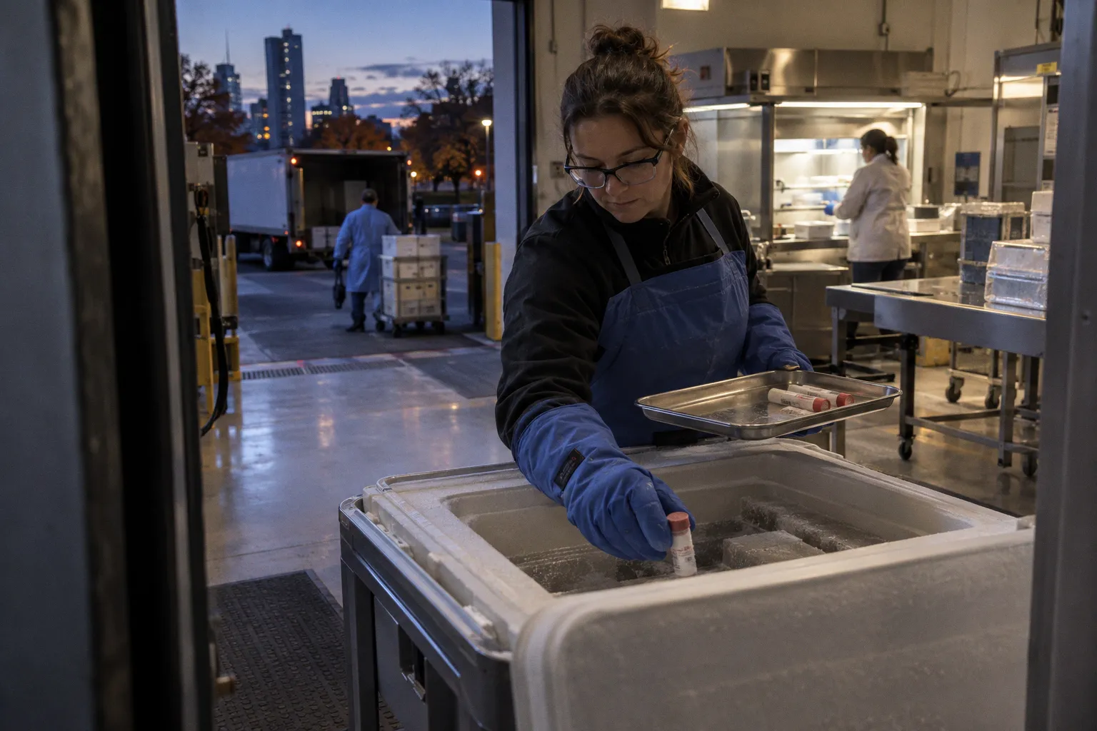
Between recovery and research placement, a donor cornea traverses a sequence of cold-chain nodes — packing, courier transport…
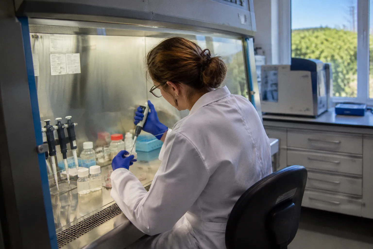
Intravitreal AAV delivery is often treated as if the main problem were choosing the right capsid. The vector is injected, the…
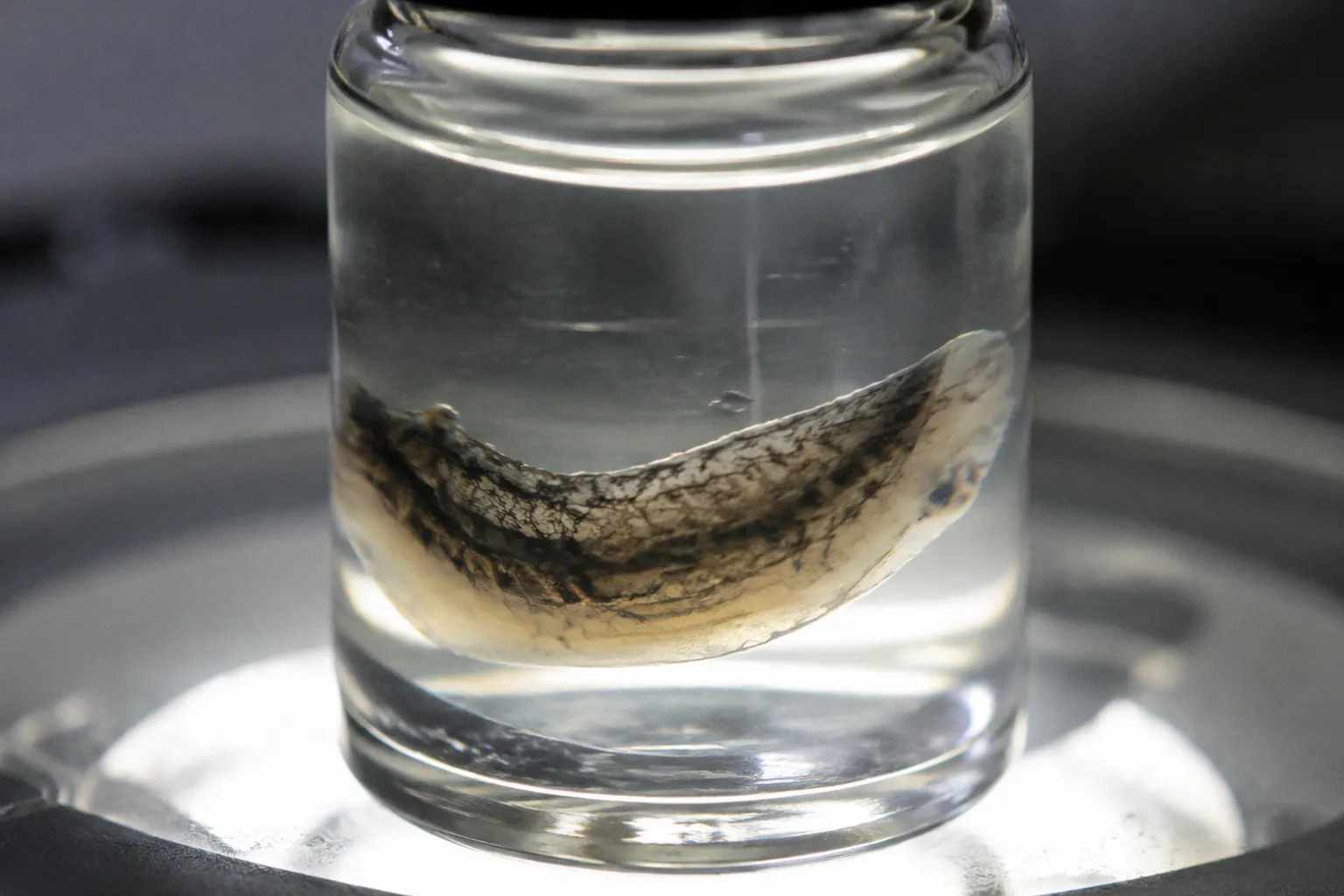
Human choroid gene expression profiling from donor eyes is moving beyond the question of which genes are present. The more useful…
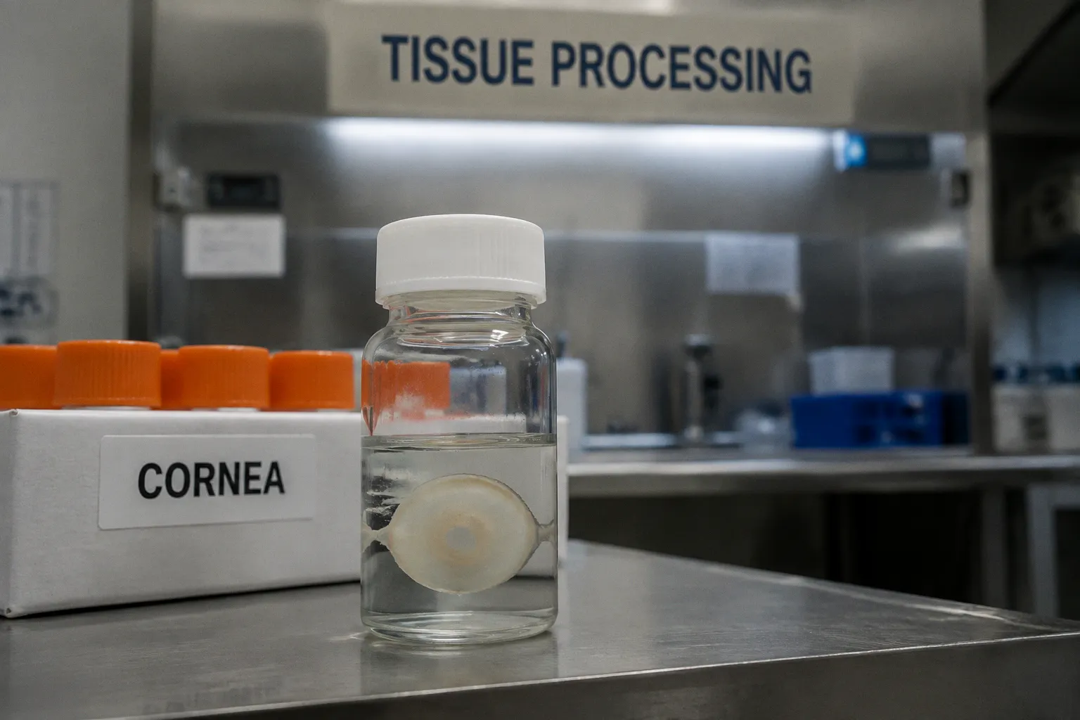
The contradiction appears at the edge of the cornea, where the limbal epithelium should preserve its regenerative reserve. In…
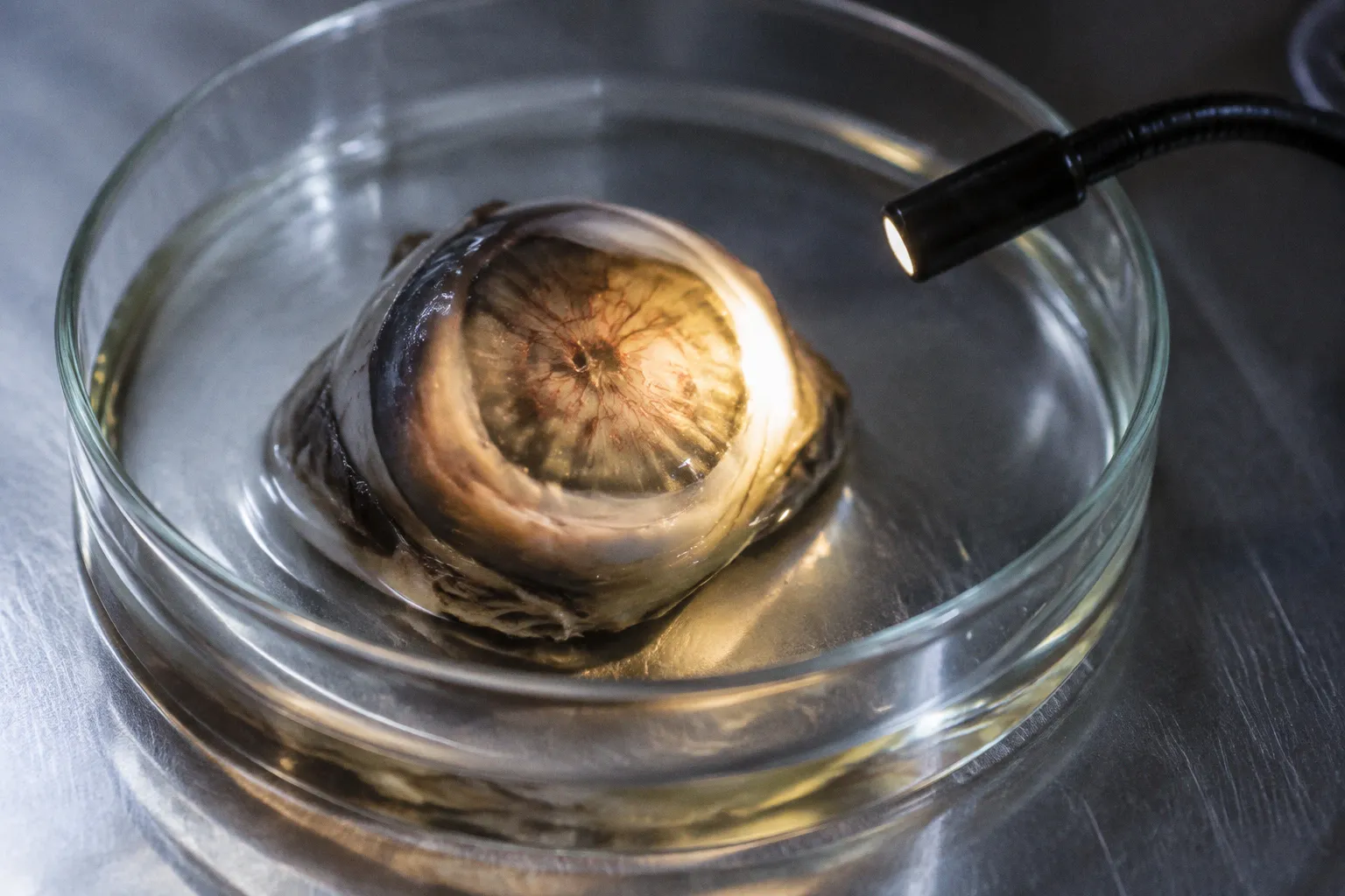
The main analytical problem in studying choroidal neovascularization in donor tissue is not lesion detection. It is spatial and…
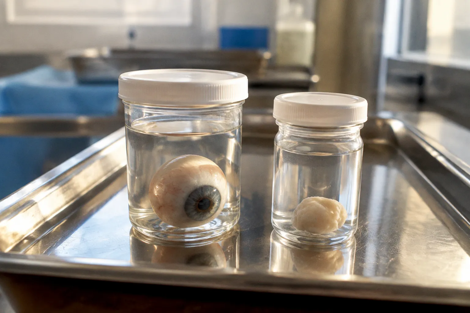
The corneal transplantation community has long operated under a quiet assumption: that whole globe enucleation is the gold…
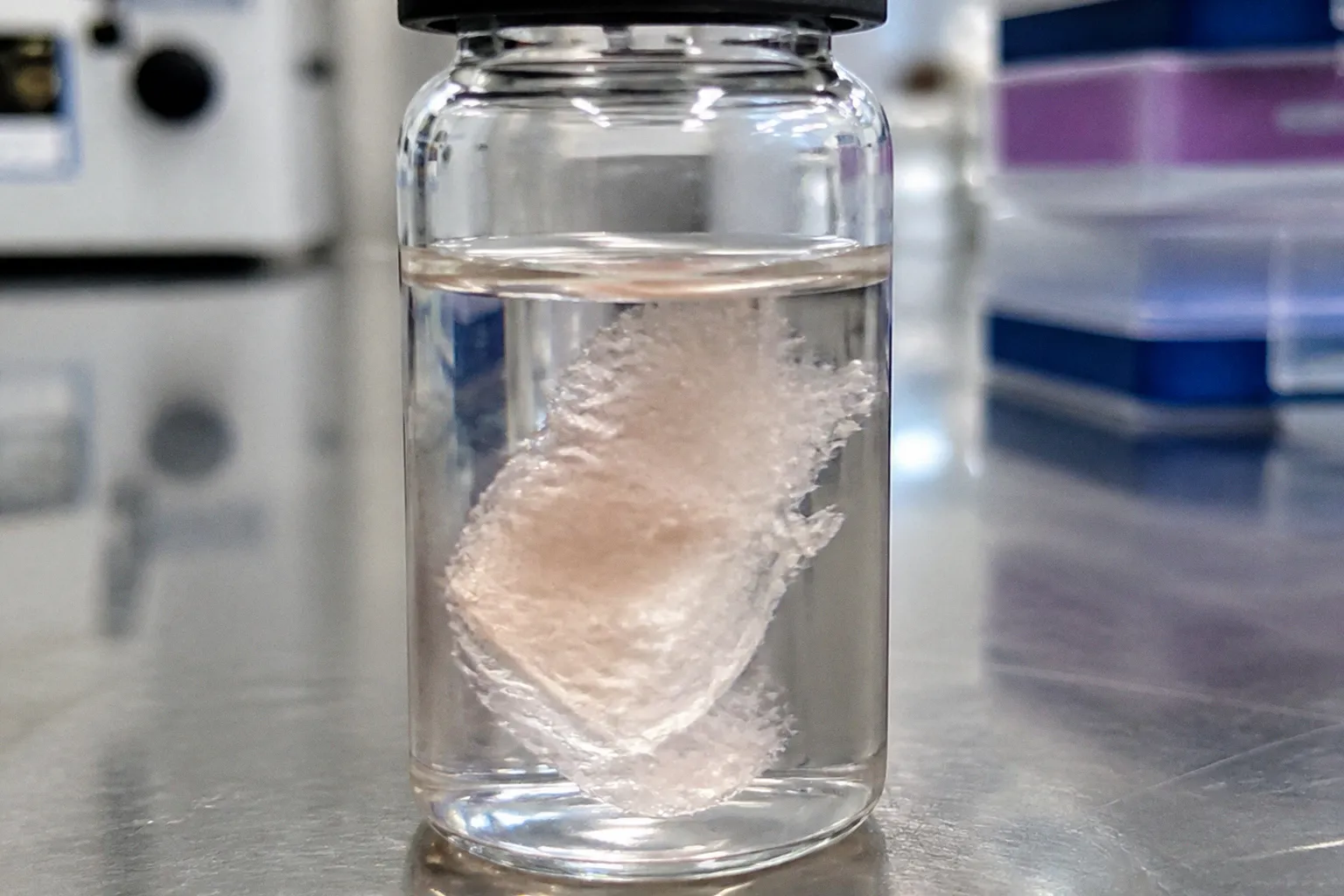
Subretinal delivery of a retinal progenitor cell suspension sounds straightforward on paper. You draw the cells into a syringe…
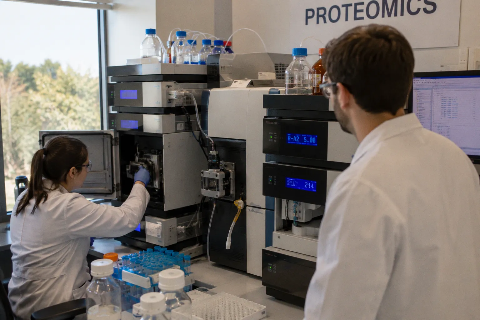
When human lamina cribrosa cells are pulled — 3% cyclic equiaxial stretch for two hours, then again for twenty-four — the…
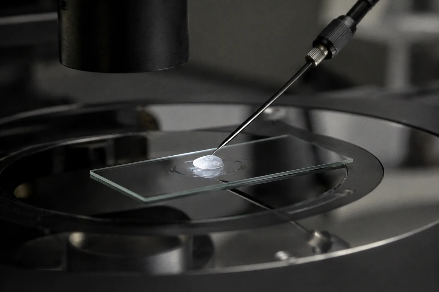
A twenty-fold increase in elastic modulus. That is the documented mechanical shift between healthy human trabecular meshwork (TM)…
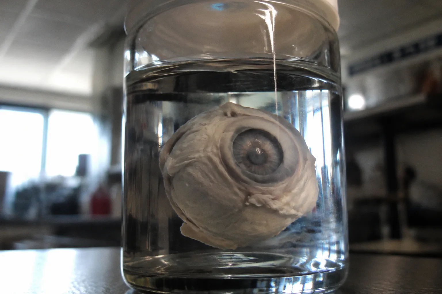
Every ocular pathologist has, at some point, encountered the assumption that donor retinal tissue is “too autolyzed to trust.”…
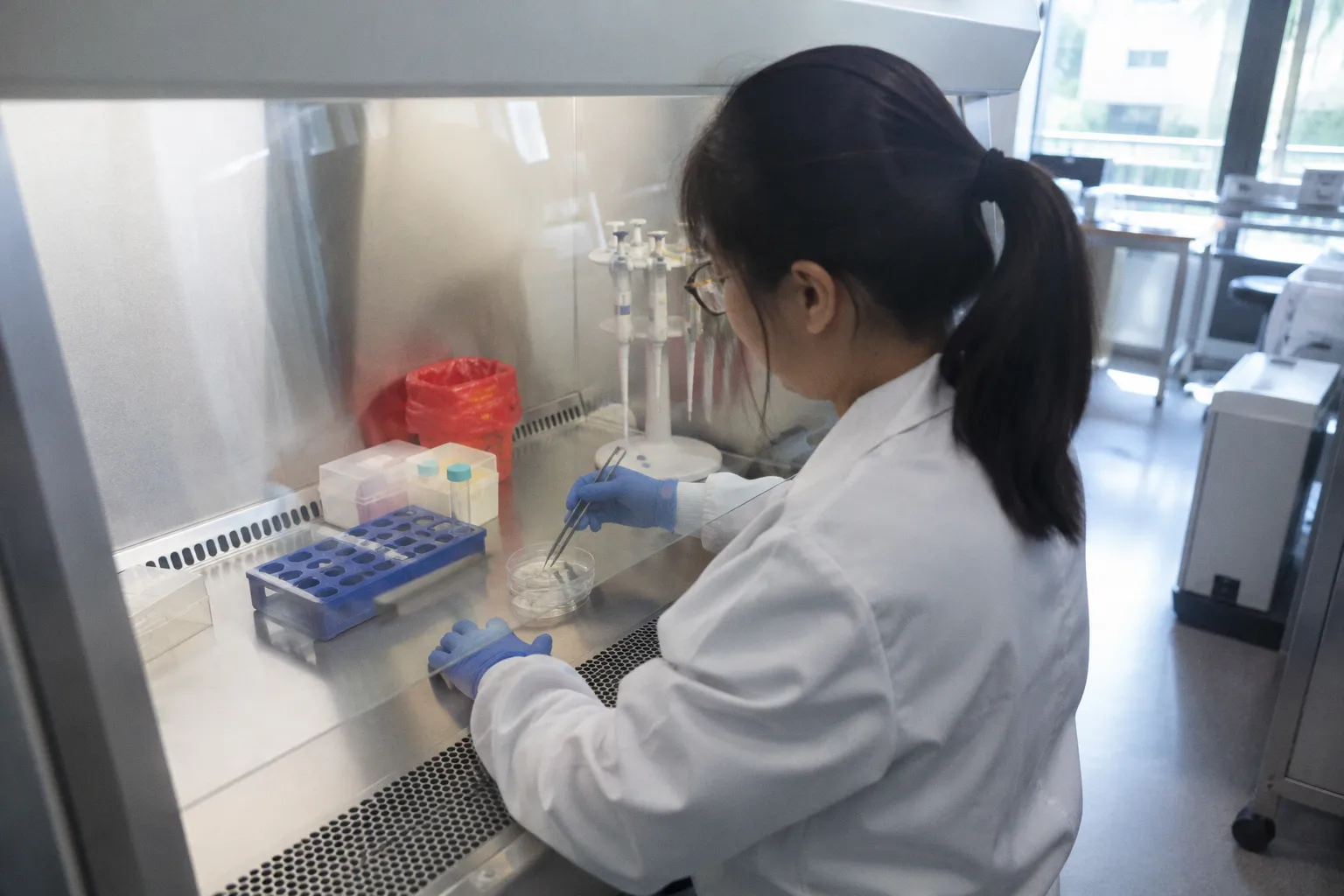
The clock starts the moment a retinal pigment epithelium patch comes off the culture substrate. Bench teams watching the…
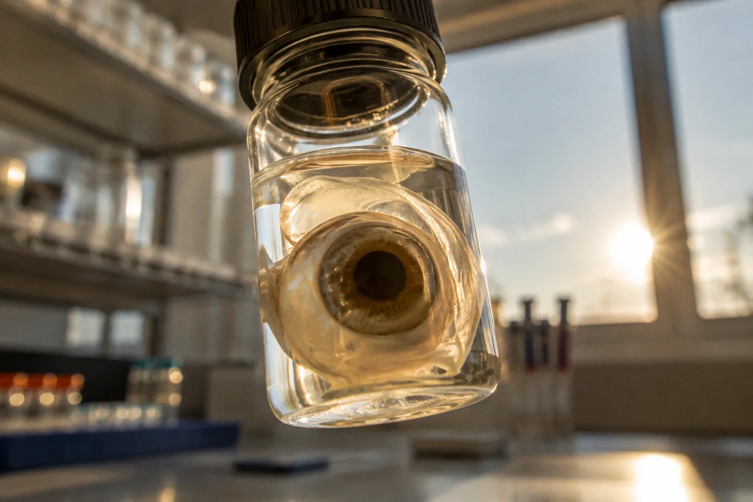
The central contradiction in donor-retina multiomics is not a disagreement between technologies. It is a disagreement between…
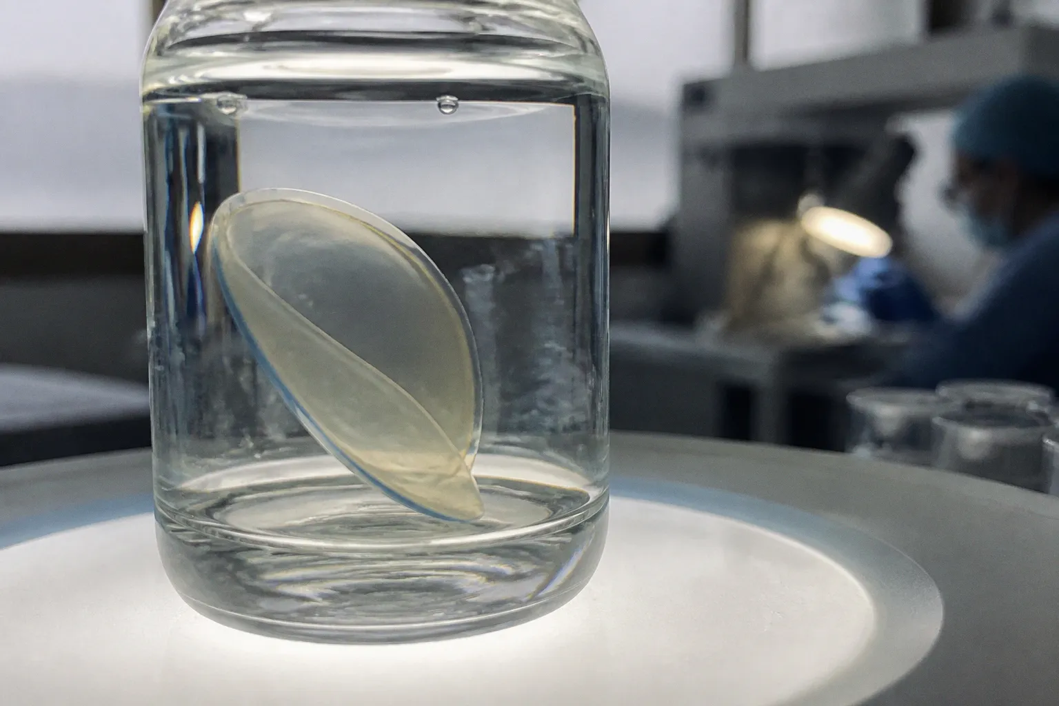
DMEK graft preparation failure is not evenly distributed across donor tissue. In a retrospective analysis of 359 corneas from 290…
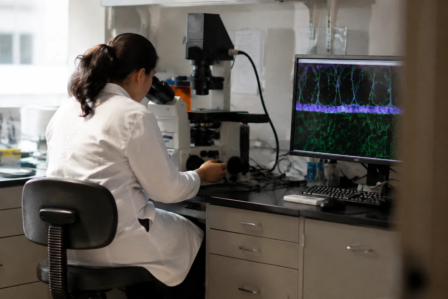
The accepted clinical shorthand is familiar: diabetic retinopathy becomes visible when microaneurysms, hemorrhages, and other…
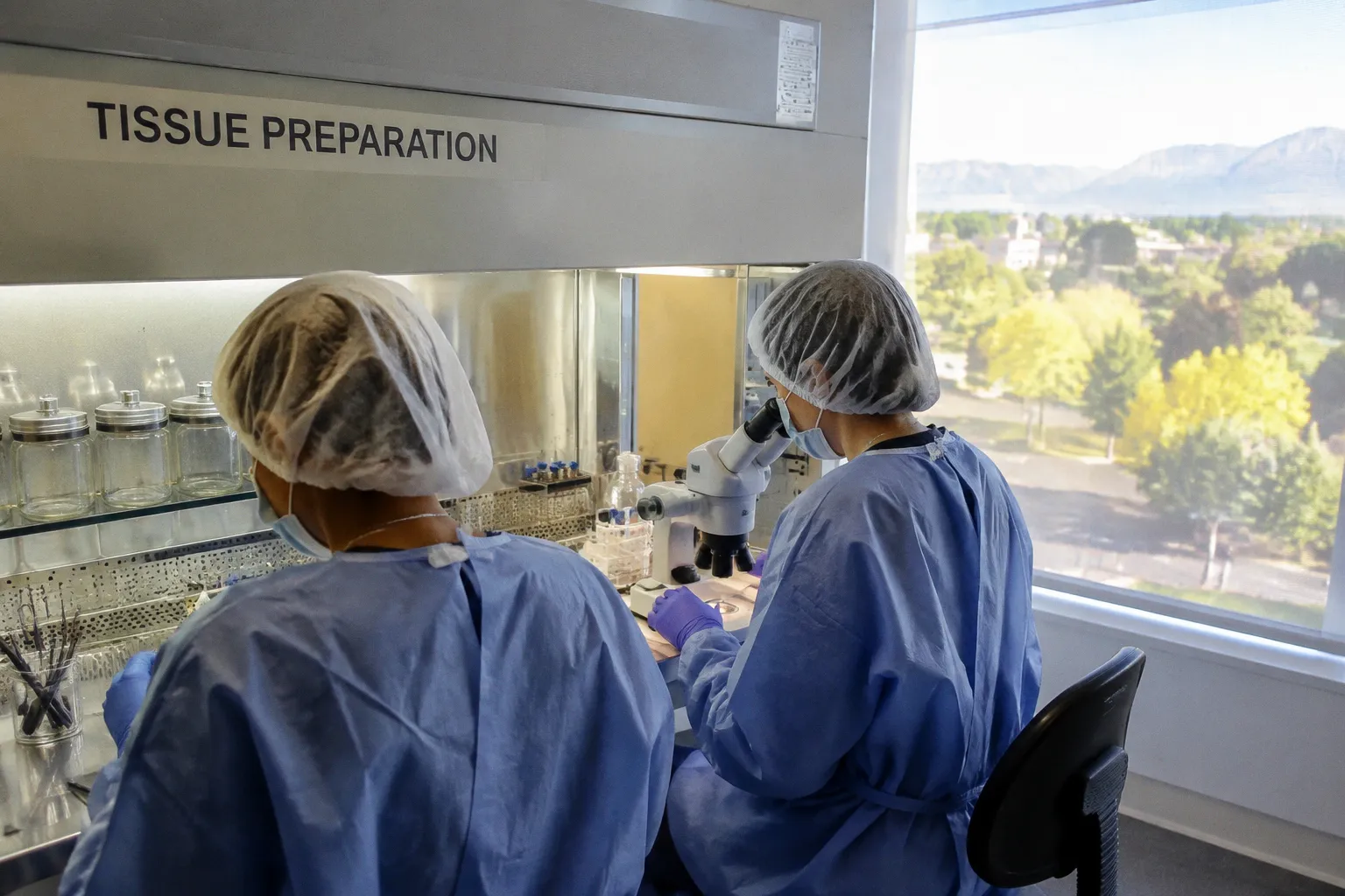
A donor cornea can look worse on the bench than it performs in the recipient. That is the uncomfortable fact behind many…
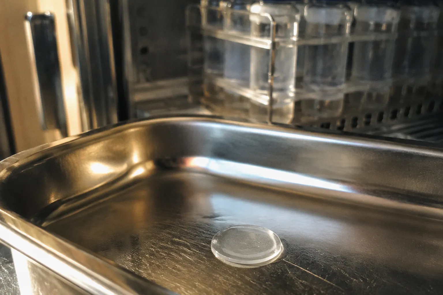
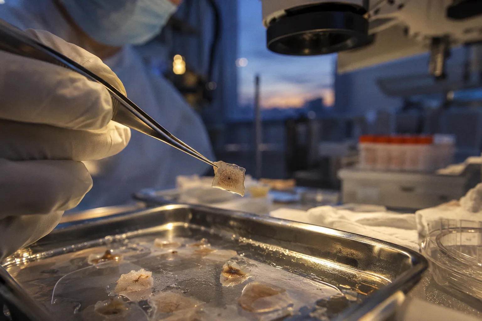
A SMILE procedure removes a thin, disc-shaped section of stromal tissue through a small corneal incision. That lenticule is…
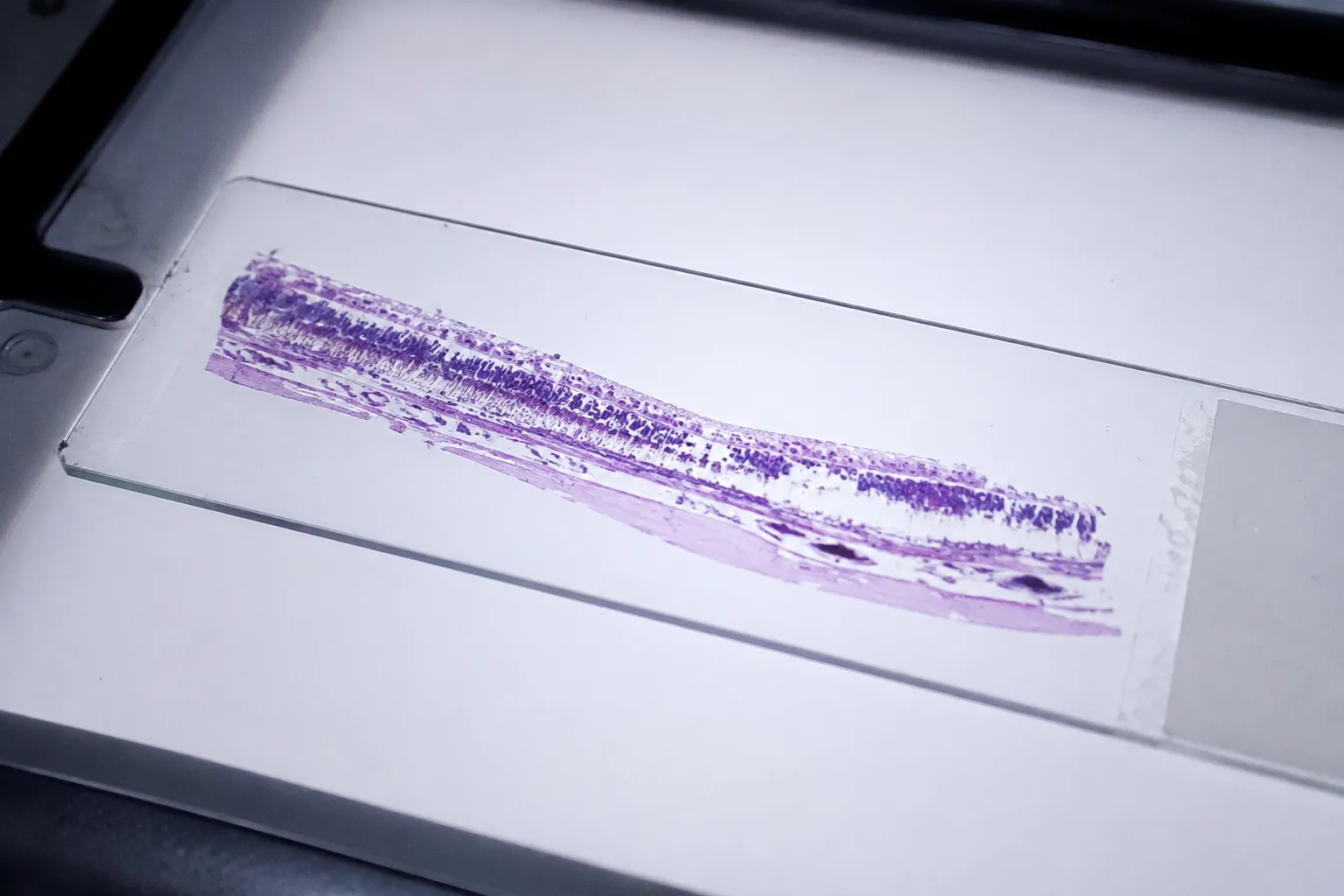
In a histopathology series of 37 donor eyes assembled through the MidAmerica, Iowa, and Utah eye banks, a quietly contradictory…
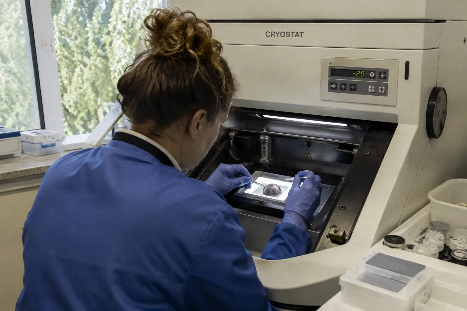
The ophthalmic research community has, for years, operated under a quietly convenient assumption: that single-cell RNA sequencing…
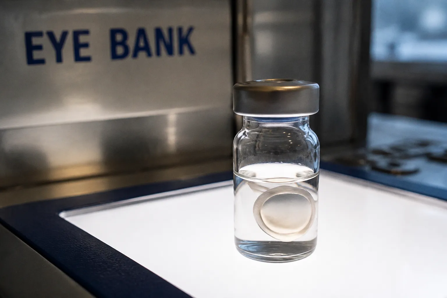
The procurement-to-preservation pipeline for donor corneal tissue operates on a compressed biological clock. Once enucleation is…
Bridging ocular biobanking logistics and vision research.