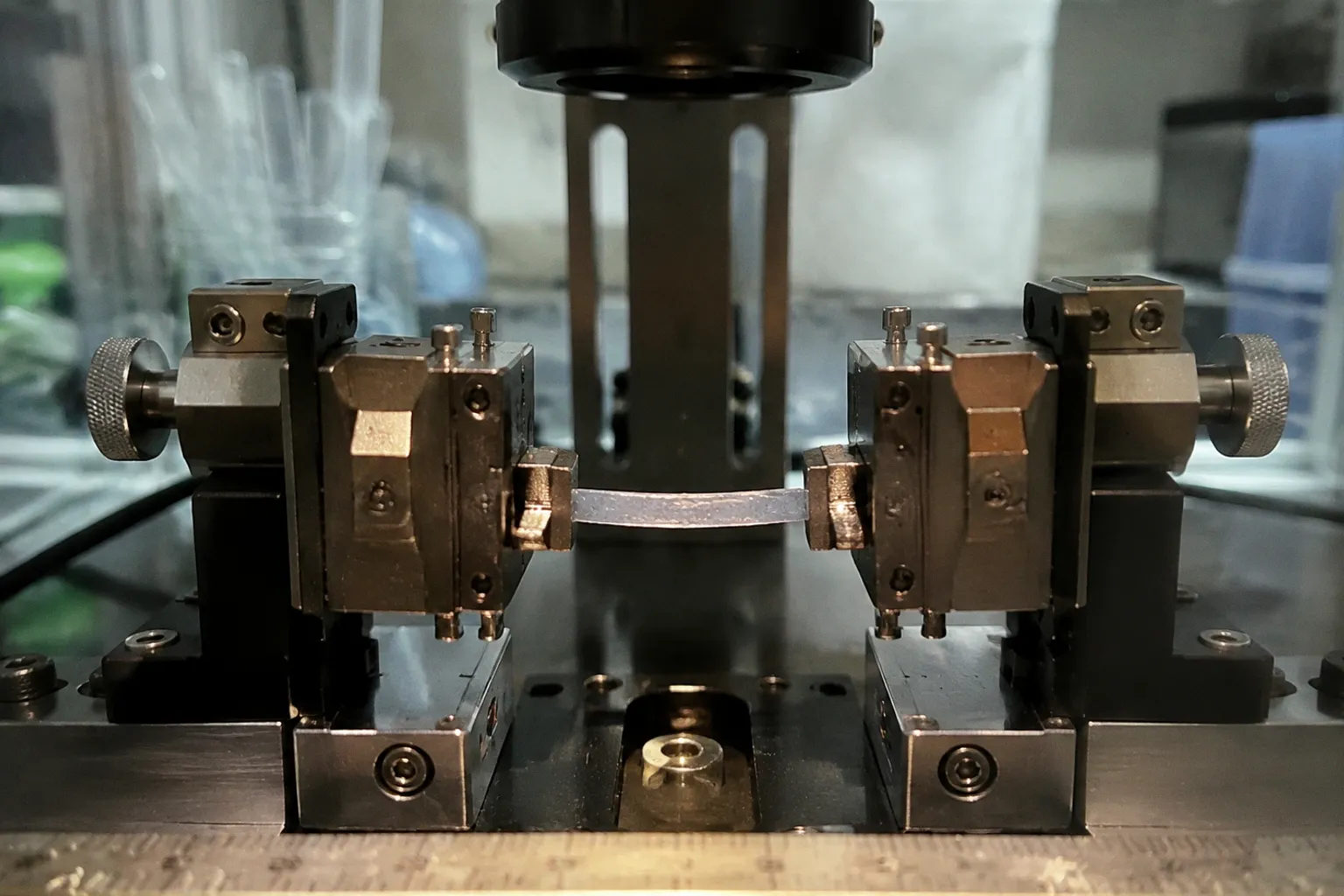
It is simple, inexpensive, and deeply embedded in the history of human corneal biomechanics.
It is also mechanically misleading when treated as a direct representation of the intact cornea.
The central problem in corneal inflation testing vs uniaxial tensile testing is not that one method is scientifically respectable and the other is obsolete. The problem is more uncomfortable: they interrogate different mechanical objects. Strip extensiometry measures a surgically altered piece of cornea. Inflation testing measures a curved tissue shell under pressure, closer to the loading condition that gives the living cornea its shape. Those outputs can be compared, but they cannot be casually substituted for one another.
That distinction matters whenever a study discusses donor cornea elastic modulus measurement, graft quality, stromal remodeling, keratoconus-like deformation, or the mechanical consequences of corneal cross-linking. A numerical modulus without a mechanically credible geometry is not a result. It is often just a very precise artifact.
The same cornea, two different mechanical questions
Uniaxial tensile testing begins with excision. A corneal strip of relatively constant width is cut from the tissue, aligned in a chosen direction, and secured in mechanical clamps. The machine then applies linear tension while recording force and displacement.
The procedure has obvious practical advantages:
- the equipment is comparatively accessible;
- the loading protocol is easy to standardize;
- the resulting curve is straightforward to plot;
- small tissue samples can be tested after regional dissection or biochemical treatment;
- multiple strips can be taken from a single donor cornea.
That convenience has encouraged a conceptual shortcut. Researchers often speak as if the strip were simply a smaller version of the intact cornea. It is not.
The native cornea is a curved, layered, anisotropic shell. Its lamellae are not arranged as a single family of parallel fibers, and its local thickness, hydration, curvature, and regional collagen architecture all influence the response to load. Cutting a strip removes the tissue from that geometry. Flattening it for mounting imposes additional deformation before the formal tensile test has even started.
Inflation testing asks a different question. An intact donor cornea, corneoscleral button, or whole globe is exposed to controlled retro-corneal fluid pressure or elevated intraocular pressure. The tissue deforms under multi-axial loading, while its curvature and much of its native boundary condition remain present.
This is not automatically a perfect simulation of physiology. Donor tissue is not living tissue, and experimental chambers introduce their own constraints. Yet the test preserves the mechanical problem that matters: pressure acts on a curved membrane, and the resulting deformation emerges from the interaction of geometry, thickness, hydration, and tissue stiffness.
The contrast is therefore not simply “simple test versus advanced test.” It is uniaxial material interrogation versus pressure-driven structural behavior.
A corneal strip is not a miniature cornea. It is a corneal specimen after geometry has already been edited by the experimenter.
Why cutting changes the result before the test begins
The most consequential artifact in strip extensiometry is often treated as a technical footnote: flattening a curved corneal strip.
A strip taken from the corneal dome has a natural curvature. Once it is cut and laid flat for clamping, the tissue is forced into a geometry it did not occupy in situ. The posterior surface experiences initial tensile strains, while the anterior surface experiences compressive strains. The stress distribution is therefore non-uniform even before the actuator begins pulling.
This matters because the machine records global force and displacement, while the tissue is experiencing a more complicated local field. The measured curve may be interpreted as a material property, but part of the curve reflects the imposed transition from a curved shell to a flattened specimen.
The edges introduce another problem. Trephination and strip cutting sever collagen lamellae and create boundaries that do not exist in the intact cornea. Clamping then concentrates load near the grips and may promote local slippage, crushing, or stress concentration. The central gauge region is not necessarily carrying a uniform tensile field, particularly when the strip is narrow, the tissue is thin, or the cut orientation intersects complex lamellar architecture.
This is why the apparent modulus from uniaxial testing cannot be assumed to equal the modulus obtained from inflation testing. The discrepancy is not necessarily evidence that one experiment was badly performed. It may be the expected consequence of asking the tissue to behave in a new geometry.
The historical appeal of strip extensiometry is understandable. Early corneal mechanical studies, including pioneering work from 1980, established a practical method for producing repeatable tensile curves. But repeatability is not the same as physiological validity. A method can be internally consistent while measuring a mechanically transformed specimen.
What inflation testing preserves
Inflation testing keeps the cornea in a pressure-bearing configuration. The tissue is mounted so that fluid pressure acts against the posterior surface, and pressure is increased according to a controlled protocol. Depending on the design, the test may use a whole globe, a donor corneal cap, or another preparation that preserves the dome.
The deformation is multi-axial. The anterior surface moves, the curvature changes, and the tissue responds through a combination of membrane tension, bending effects, collagen recruitment, regional thickness variation, and boundary constraints. The measured behavior is therefore closer to the structural mechanics of the cornea than the response of a cut strip under a single tensile axis.
Comparative work has shown several recurring differences between the methods:
- uniaxial tests can display a more extended toe region;
- strip tests can produce higher peak strains than inflation tests at equivalent pressure-derived wall stresses;
- the apparent stiffness range may differ substantially depending on geometry and analysis;
- pressure-based loading can reveal regional and full-field deformation that a clamp-to-clamp displacement cannot resolve;
- inflation results are strongly dependent on boundary conditions, thickness measurement, hydration, and pressure calibration.
The toe region deserves particular suspicion. It is tempting to interpret a long low-stiffness phase as direct evidence of collagen recruitment in the same way that one might interpret a whole-tissue physiological response. But in a flattened strip, the initial curve also incorporates the release or redistribution of pre-existing curvature-related strain and the rearrangement of tissue around the clamps. The curve is not meaningless. It is simply not a clean readout of one biological mechanism.
A pressure-based experiment has its own analytical burden. Pressure is not stiffness. To infer mechanical properties, the investigator must connect pressure, geometry, thickness, and deformation through an appropriate model. If the cornea is treated as a uniform isotropic membrane when its structure is layered and directionally organized, the resulting modulus may look authoritative while hiding a paradigm deficit in the model itself.
The comparison researchers actually need
The most useful way to compare the two methods is not to ask which one produces the “true” modulus. That question assumes a single number exists independently of the test configuration. It does not.
| Parameter | Inflation testing | Uniaxial tensile testing |
|---|---|---|
| Specimen geometry | Intact or near-intact curved cornea, cap, or globe | Excised strip with cut edges |
| Primary loading | Fluid pressure and multi-axial deformation | Linear tension along one axis |
| Physiological relevance | Closer to pressure-bearing corneal behavior | Closer to directional tensile response of a specimen |
| Main strength | Preserves curvature and enables full-field deformation analysis | Simple setup, controlled orientation, and convenient sampling |
| Main artifact | Boundary conditions, hydration, and model assumptions | Flattening, edge cutting, clamping, and non-uniform strain |
| Typical data | Pressure–deformation relationship and spatial strain maps | Force–displacement or stress–strain curve |
| Regional information | Available with imaging, especially 3D-DIC | Limited to the selected strip and gauge region |
| Best use | Structural behavior, deformation mapping, model validation | Material comparison, directional effects, local treatment studies |
| Main interpretive risk | Mistaking model-derived stiffness for a direct material constant | Treating strip modulus as intact-corneal modulus |
That table is not an argument for abandoning tensile testing. It is an argument for stopping the methods from impersonating one another.
If the research question concerns how a local stromal treatment changes tissue resistance along a defined direction, a strip test can be entirely appropriate. If the question concerns how a donor cornea deforms under pressure, whether a graft preserves its dome, or how mechanical heterogeneity appears across the anterior surface, inflation testing is the more coherent design.
The method should follow the question. Instead, the field often chooses the method first because the apparatus is already available, then retrofits the biological question to match the data.
Stress, strain, and the dangerous comfort of a single modulus
A corneal mechanical curve is often reduced to a stiffness value. This is convenient for statistical comparison and disastrous when the reduction erases the conditions under which the value was obtained.
In uniaxial testing, stress is commonly derived from force divided by an estimated cross-sectional area, while strain is derived from displacement relative to gauge length. Both calculations become vulnerable when the strip changes width, slips in the grips, deforms outside the intended gauge region, or begins with residual curvature-related strain.
In inflation testing, the investigator must estimate how pressure translates into wall stress. That conversion depends on the geometry of the tissue and the assumptions used in the model. A thick-wall formulation, a thin-shell approximation, or a finite-element reconstruction may produce different results from the same pressure–deformation record.
This is not a minor mathematical disagreement. It determines whether the reported value describes:
1. a local material response;
2. a structural response of the whole cornea;
3. a model-dependent effective parameter;
4. or a mixture of all three.
Validation studies comparing inflation and tensile approaches have reported stiffness values in the broad range of approximately 0.2 to 1 MPa, depending on the preparation, protocol, and analytical framework. The range itself is a warning. It should not be compressed into a universal donor cornea modulus and repeated as if tissue mechanics were a laboratory constant like temperature.
A peak pressure of 150 mmHg has been used in whole-globe comparative inflation experiments, while uniaxial counterparts have applied peak stresses around 0.13 MPa. These values are useful for comparing protocols, not for claiming equivalence between loading modes. Pressure and uniaxial stress are not interchangeable labels. They arise from different geometries and different stress fields.
Why 3D-DIC changes the argument
Inflation testing becomes considerably more informative when paired with three-dimensional digital image correlation, or 3D-DIC. The method tracks surface features during pressure elevation and calculates pointwise deformation across the anterior cornea.
A protocol targeting controlled intraocular pressure elevation to 40 mmHg can produce a full-field view of how the surface moves rather than a single displacement value at one central point. That distinction is critical. The cornea is not mechanically uniform, and a central average can conceal regional behavior near the limbus, around a graft interface, or across areas with altered stromal architecture.
Full-field measurements can reveal:
- localized zones of greater strain;
- directional differences in deformation;
- asymmetric behavior across the corneal dome;
- changes in deformation patterns after tissue treatment;
- disagreement between geometric assumptions and the actual surface response.
This does not make 3D-DIC a magic instrument. Surface tracking depends on image quality, speckle preparation, camera calibration, and stable experimental conditions. It also measures the anterior surface, not every internal lamellar movement. Still, it addresses a basic weakness of strip testing: the assumption that one axis and one averaged displacement can stand in for the entire cornea.
Paradoxically, a more detailed inflation experiment may produce a less tidy result. Instead of one attractive modulus, the investigator may find a spatially variable deformation field that resists reduction. That is not experimental failure. It is often the biological signal that a single-number paradigm was trying to suppress.
Donor tissue turns protocol details into biological variables
Human corneal biomechanics testing is unusually sensitive to specimen history. Donor tissue is not mechanically neutral material waiting to be assigned a number. Time from recovery, storage conditions, hydration, epithelial status, endothelial integrity, scleral attachment, and preparation geometry can all influence the response.
For corneal tissue mechanical characterization, the minimum useful description should include:
- whether the sample was a whole globe, corneoscleral button, corneal cap, or strip;
- the time and conditions of preservation;
- whether the epithelium and endothelium were retained;
- the hydration or swelling state at testing;
- the pressure or displacement ramp;
- the orientation of any strip relative to the corneal axes;
- the method used to measure thickness;
- the boundary conditions at the limbus or clamps;
- the point at which tissue failure or test termination was defined.
Without those details, comparisons across donor cohorts become fragile. A study may report that one group is stiffer than another when the difference actually reflects storage duration, swelling, or the distribution of tissue samples across central and peripheral regions.
The issue is particularly sharp in Fuchs dystrophy donor corneas, graft-quality assessment, and studies involving endothelial or Descemet membrane preparations. These tissues may be valuable precisely because they carry disease-associated or transplant-relevant features. Removing them from their native configuration and reducing the test to a narrow strip can discard the structural context that makes the specimen scientifically important.
A DMEK-related preparation, for example, is not mechanically equivalent to a full-thickness corneal button. A study focused on donor corneal endothelial cells may need to preserve the endothelial–Descemet interface for biological reasons, while a biomechanics study may be tempted to remove layers for convenience. Those are different priorities. They should not be quietly merged under the phrase “corneal stiffness.”
When uniaxial testing remains the right tool
The critique of strip extensiometry becomes unhelpful if it turns into another dogma. Uniaxial testing remains useful when the experimental question is explicitly directional and specimen-level.
It is well suited to:
- comparing treated and untreated strips from the same donor;
- examining regional stromal differences;
- testing the effect of enzymatic digestion, cross-linking, or biochemical modification;
- measuring directional tensile response;
- generating controlled data for constitutive model development;
- working with limited tissue where intact inflation is impractical.
Its strongest feature is not physiological realism. It is experimental control. The investigator can define strip orientation, width, gauge length, loading rate, and endpoint. That can be valuable when the study needs relative changes rather than an absolute description of intact-corneal behavior.
The correct language, however, must remain narrow. A strip result can support a statement about the tensile response of the prepared specimen under the reported conditions. It does not automatically support a statement about how an intact donor cornea will deform under intraocular pressure.
That distinction becomes especially important when comparing cross-linking protocols. A treated strip may show increased resistance along the loading direction, while an inflated cornea may display a more complex change in surface deformation. Neither result invalidates the other. They answer different questions about how treatment modifies tissue mechanics.
The inflation test is not innocent either
Inflation testing is closer to the native mechanical problem, but “closer” is not the same as “pure.”
The preparation may be mounted in a chamber that fixes the limbus more rigidly than physiological anatomy would. A corneoscleral button may not reproduce the support provided by the globe. The pressure sensor may not reflect the exact pressure at the posterior corneal surface. Tissue hydration can change during the experiment. The cornea may also undergo viscoelastic, time-dependent behavior that is obscured by a rapid pressure ramp.
There is a second analytical danger: inflation data can encourage overconfident inverse modeling. If the model contains assumptions about isotropy, incompressibility, thickness uniformity, or collagen architecture that are not independently verified, the fitted stiffness becomes a compromise among the data and the assumptions.
A complex model is not necessarily a better model. Sometimes it is simply a more elaborate way to hide uncertain boundary conditions.
The strongest inflation studies therefore report the geometry and constraints with unusual care, use imaging rather than a single central displacement, and test whether the inferred parameters remain stable when reasonable modeling assumptions change. The goal is not to produce the most impressive modulus. It is to determine which aspects of the deformation are genuinely supported by the experiment.
Choosing the method for the claim
A useful decision begins with the claim, not the instrument.
If the claim is about intact corneal deformation under pressure, inflation testing is the defensible primary method. Add 3D-DIC when regional behavior matters.
If the claim is about directional tensile properties of stromal tissue, uniaxial testing may be appropriate, provided the paper clearly describes the specimen geometry and avoids presenting the result as a direct in vivo modulus.
If the goal is cross-method comparison, paired testing is essential. Ideally, the investigator should characterize matched tissue preparations and report how the stress–strain response changes when the same biological material is moved from curved inflation geometry to a flattened strip.
If the study concerns graft mechanics, preserving the graft-relevant structure should take priority over laboratory convenience. A strip cut from a donor cornea may provide useful material data while eliminating the very interface or curvature that determines surgical behavior.
And if the objective is model validation, neither method should stand alone. Inflation provides structural deformation under pressure; uniaxial testing provides directional material constraints. Used together, they can prevent a finite-element model from fitting one experiment while failing the other.
The serious comparison is not inflation versus tensile testing. It is which method leaves fewer unanswered questions for the claim being made.
What a credible paper should disclose
A mechanically literate paper does not need to pretend that its method is universal. It needs to make the limits visible.
For inflation experiments, readers should be able to reconstruct:
- the specimen configuration and support at the limbus;
- the pressure range and ramp rate;
- the method of thickness and curvature measurement;
- the imaging system and deformation metric;
- the assumptions used to infer stress or stiffness;
- the handling of hydration and temperature;
- the criteria for excluding damaged or leaking specimens.
For uniaxial experiments, the essential details include:
- strip dimensions and orientation;
- the location from which strips were taken;
- the method of cutting and edge preparation;
- grip design and any pretension;
- gauge-length definition;
- strain calculation method;
- evidence of slippage or grip damage;
- whether corrections were applied for curvature and cutting artifacts;
- the anatomical and preservation history of the donor tissue.
Without these details, the apparent precision of the reported modulus is cosmetic. A value carried to several decimal places does not compensate for an unreported hydration state or an undefined gauge region.
The remaining paradigm deficit
Corneal biomechanics has spent decades translating a curved, layered, viscoelastic organ into convenient laboratory geometries. That translation has produced useful knowledge, but it has also created a persistent habit: treating the easiest measurable quantity as the most biologically meaningful one.
Uniaxial tensile testing is not the villain. Inflation testing is not the final authority. Both methods are abstractions, and both can mislead when their outputs are treated as interchangeable measures of a single intrinsic stiffness.
The practical conclusion is more demanding than choosing a winner. Researchers should align the loading mode with the biological question, disclose how donor tissue was transformed before testing, and resist the seduction of a universal modulus. Inflation testing better preserves the cornea’s pressure-bearing architecture. Uniaxial testing offers controlled directional interrogation. Their results become genuinely informative only when the geometry, stress field, and analytical assumptions remain visible.
The field does not need another triumphant method. It needs fewer claims that exceed the experiment. Until then, the most important number in a corneal biomechanics paper may not be the modulus at all. It may be the distance between what the specimen was asked to do and what the authors say the living cornea does.