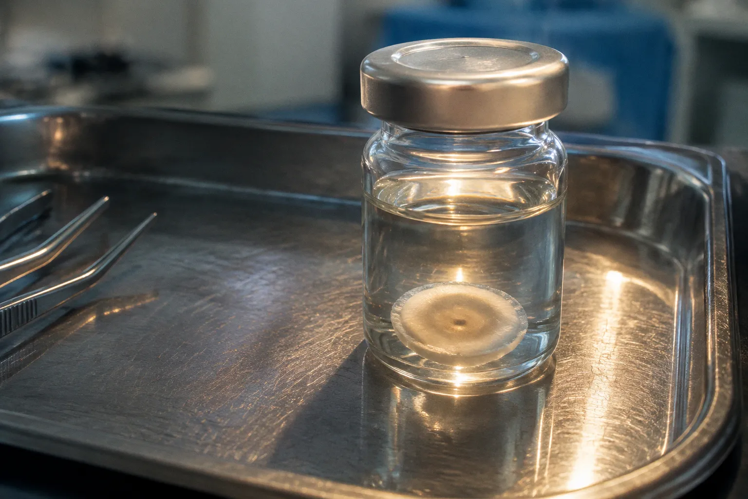
Early disease may not be obvious on macroscopic inspection or routine specular microscopy, while the underlying Descemet’s membrane–endothelial complex is already thickening and developing guttae.
That is the central difficulty in fuchs dystrophy donor cornea tissue assessment: the specimen must be treated as more than a transparent disc with a cell-density number attached. Its clinical history, endothelial architecture, guttae distribution, effective endothelial cell density, and intended use all affect whether the tissue is suitable for a particular study.
For translational work, the bench question is not simply whether endothelial cells are present. It is whether the tissue can support a reliable experiment, whether its pathological features are documented well enough to interpret the result, and whether the findings can be connected to surgical realities such as graft survival, tissue handling, and functional recovery.
Start with the tissue question, not the microscope
Before imaging begins, define what the donor cornea is expected to answer.
A specimen intended for endothelial cell stress studies needs a different level of characterization from one being used for broad histopathology. A cornea selected for Descemet membrane work may be valuable precisely because it contains guttae and altered matrix architecture. The same features could make it unsuitable for a study that requires a relatively uniform endothelial surface.
This distinction is particularly important in Fuchs endothelial corneal dystrophy, where pathological change is not distributed evenly across the cornea. Central guttae may occupy the region most relevant to clinical disease, while the peripheral endothelium can retain a different cell pattern. A single endothelial cell-density measurement may therefore give a misleading impression of tissue quality if it does not identify where the image was taken and whether the analyzed region is free of guttae.
A useful intake record should capture:
- The donor’s known clinical diagnosis, when available, and the source of that information.
- Whether the cornea is being evaluated for transplantation research, explant culture, imaging, molecular work, or histopathology.
- The preservation method and time in organ culture before analysis.
- The location of each image or tissue section, particularly whether it is central, paracentral, or peripheral.
- The presence, density, and morphology of guttae.
- Whether the endothelial surface is continuous, irregular, or visibly disrupted.
- The planned endpoint: cell density, cell stress, membrane thickness, ultrastructure, or graft-related assessment.
This is not administrative overhead. It is how a laboratory prevents a pathological specimen from being treated as an interchangeable control.
The clinical context changes the meaning of the number
Fuchs dystrophy is associated with progressive endothelial cell loss and guttae formation on Descemet’s membrane. The endothelial layer is responsible for maintaining corneal deturgescence, so a reduction in functional endothelial reserve has direct implications for stromal clarity and postoperative recovery.
For research, however, “endothelial cell density” must be handled carefully. In regions containing central guttae, the apparent cell count may not represent the healthy cell population. The effective density should account for the guttae-free area fraction rather than treating the entire image as a uniform field.
This matters when comparing donor tissue groups. A specimen with relatively preserved peripheral cells can appear stronger than it is if the central pathological area is excluded without being reported. Conversely, a low count from a guttae-heavy central region may reflect the geometry of the disease rather than a uniform loss across the entire cornea.
A donor cornea is not validated by one cell-density figure. It is validated by showing where that figure came from, what the image contains, and how the disease changes its interpretation.
Quantifying Descemet’s membrane and the endothelial complex
One of the most reproducible bench observations in Fuchs dystrophy is thickening of Descemet’s membrane and the associated endothelial complex. In one reported comparison, the combined thickness averaged 34.8 ± 11.6 µm in FECD eyes, compared with 19.3 ± 2.3 µm in healthy controls, a statistically significant difference.
That finding gives the laboratory a useful structural endpoint, but thickness should not be treated as a surrogate for every aspect of tissue quality. A thicker complex may reflect disease-associated matrix deposition and guttae formation, yet it does not by itself describe the viability, stress state, or surgical behavior of the endothelial cells.
A practical measurement sequence
A robust assessment generally follows the anatomy from broad structure to cellular detail:
1. Orient the specimen and document the sampling region.
The central cornea, the mid-periphery, and the outer peripheral region may not carry the same disease burden. Record the location before imaging or sectioning.
2. Use optical imaging to establish the overall architecture.
Optical coherence tomography can help define the contour and thickness of the cornea and provide a non-destructive structural overview. It is especially useful when the laboratory needs to compare tissue regions before committing the specimen to destructive analysis.
3. Measure the Descemet’s membrane–endothelial complex consistently.
The measurement boundary should be defined in advance. A protocol that changes the boundary from one image to another will create apparent biological variation that is actually a measurement problem.
4. Map guttae separately from membrane thickness.
Guttae density, size, clustering, and distribution should not be folded into a single thickness value. They represent related but distinct features of the disease.
5. Confirm selected findings with higher-resolution methods.
Electron microscopy can provide ultrastructural detail when the question concerns membrane architecture, cell morphology, or the relationship between guttae and the endothelial surface.
The goal is not to force every specimen through every imaging platform. It is to match the technique to the question and preserve enough spatial information that another investigator can understand what was actually measured.
What thickness can—and cannot—tell you
Thickness is valuable for group-level comparisons and for identifying disease-associated structural change. It can support a distinction between healthy control tissue and affected donor cornea, particularly when paired with endothelial imaging and histopathology.
It does not, on its own, establish that a graft will fail, that the cells are non-viable, or that the tissue is unsuitable for all forms of research. A cornea with substantial Descemet membrane pathology may be highly informative for a guttae or matrix study. A cornea with the same structural finding may be inappropriate for a study requiring a stable, confluent endothelial monolayer.
That is why tissue characterization should end with a use-specific disposition rather than a generic pass-or-fail label.
Effective endothelial cell density follows disease severity
In Fuchs dystrophy, the relationship between clinical severity and endothelial cell loss is one of the more clinically useful links between the examination room and the research bench. Confocal imaging has shown a strong linear relationship between effective endothelial cell density and subjective clinical severity grade, with a reported correlation of r = −0.93.
The direction is clinically intuitive: as disease severity increases, effective endothelial cell density decreases. The strength of the relationship is useful because it supports the idea that a well-documented clinical grade can help organize tissue cohorts for laboratory work.
But the word effective matters. A nominal density calculated across an image that includes guttae may not reflect the density of functioning cells in the usable area. Central guttae can distort the apparent cell geometry, obscure cell borders, and create regions that should not be counted in the same way as guttae-free endothelium.
Building a defensible cell-density assessment
For fuchs endothelial cell loss bench validation, the laboratory should make the counting method visible rather than presenting the result as a black box.
A sound record should include:
- The imaging modality used, such as specular or confocal microscopy.
- The region sampled and its distance from the corneal center, when known.
- The number and quality of usable fields.
- Whether guttae were included, excluded, or analyzed separately.
- The method used to identify cell borders.
- The distinction between nominal density and effective density.
- Any areas where cell borders could not be resolved reliably.
- The clinical severity grade used for comparison, if available.
This documentation is particularly important when tissue is being compared across eye-bank sources. Different preservation intervals, imaging settings, and operator decisions can produce differences that look biological but are actually procedural.
Peripheral tissue and DSO-related selection
The peripheral endothelium may retain greater cell density than the central diseased area. That observation has practical relevance for studies of Descemet stripping only, where selection criteria may include a minimum peripheral endothelial cell density of 1,000 cells/mm².
The number should not be treated as a universal suitability threshold for every experiment or procedure. It belongs to a specific selection context and must be interpreted alongside guttae distribution, cell morphology, wound-healing capacity, and the intended surgical approach.
For a research team studying anterior segment donor graft selection, the key point is simple: central and peripheral measurements answer different questions. Central data describe the most visibly affected region. Peripheral data may help estimate the reserve available for a procedure that relies on remaining host endothelial cells or on repopulation from healthier margins.
In Fuchs tissue, the location of the measurement can be as important as the measurement itself.
Advanced imaging and histopathological bench validation
Routine eye-bank evaluation remains essential, but it is not designed to answer every research question. Macroscopic inspection and specular microscopy can identify obvious abnormalities, yet they may fail to detect early Fuchs dystrophy in donor corneas.
That limitation has direct consequences for tissue distribution. A specimen classified as acceptable by routine screening may still contain early disease-associated changes that affect a cell-culture experiment, a matrix study, or the interpretation of a control group.
This does not make routine screening ineffective. It means that screening and research characterization serve different purposes.
Match the method to the biological question
| Research question | Useful assessment approach | Main limitation |
|---|---|---|
| Is the endothelial surface visibly intact? | Macroscopic inspection and specular microscopy | Early disease may remain undetected |
| How does disease affect cell density? | Confocal or specular imaging with regional analysis | Guttae can distort effective counts |
| How thick is the Descemet membrane–endothelial complex? | OCT and defined histological or ultrastructural measurements | Boundary selection must be standardized |
| What is the arrangement of guttae and nearby cells? | High-resolution imaging and histopathology | Destructive methods limit later use |
| What are the fine structural changes in archived tissue? | Electron microscopy, including serial block face SEM when appropriate | Sample preparation is technically demanding |
| How do cells respond to culture conditions? | Explant handling followed by cell-stress and guttae analysis | Handling itself can alter the stress profile |
For archived resin-embedded human corneal tissue, three-dimensional volume electron microscopy can provide detailed structural information. Serial block face scanning electron microscopy requires de-plastination followed by additional staining with uranyl acetate and lead acetate. Those preparation steps are not minor refinements; they determine whether the preserved tissue can be interpreted at the intended resolution.
A laboratory planning this type of work should decide early whether the specimen must remain available for other endpoints. Once tissue has undergone destructive processing, it cannot return to a live explant workflow or a routine graft-preparation pathway.
Histology should answer a defined question
Histopathology is most useful when the section is tied to a specific endpoint. A general statement that the tissue is abnormal is less informative than a structured description of:
- Descemet membrane thickness and layering.
- The distribution and morphology of guttae.
- Endothelial cell loss or irregularity.
- Areas of denudation or disrupted cell coverage.
- Stromal architecture relevant to the planned experiment.
- The relationship between imaging findings and tissue sections.
This paired approach—imaging before sectioning, histology afterward—allows the laboratory to connect two-dimensional or non-destructive observations with the actual tissue architecture. It also helps explain why two specimens with similar overall clinical grades may behave differently in culture.
Standardizing explant handling before stress analysis
Cellular stress markers are highly sensitive to handling. In research explants, the period between tissue preparation and assay can become a major source of variation if it is not standardized.
A reported handling approach keeps corneal endothelial explants on Descemet’s membrane overnight at 37°C in growth medium before evaluating cell-stress markers and guttae density. The purpose is not to recreate a clinical graft environment. It is to establish a consistent pre-assay condition so that samples are compared after a defined recovery or equilibration interval.
Why the overnight step matters
An explant does not enter the laboratory in a neutral state. Donor age, disease burden, preservation history, dissection trauma, temperature changes, and time outside controlled conditions can all influence the cells. If one specimen is analyzed immediately while another spends many hours in culture medium, the resulting differences may reflect handling rather than Fuchs pathology.
Keeping the endothelium attached to Descemet’s membrane also preserves the local relationship between cells and their extracellular matrix during the standardization period. That is relevant when the study examines guttae, adhesion, cell spreading, or stress responses linked to the diseased membrane.
A practical workflow might include the following sequence:
1. Prepare the explant using a consistent dissection approach and document any visible mechanical damage.
2. Keep the endothelial surface oriented consistently in the growth medium.
3. Maintain the defined temperature and overnight interval for all comparable specimens.
4. Record whether the tissue remained attached to Descemet’s membrane throughout handling.
5. Image or sample the explant using the same order of operations across the study group.
6. Analyze cell-stress markers alongside guttae density rather than treating either endpoint as a complete description of tissue health.
The last point is important. A high guttae burden does not automatically predict the same cellular stress profile in every specimen, and a preserved cell count does not demonstrate normal function. The value comes from combining structural and cellular endpoints.
Avoid turning culture into a second source of uncertainty
Growth medium composition, temperature, incubation time, and tissue orientation should be recorded with the same care as the imaging settings. If those parameters change between batches, the laboratory may lose the ability to distinguish donor-related biology from culture-related variation.
The most useful standard operating procedure is usually not the longest one. It is the one that identifies the variables capable of changing the endpoint and keeps them stable. In endothelial research, those variables include the time from dissection to culture, the duration of the pre-assay interval, the condition of Descemet’s membrane, and the criteria for excluding damaged explants.
Where traditional eye-bank screening stops
Traditional screening is optimized for safety, basic tissue suitability, and operational decisions. Research-grade characterization asks a narrower and more demanding question: what biological information is hidden inside a specimen that appears acceptable at routine inspection?
Early Fuchs dystrophy is a good example. Macroscopic findings and standard specular microscopy may not reliably identify early disease. If affected tissue enters a control group, the consequences can extend beyond one mislabeled sample. It can alter cell-density distributions, increase apparent variability, weaken a treatment comparison, and make a promising intervention look less consistent than it is.
At the same time, not every donor cornea requires genetic testing or advanced microscopy. The appropriate level of assessment depends on the study’s endpoint and the cost of a false classification.
Early-onset Fuchs dystrophy has been linked to point mutations in COL8A2, which encodes the alpha-2 subtype of collagen VIII. Most late-onset cases are associated with non-coding repeat expansions in TCF4. These genetic associations are clinically and biologically important, but they do not establish a universal eye-bank protocol for genetic testing of all donor tissues before research distribution.
The practical response is stratification. A laboratory may choose to:
- Use routine screening for exploratory work where early disease would not invalidate the endpoint.
- Add regional endothelial imaging when cell density or cell stress is central to the study.
- Use OCT or histopathology when membrane thickness and architecture are key outcomes.
- Reserve genetic or advanced molecular characterization for studies specifically addressing disease mechanism, donor classification, or genotype–phenotype relationships.
- Separate tissue into clearly documented control, suspected disease, and confirmed disease groups rather than forcing uncertain specimens into a binary label.
Research controls need a stronger definition
A control cornea is not necessarily a cornea that passed routine eye-bank screening. It is a specimen whose relevant disease features have been assessed to a level appropriate for the experiment.
For a study of donor corneal guttae density scoring, the control must be free of the pathological features being measured or must be analyzed in a way that makes the distinction explicit. For a cell-stress experiment, the control should have documented handling conditions and a comparable preservation history. For a study of keratoplasty graft quality, endothelial cell density must be interpreted together with morphology, regional distribution, and the mechanical or surgical demands of the graft.
This is where tissue logistics become part of the science. A specimen that arrives without preservation timing, imaging location, or dissection notes carries an uncertainty that no later statistical analysis can completely remove.
Connecting bench findings to anterior segment surgery
The value of Fuchs dystrophy tissue research ultimately depends on whether it improves clinical decisions or makes future interventions more predictable. That connection is easiest to see in anterior segment surgery.
Endothelial keratoplasty depends on the interaction between donor tissue quality, recipient anatomy, graft preparation, surgical delivery, and postoperative cell survival. Descemet membrane endothelial keratoplasty uses a very thin tissue construct that must be prepared, loaded, delivered, unfolded, and positioned without creating unnecessary endothelial trauma. A laboratory model that ignores those physical steps may produce elegant cellular data with limited surgical relevance.
For translational interpretation, ask four practical questions:
1. Will the measured cell population survive the preparation and delivery process?
2. Does the assay reflect the surface or region that will matter clinically?
3. Can the tissue recover its functional role after manipulation, not merely retain a positive viability marker?
4. Does the disease feature being studied affect graft integration, postoperative clearing, or long-term cell reserve?
These questions do not turn a bench experiment into a clinical trial. They keep the experiment connected to the conditions under which a graft must function.
Fuchs tissue can also inform the biology of limbal and anterior segment repair more broadly. The corneal stroma, trabecular meshwork, anterior chamber drainage structures, and endothelial layer are not interchangeable research materials, but they share a translational problem: tissue architecture matters. Cells behave differently when removed from their native matrix, and the degree of that difference should be visible in the study design.
A practical endpoint for each donor cornea
At the end of assessment, the specimen should have more than a diagnosis label. It should have a research profile that explains what can be trusted.
A concise profile might include:
- Structural status of Descemet’s membrane and the endothelial complex.
- Central and peripheral effective endothelial cell density, where feasible.
- Guttae distribution and density.
- Imaging quality and unresolved regions.
- Preservation and explant-handling history.
- Histopathological or ultrastructural findings, if performed.
- Recommended uses and limitations.
- Whether the tissue is suitable as a control, disease model, or exploratory specimen.
This approach supports better coordination between eye banks, laboratories, and clinical investigators. It also makes later analysis more honest. A specimen can be valuable even when it is not suitable for transplantation or for a tightly controlled cell-density study. The important point is to describe its value accurately.
The clinical standard is useful, but it is not the whole standard
Fuchs dystrophy donor cornea tissue assessment works best when it combines the discipline of eye-bank screening with the specificity of research pathology. Routine inspection can identify major abnormalities. Specular and confocal imaging can define regional cell loss. OCT and histology can expose membrane thickening. Electron microscopy can resolve structure that routine methods cannot. Standardized explant handling can reduce avoidable variation before stress and guttae analyses begin.
No single technique replaces the others. The strongest workflow is layered, with each method answering a different question.
The cautious optimism here is justified. Better characterization does not eliminate the biological variability of donor tissue, and it does not create a universal protocol for every eye bank or every experiment. It does, however, make that variability visible. Once the laboratory knows whether it is looking at early guttae, advanced cell loss, membrane thickening, handling injury, or a mixture of all four, the research becomes more interpretable—and the path from donor tissue to surgical relevance becomes much clearer.