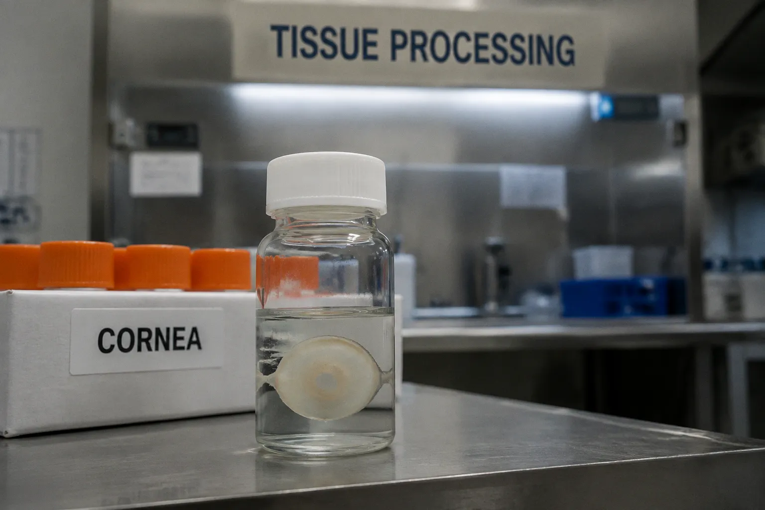
In older donor tissue, limbal epithelial stem cells retain telomere length and telomerase activity at broadly comparable levels to cells from younger donors, yet their functional performance declines: colony-forming efficiency falls, explant outgrowth slows, and the crypt-like architecture of the limbal niche becomes visibly less pronounced.
That distinction matters. The evidence does not describe a simple case of aged cells becoming biologically inert. It describes a more exacting failure of tissue organization and regenerative execution. In donors older than 65, the colony-forming efficiency ratio falls to 0.3 ± 0.06 compared with younger donor groups, while the proportion of p63α-positive outgrowth cells decreases from 18.2 ± 3.6% in donors aged 45 or younger to 14.1 ± 4.6%. The cells are still present. The question is whether the niche can still instruct them to behave like a durable epithelial reserve.
For researchers working with human limbal tissue, this is the central issue behind limbal stem cell potency in older donor eyes. Donor age is not an absolute exclusion criterion, but it is a measurable variable that can shift the starting biology of the specimen before culture, expansion, or transplantation begins.
Morphological shifts in the aged limbal niche
The limbus is not merely a border between cornea and conjunctiva. It is a structured epithelial compartment containing the microanatomical features that support limbal epithelial stem-cell maintenance, including crypt-like topography and localized cellular organization. When that architecture changes, the effect is not cosmetic. It alters the physical and biochemical context in which stem and progenitor cells divide, migrate, and replenish the corneal surface.
Cadaveric corneal imaging shows that the microstructural area occupied by limbal epithelial crypts is sharply reduced after 60 years of age. The niche becomes smoother, less deeply articulated, and apparently less capable of preserving the spatial heterogeneity associated with a functional stem-cell compartment.
This architectural smoothing provides one plausible explanation for why cellular output can decline even when intrinsic chromosomal-age markers remain comparatively stable. A stem cell does not operate in isolation. Its behavior is shaped by epithelial neighbors, basement-membrane interactions, extracellular matrix composition, local growth-factor gradients, and the geometry of the compartment itself. Damage or simplification at any of these levels can reduce regenerative performance without producing immediate telomere shortening.
The distinction is especially important when interpreting tissue harvested after death. A limbal specimen carries two histories:
1. The donor’s biological history, including age-related changes in the limbal niche and epithelial cell population.
2. The specimen’s post-mortem and preservation history, including the interval before recovery, transport conditions, dissection quality, and organ-culture storage.
These histories converge in the assay. A lower cell yield or weaker outgrowth cannot automatically be assigned to age unless the procurement and preservation variables are recorded with enough precision to separate donor biology from tissue handling.
The older limbus does not necessarily lose its stem cells first; it loses some of the architecture that tells those cells how to behave.
The observed morphological change therefore functions as more than a descriptive endpoint. It may be a tissue-level explanation for the age effect: a less complex niche, fewer protected microdomains, and a weaker local environment for maintaining clonogenic epithelial populations.
What the colony-forming data actually show
Colony-forming efficiency is one of the more informative functional readouts for human limbal epithelial cells because it tests whether isolated cells can generate proliferative colonies under defined culture conditions. It is not a complete measure of clinical potency, but it moves the analysis beyond simply counting viable cells.
In the available donor comparisons, the CFE ratio declines significantly in older tissue, reaching 0.3 ± 0.06 in donors older than 65 relative to the younger reference group. That is a substantial reduction in colony-forming performance. It suggests that fewer harvested cells retain the combination of survival, proliferative capacity, and clonogenic behavior required to establish colonies after isolation.
The result should not be translated into the claim that older donor cells are non-viable. That would confuse a reduction in functional frequency with total loss of function. The aged limbus may still yield cells capable of expansion, but the proportion of cells that behave as robust colony-forming progenitors is lower.
Several biological states may be mixed inside the same cell suspension:
- Cells with relatively preserved stem-like behavior and strong proliferative potential.
- Early progenitors that can divide but have limited long-term clonogenic capacity.
- More differentiated epithelial cells that survive isolation but contribute little to sustained colony formation.
- Cells stressed by dissection, enzymatic release, post-mortem delay, or storage.
The assay reports the resulting population-level performance. It does not, by itself, identify which pathway caused each cell to fail. This is why CFE should be interpreted alongside tissue morphology, marker expression, isolation yield, and the details of organ-culture preservation.
The p63α data add another layer. Outgrowth cells from donors aged 45 or younger contain 18.2 ± 3.6% p63α-positive cells, compared with 14.1 ± 4.6% in donors older than 65. The difference is statistically significant, but p63α should not be treated as a solitary molecular password for transplant success. It is a marker within a broader phenotype, and marker abundance does not automatically equal durable regenerative function.
Still, the direction of the signal is coherent. Older tissue shows:
- Less efficient colony formation.
- A lower proportion of p63α-positive outgrowth cells.
- Reduced explant outgrowth.
- A more weakly structured limbal niche.
When independent readouts move in the same direction, the age effect becomes difficult to dismiss as an artifact of a single assay.
The paradox of stable telomeres and declining potency
The most revealing finding is also the one most likely to be oversimplified. Telomere length and telomerase activity remain relatively stable across donor age groups, even as clonogenic capacity declines.
This weakens the idea that telomere shortening is the primary driver of age-associated limbal stem-cell impairment in these specimens. The cells do not appear to follow a straightforward model in which replicative exhaustion is directly reflected by progressively shortened telomeres. Instead, the decline may arise from changes in cellular state, tissue organization, signal reception, or the ability to respond to injury and culture conditions.
Several mechanisms remain plausible, but the exact molecular explanation is unresolved. The available evidence does not establish whether the dominant defect lies in:
- Altered communication between limbal epithelial cells and their supporting niche.
- Changes in extracellular matrix or basement-membrane signaling.
- Accumulation of senescent or partially differentiated epithelial cells.
- Reduced stress tolerance during post-mortem recovery and tissue isolation.
- A shift in the balance between self-renewal and lineage commitment.
- Age-related changes in stromal, vascular, immune, or neural components adjacent to the limbal epithelium.
That uncertainty is not a weakness of the data. It is the precise boundary of what the data can support. A stable telomere profile paired with lower CFE tells us that the functional decline is real but not explained by telomere metrics alone.
For laboratories, this has a practical consequence: a donor-screening panel built around telomere length would miss a central component of the age-related phenotype. A tissue may display preserved telomeric parameters while producing fewer colonies, slower outgrowth, and a lower fraction of p63α-positive cells.
Which readouts move with donor age?
| Readout | Younger donor tissue | Older donor tissue | Interpretation |
|---|---|---|---|
| Limbal explant outgrowth, day 9 | 296 ± 54.7 mm² in donors aged ≤45 | 257 ± 44.0 mm² in donors aged >65 | Slower or less extensive epithelial expansion |
| Colony-forming efficiency | Higher reference level | 0.3 ± 0.06 in donors aged >65 | Reduced clonogenic performance |
| p63α-positive outgrowth cells | 18.2 ± 3.6% | 14.1 ± 4.6% | Lower representation of a relevant stem/progenitor-associated phenotype |
| Limbal crypt architecture | More developed microstructural area | Sharply reduced after age 60 | Smoother, less complex niche topography |
| Telomere length and telomerase activity | Relatively stable | Relatively stable | Does not explain the functional decline on its own |
| Total isolated limbal cell yield | Higher tendency | Inversely associated with age | Lower recovery from older stored tissue |
The table exposes the central pattern: age-associated impairment is visible in function, architecture, and recovery, but not in every intrinsic cellular metric. Biology has not produced a single failing switch. It has produced a cascade with several partially independent branches.
Explant outgrowth and the hidden cost of storage
Limbal explant outgrowth provides a more tissue-level view than CFE. Instead of asking only whether individual isolated cells can form colonies, it examines how a piece of limbal tissue expands across a scaffold under culture conditions.
On amniotic membrane scaffolds, limbal explant outgrowth measured on day 9 reaches 296 ± 54.7 mm² in donors aged 45 or younger, compared with 257 ± 44.0 mm² in donors older than 65. The difference is significant, and it aligns with the CFE findings: older tissue does not expand as aggressively from the explant.
The mechanism may involve a reduced number of competent cells, a slower proliferative response, impaired migration, or some combination of these events. Explant outgrowth is not simply a cell-count assay. It depends on the ability of the tissue fragment to release viable epithelial populations, establish adhesion to the scaffold, migrate across the surface, and maintain proliferation without prematurely differentiating.
The storage context becomes critical here. In organ-culture stored tissue, total isolated limbal cell yield shows an inverse relationship with donor age, with an association reported as R² = 0.27 and p < 0.001. The result indicates that older donor tissue tends to produce fewer isolated limbal cells, but it does not mean that age explains every difference between specimens. An R² of 0.27 leaves substantial variation attributable to other factors.
Those factors are not incidental details in an ocular biobank. They are part of the biological record of the sample. A meaningful human limbal stem cell harvest criteria framework should therefore treat donor age as one axis within a larger matrix that includes:
- Time from death to recovery, where available.
- Time and conditions between recovery and organ-culture placement.
- Duration of storage before dissection or isolation.
- Anatomical precision of the limbal harvest.
- Presence of peripheral corneal or conjunctival contamination.
- Scaffold type and preparation.
- Enzymatic digestion conditions.
- The passage or expansion stage at which cells are assessed.
- The precise definition used for viability, outgrowth, and colony formation.
Without these fields, age can become a misleading proxy for tissue quality. An older donor specimen recovered promptly and handled consistently may outperform a younger specimen compromised by delay or poor dissection. The research question is not whether age matters in the abstract. It is how much of the observed phenotype belongs to age, how much belongs to preservation, and how the two interact.
What donor age means for tissue selection
The evidence supports a graded selection model rather than a binary rule. Donor age should influence how limbal tissue is allocated, characterized, and compared, but it should not be used to declare all older tissue unsuitable.
For studies focused on regenerative biology, younger donor tissue may offer a more favorable starting population because it tends to show larger explant outgrowth, higher CFE, greater p63α representation, and higher isolated-cell yield. That makes it valuable when the experimental question depends on maximal expansion or on preserving a high-frequency clonogenic compartment.
Older donor tissue, however, remains scientifically important. It may be the more appropriate specimen for studying:
- Age-related remodeling of the limbal niche.
- The relationship between crypt architecture and epithelial regeneration.
- The separation of intrinsic cellular aging from microenvironmental decline.
- The effects of organ-culture preservation on compromised tissue.
- Mechanisms that preserve telomeres while reducing clonogenic output.
- The biology of donor corneas with reduced regenerative reserve.
The correct specimen therefore depends on the question. A laboratory attempting to optimize limbal epithelial expansion may stratify samples by age and prioritize younger tissue for baseline production runs. A laboratory investigating epithelial aging should not remove older specimens from the collection; doing so would erase the phenotype under study.
For transplant-oriented research, donor age should be reported alongside functional measures rather than used as a surrogate for them. A tissue bank or research coordinator handling limbal material should be able to distinguish at least three separate statements:
1. The donor was older.
2. The tissue yielded fewer cells or expanded more slowly.
3. The tissue displayed a lower frequency of cells with the selected functional or phenotypic markers.
These statements may be related, but they are not interchangeable. Age is a biological variable. CFE, outgrowth area, p63α expression, and total cell yield are measured properties of the recovered specimen. The strongest selection decisions preserve that distinction.
A practical comparison for research allocation
| Research objective | More informative donor-age strategy | Core measurements |
|---|---|---|
| Maximize epithelial expansion | Enrich for younger donors while retaining age-matched controls | Outgrowth area, CFE, cell yield |
| Study limbal aging | Include younger and older donors with standardized recovery and storage records | Crypt architecture, p63α, CFE, telomeres |
| Examine preservation injury | Match tissue-handling variables across age groups | Viability, yield, outgrowth, storage duration |
| Investigate niche biology | Pair structural imaging with epithelial functional assays | Crypt area, morphology, colony formation |
| Evaluate translational potential | Avoid age as the sole exclusion criterion; compare functional quality directly | Marker profile, expansion performance, tissue provenance |
The most useful collection is not the one with the youngest donors. It is the one with enough metadata to reveal why one specimen performs differently from another.
Does age compromise limbal stem cells?
Yes, donor age compromises several measurable aspects of limbal stem-cell performance, but the word “compromise” must be used precisely.
In donors older than 65, limbal tissue shows lower colony-forming efficiency, reduced p63α-positive outgrowth, smaller day-9 explant expansion on amniotic membrane, and lower total isolated-cell yield in organ-culture stored specimens. After 60, the limbal crypt architecture also becomes markedly reduced. Taken together, these findings indicate that the aged limbal niche has a weaker regenerative profile than younger donor tissue.
But the decline is not equivalent to complete loss of stem-cell capacity. Telomere length and telomerase activity remain relatively stable, and the available evidence does not establish that older donor limbal cells are incapable of expansion or therapeutic use. Nor does it establish that donor age alone determines long-term clinical graft survival after allogeneic limbal stem-cell transplantation.
The more accurate conclusion is narrower and more useful: older donor age lowers the probability that recovered limbal tissue will contain a highly clonogenic, rapidly expanding epithelial population, while leaving the cellular mechanism of that decline only partly resolved.
That unresolved mechanism sits between the cell and its niche. The cells retain some intrinsic markers of replicative reserve, yet the tissue loses architectural complexity and functional output. The next question is therefore not simply whether the older limbus contains stem cells. It is whether the aged microenvironment still provides the signals, geometry, and matrix context required to keep those cells in a regenerative state after recovery, storage, and culture.