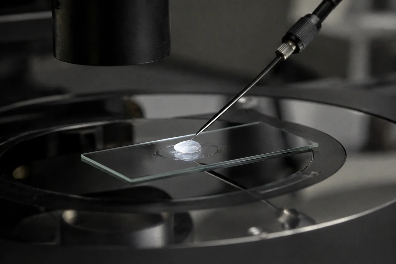
The figure collapses what was long treated as an age-related outflow decline into a reproducible, quantitative failure mode. For eye banks, the consequence is operational: donor eyes are not interchangeable substrates. They are stratified reservoirs of disease-stage biomechanics, and the procurement pipeline has to price that variance into every downstream assumption about research utility.
The bottleneck sits at the juxtacanalicular region of the outflow pathway. When normal donor tissue registers a mean modulus of approximately 4.0 kPa on micro-indentation, advanced POAG samples routinely exceed 80 kPa, with a fold-change large enough to make the disease a load-bearing variable in any tissue-quality model.
Quantifying Pathological Stiffening: AFM vs. Finite Element Modeling
The first analytical decision in any donor study is methodological. Two principal modalities disagree on absolute stiffness by nearly an order of magnitude, and the disagreement is not noise—it is a signature of the scale each technique interrogates.
Direct micro-indentation via atomic force microscopy remains the reference standard for human TM tissue. The seminal Last et al. study, published in Investigative Ophthalmology & Visual Science in 2011, established a baseline mean elastic modulus of 4.0 ± 2.2 kPa in normal juxtacanalicular tissue against a glaucomatous mean of 80.8 ± 32.5 kPa. Subsequent donor-cohort replications have held the lower bound but widened the variance band on the disease end, with local moduli in advanced POAG samples crossing 100 kPa in some specimens.
Finite element modeling (FEM), constrained by anterior segment OCT imaging and inverse calibration against clinical outflow facility, returns different magnitudes. Published bulk stiffness values under this framework cluster at 70 ± 20 kPa for normal donor TM and 98 ± 19 kPa for glaucomatous donor TM. The FEM "normal" figure sits inside the AFM "glaucomatous" range, and the FEM "glaucomatous" figure sits only modestly above it.
| Parameter | AFM (micro-indentation) | FEM (inverse OCT-based modeling) |
|---|---|---|
| Normal TM mean modulus | 4.0 ± 2.2 kPa | 70 ± 20 kPa |
| Glaucomatous TM mean modulus | 80.8 ± 32.5 kPa | 98 ± 19 kPa |
| Volume sampled | Micrometer-scale tissue | Whole-angle bulk response |
| Modality signature | Local juxtacanalicular stiffness | Integrated hydraulic resistance |
| Sensitivity to ECM cross-linking | High | Moderate |
The discrepancy is structural, not contradictory. AFM reads the intrinsic elasticity of the extracellular matrix at the local cell–matrix interface; FEM captures the combined mechanical and hydraulic behavior of the entire conventional outflow pathway, including collector channels and episcleral venous pressure contributions. Any cross-modality comparison has to acknowledge that the two numbers describe different physical observables. They cannot be averaged, ratio-transformed, or treated as interchangeable inputs into a single model.
Direct AFM measures intrinsic matrix stiffness; inverse FEM measures effective outflow resistance. The two diverge by roughly an order of magnitude—and the gap is diagnostically informative.
Cellular Depletion and the Decline of the TMSC Population
Stiffening arrives in donor eyes as a population collapse. TM cellularity declines with both age and disease, but the disease signal dominates. Section-count data from human donor eyes place young donors at 72.17 ± 3.8 TM cells per histological section, healthy older donors at 58.84 ± 6.2 cells, and glaucomatous donors at 48.22 ± 2.3 cells. The drop between the healthy older cohort and the glaucomatous cohort—roughly 18%—is disproportionate to the age gap between the two groups and tracks with disease progression rather than chronological drift.
The decline is sharper inside the stem cell compartment. Trabecular meshwork stem cells (TMSCs) are documented at 6.92 ± 0.83 per section in young donor eyes and 1.11 ± 0.43 in glaucomatous donor eyes—a roughly 84% reduction. Healthy older donors retain 3.07 ± 1.10 TMSCs per section, which positions the glaucomatous loss as an accelerated trajectory rather than an extension of normal aging.
For biobankers, the implication is procurement-tier specific. Donor corneas harvested for anterior segment research now have to be classified not only by endothelial count and stromal clarity but by the anticipated TM reserve of the originating globe. Eyes from donors with documented POAG, even in early stages, will yield TM tissues with depleted cellular machinery and a stiffened matrix. Eyes from older healthy donors will yield TM tissues with reduced—but qualitatively intact—cell populations whose biomechanical readouts still cluster near the normal AFM range.
Three operational variables follow:
1. Procurement lead time postmortem. TMSCs and TM cellularity fall further as death-to-preservation interval extends; downstream TM studies require tighter cold-chain controls than corneal endothelial workups.
2. Donor chart stratification. Glaucoma status in the donor record is no longer background metadata for TM studies—it is a primary classifier.
3. Yield metrics per donor class. Expected TMSC yield from a POAG donor is a fraction of what a young healthy donor supplies, and this gap compounds downstream in any expansion or differentiation protocol.
Senescence-Associated ECM Remodeling in the Juxtacanalicular Region
Stiffening without cell loss is a rheological event; stiffening with cell loss and senescence is a tissue-identity event. Donor-derived TM cells cultured to senescence register 1.88-fold to 2.57-fold higher stiffness than early-passage proliferative controls, depending on serum conditions in the culture system. That cell-autonomous shift is smaller than the 20-fold jump observed in whole donor tissue, which points to the extracellular matrix as the dominant mechanical substrate in advanced disease.
The juxtacanalicular region is where the matrix deposits accumulate. Cross-linking of fibrillar collagen, accumulation of basement membrane components, and increased deposition of matricellular proteins all reduce the compliant response of the tissue under physiological outflow loads. The result, recorded by AFM in human donor tissue, is a resistance to deformation that no longer tracks with cellular tone or cytoskeletal contractility.
This is the cell–matrix feedback loop that donor tissue finally made visible. Senescent cells secrete a stiffer, more cross-linked matrix. The stiffer matrix accelerates senescence in the remaining cells. Donor AFM data quantify both halves of that loop from the same specimens, and the consistency across cohorts is what makes the 20-fold figure a load-bearing number for translational research.
The do-not-claim boundary here is precisely stated: cell loss is not "caused solely by aging." The reduction observed in glaucomatous donor tissue exceeds what age-matched healthy controls show, and the histopathological signature includes features that aging alone does not produce.
Biomechanical Disparities in the Inner Wall of Schlemm's Canal
The drainage pathway does not end at the TM. Donor tissue studies have extended the stiffness analysis downstream to the inner wall of Schlemm's canal, and the numbers change the troubleshooting map.
Direct measurements on human donor eyes place the inner wall of Schlemm's canal at 0.4 kPa in normal tissue and 0.7 kPa in glaucomatous tissue—an absolute change that looks small but a relative change of 75%. The inner wall is more compliant than the juxtacanalicular TM by roughly an order of magnitude even after disease, which is mechanically consistent with its role as a thin endothelial barrier rather than a load-bearing mesh.
The implication for outflow resistance modeling is significant. If the juxtacanalicular TM stiffens 20-fold while the inner wall stiffens less than 2-fold, the dominant resistance locus stays in the TM. Pharmacological interventions that successfully soften the TM would, on the donor data, restore most of the physiological pressure drop. Interventions targeted at the inner wall would address a smaller, though still real, fraction of the pathology.
This is where donor procurement intersects with preclinical pharmacology. Tissue-engineered TM models calibrated to the 4.0 kPa normal baseline can use AFM readouts as a quality-control gate before drug screening. Models calibrated to the FEM bulk range will systematically register as "stiff" against any local AFM measurement.
Clinical Implications of Outflow Resistance and Tissue Elasticity
The clinical translation of these donor measurements runs through three channels: diagnostic thresholds, surgical planning, and pharmacological development.
Diagnostic thresholds. OCT-derived biomechanical indices are entering clinical workflows, and their reference intervals are being calibrated against the same FEM and AFM donor datasets that produce the figures above. The wide variance band on glaucomatous TM stiffness—standard deviations of ±32.5 kPa on AFM, ±19 kPa on FEM—means that population-level cutoffs will work for screening but individual staging needs longitudinal tracking rather than single-timepoint classification.
Surgical planning. Angle-based and trabecular-bypass procedures depend on tissue compliance assumptions that the donor data now calibrate more precisely. A donor eye with end-stage POAG carries TM tissue that, on AFM, responds as a rigid scaffold rather than a viscoelastic mesh. Surgical interventions that assume residual compliance will underperform in these eyes, and donor-side pre-screening flags this in advance.
Pharmacological development. The molecular trigger initiating ECM cross-linking and elevated stiffness before observable IOP rise remains unresolved—donor tissue studies have not closed that loop, and postmortem pharmacological reversal of stiffness in human donor eyes has not been demonstrated as durable. Until those gaps close, drug development targets the downstream matrix and cellular senescence pathways, with donor tissue as the assay substrate rather than the therapeutic endpoint.
The procurement side of this picture is settled. Donor eyes deliver the only direct read on human TM mechanics. Cultured cells, animal models, and engineered hydrogels each contribute, but the donor-derived AFM and FEM values are the reference grid. Eye banks that annotate incoming globes with POAG status, donor age, and death-to-preservation interval produce research tissues that resolve cleanly into the normal, age-shift, and disease-shift bands. The logistics of that annotation determine how cleanly any individual study can read stiffness against baseline.
What remains unresolved is whether the 20-fold stiffening can be pharmacologically reversed in human tissue under any preservation or treatment window. Donor studies have not yet produced that answer, and it remains the highest-value open variable in the entire translational grid.
Researched and written by Joseph McCarthy
orbsproject.org · Corneal & Anterior Biology · Evening long-read