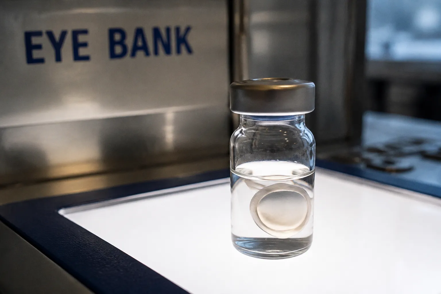
Once enucleation is complete, endothelial cell viability begins a measurable decline governed by storage temperature, medium formulation, and elapsed time from recovery. The choice of preservation protocol—and the corneal storage media that implements it—determines whether a recovered cornea reaches the operating theater as viable graft material or is reclassified as a research-grade specimen.
Two methodologies dominate the global landscape: hypothermic cold storage at 2°C to 8°C, and organ culture at 31°C to 37°C. Each carries distinct trade-offs in duration, handling complexity, and endothelial yield. The data behind these preservation protocols reveals not a single superior method, but a bifurcated operational grid tuned to regional infrastructure, regulatory frameworks, and clinical demand patterns.
The Mechanics of Hypothermic Cold Storage: From Optisol-GS to Life4C
Hypothermic storage suspends the cornea in a metabolically depressed state. Tissue is maintained at approximately 4°C, within the prescribed 2°C to 8°C range, slowing enzymatic degradation and reducing cellular demand for oxygen. Under these conditions, donor corneas remain viable for up to 14 days, making hypothermic storage the dominant preservation method in North America and Asia.
Optisol-GS remains the reference intermediate-term cold storage medium across U.S. eye banks. Its formulation combines chondroitin sulfate and dextran to osmotically regulate stromal hydration, preventing the edema that would otherwise compromise endothelial morphology during cold incubation. The medium also incorporates buffer systems and antioxidant agents to mitigate free-radical accumulation across the hypothermic interval. Dextran concentrations in Optisol-GS are calibrated to maintain stromal thickness within a surgically acceptable range at the point of graft delivery, though final corneal thickness at the time of transplant depends on the cumulative effects of storage duration and donor tissue characteristics.
A 2007 FDA approval introduced Life4C, an intermediate-term cold storage medium with a modified additive profile. Recombinant human insulin and glutathione appear in its composition—compounds intended to support metabolic stabilization of the endothelium during cold storage. The rationale is that insulin supports glucose uptake pathways in hypothermically suppressed endothelial cells, while glutathione provides an additional layer of antioxidant defense against reactive oxygen species that accumulate even at reduced temperatures. Clinical adoption has been gradual, constrained by validation requirements, procurement workflow reconfiguration costs, and the operational entrenchment of Optisol-GS across established tissue banks.
Additional cold storage formulations exist in regional markets. Eusol-C, used primarily in European programs that employ cold storage as a preliminary step before organ culture or for short-duration protocols, incorporates a different balance of osmotic agents and nutrients. The proliferation of formulations reflects a broader trend: no single medium has achieved universal dominance, and procurement programs frequently select storage media based on validated protocols within their regulatory jurisdiction, surgeon preference, and the logistical profile of their recovery-to-transplant pipeline.
The logistical advantage of hypothermic storage is infrastructure compatibility. Cold-chain transport, sealed vials, and standard refrigeration units align with existing blood-bank and tissue-procurement systems. For high-throughput eye banks with limited incubator capacity or constrained laboratory personnel, this remains the default operational pipeline. The handling requirements are minimal: tissue arrives in sealed containers, is inspected, and is stored until allocation or expiry. Each step is documented, but the chain-of-custody complexity is lower than organ culture workflows.
Hypothermic storage trades extended duration for infrastructure simplicity. The trade-off is endothelial yield measured in single-digit percentage losses over the first week.
Organ Culture Dynamics: Extending Viability to 35 Days
Organ culture operates at 31°C to 37°C, closer to physiological temperature. This warmer environment maintains corneas in a metabolically active state, supported by culture media supplemented with fetal bovine serum, antibiotics, and antimycotics. Under organ culture conditions, preservation extends to 30 to 35 days, more than double the hypothermic ceiling, and the method is widely adopted across European eye banks.
The extended duration creates downstream operational flexibility. Serological testing can be completed without compressing the tissue's remaining viability window. Surgical scheduling gains a buffer for recipient matching, transport coordination, and elective procedure timing. Tissue that fails clinical screening can be redirected to research biorepositories without the urgency that a 14-day hypothermic clock imposes. For biobank administrators managing dual-use procurement pipelines—clinical allocation and research distribution—the organ culture window provides a critical scheduling margin that hypothermic storage cannot match.
The operational cost is correspondingly higher. Incubators, sterile laminar-flow handling, and media exchange procedures at defined intervals demand infrastructure and trained personnel that many high-volume North American programs have not adopted at scale. Corneas stored in organ culture also undergo a dextran-based deswelling step prior to surgical use—a protocol-dependent procedure typically performed under controlled cold conditions, with the specific parameters (duration, dextran concentration, temperature) varying between eye banking programs. This deswelling phase adds procedural complexity and handling exposure to the chain of custody, introducing additional points where protocol deviation can affect tissue quality.
Microbiological surveillance is integrated into organ culture protocols, precisely because warm-temperature incubation supports bacterial and fungal proliferation that cold storage suppresses. Contamination events that would be arrested at 4°C can compromise tissue within days at 31°C. The baseline contamination rate reported across organ culture programs varies, but the risk is structurally inherent to the methodology: maintaining tissue at near-physiological temperatures in nutrient-rich media creates conditions favorable to microbial growth. Mitigation depends on rigorous aseptic technique during procurement and handling, prophylactic antibiotic supplementation in the culture medium, and scheduled microbiological sampling at defined intervals throughout the storage period. Programs with lower contamination rates typically correlate with higher investment in laminar-flow infrastructure and staff training, underscoring that organ culture's advantages in storage duration are inseparable from its demands on operational discipline.
The handling risk is not limited to microbiological contamination. Repeated media exchanges, deswelling protocols, and the physical transfer of tissue between containers at each procedural checkpoint increase the cumulative handling exposure relative to a single-step cold storage workflow. Each handling event carries a non-zero probability of endothelial trauma, mechanical damage to the corneal rim, or documentation error in the chain-of-custody record. These are manageable risks within well-resourced programs, but they are risks nonetheless, and they factor into why organ culture has not displaced hypothermic storage universally despite its duration advantage.
Comparative Endothelial Performance: Analyzing Cell Loss Metrics
Endothelial cell density (ECD), central corneal thickness (CCT), and endothelial cell loss (ECL) constitute the standard evaluation framework for assessing cornea preservation media performance. These parameters determine whether a cornea meets minimum thresholds for penetrating keratoplasty, endothelial keratoplasty, or lamellar procedures. The clinically relevant thresholds differ by procedure type: endothelial keratoplasty, which transplants only the endothelial cell layer and posterior stroma, is more sensitive to ECD loss than full-thickness penetrating keratoplasty, where the broader graft compensates to some degree for localized endothelial deficits.
A direct comparative evaluation of two cold storage media, Optisol-GS and Eusol-C, measured endothelial cell loss at day 5 post-procurement. Optisol-GS yielded 8.149% cell loss; Eusol-C yielded 7.614%. The differential is narrow, suggesting that within the hypothermic class, formulation differences produce measurable but modest variation in early-stage degradation kinetics. The ranking, however, varies by study and donor population; no single medium dominates across all measured parameters. Donor age, time from death to preservation, and cause of death introduce confounding variables that complicate direct medium-to-medium comparison even within controlled study designs.
Organ culture data exhibits different kinetics. Because the tissue remains metabolically active, endothelial cell loss accumulates more gradually but continues throughout the longer storage window. Direct comparison between methodologies is complicated by the endpoint mismatch: a 5-day hypothermic cornea and a 25-day organ-cultured cornea are not equivalent units. Normalized to days, organ culture cell loss rates often appear lower; normalized to total storage yield, the cumulative attrition can exceed hypothermic benchmarks. The relevant metric for clinical allocation is not rate but absolute cell density at the point of transplantation, and this is where the two methodologies converge on a shared constraint.
What the data establishes unambiguously is that neither method eliminates endothelial attrition. Both impose a measurable biological cost over time. The operational question becomes which degradation profile aligns with the clinical scheduling window and the available chain-of-custody infrastructure at each procurement site. For programs operating at high volume with rapid allocation cycles, the predictable, linear degradation of hypothermic storage may represent a lower total risk profile than the longer but higher-complexity organ culture pathway.
| Parameter | Hypothermic Cold Storage | Organ Culture |
|---|---|---|
| Temperature range | 2°C–8°C | 31°C–37°C |
| Maximum storage duration | Up to 14 days | 30–35 days |
| Endothelial cell loss trajectory | Faster early loss, linear | Slower initial loss, cumulative |
| Infrastructure requirement | Standard refrigeration | Incubators, laminar-flow, media exchange |
| Deswelling step required | Formulation-dependent (dextran in medium) | Protocol-dependent, typically cold dextran bath |
| Contamination risk profile | Low (cold suppresses growth) | Higher (warm, nutrient-rich environment) |
| Dominant regional adoption | North America, Asia | Europe |
Cell loss is the throughput constraint. Every percentage point of avoidable endothelial attrition translates to corneas reclassified from surgical to research grade.
Redefining the Time Window: Insights from the Cornea Preservation Time Study
The Cornea Preservation Time Study (CPTS) addressed a practical operational question that had constrained biobanking throughput for years: how long can donor tissue be stored under hypothermic conditions without compromising graft success? Published findings validated the use of hypothermically stored donor corneas for up to 11 days, without significant compromise to clinical outcomes across the studied keratoplasty indications. The primary outcome measure was graft success at three years post-transplant, with secondary endpoints including endothelial cell loss and the incidence of graft failure or regraft.
This was not a trivial finding. Before the study, many surgeons operated under more conservative time limits, discarding or reclassifying tissue that exceeded tighter internal thresholds despite no evidence-based justification for the cutoff. These self-imposed limits varied widely between programs—some institutions imposed 7-day limits, others 10—with the variation reflecting institutional habit rather than data-driven risk assessment. The study data provided an evidence-based expansion of the viable hypothermic envelope, increasing the effective throughput of donor corneas without proportionally increasing procurement volume.
For biobank logistics, this translates to a wider scheduling buffer. Procurement teams gain time to coordinate serological clearance, tissue typing, and recipient matching. Transport legs can extend, expanding the geographic catchment area of each recovery center and improving donor-to-recipient allocation efficiency. The result is a measurable reduction in procurement-to-implantation latency at the operational level, though exact figures vary by program. The downstream effect cascades through the allocation chain: longer validated storage reduces the probability that a suitable donor cornea goes unallocated due to scheduling misalignment or transport time constraints.
The CPTS findings also carry implications for research biorepositories. Tissue that falls outside the validated clinical window—or that fails allocation due to borderline ECD counts—can be redirected to research pipelines with a clearer understanding of its degradation state. Biorepository administrators can use CPTS data to set internal thresholds for research-grade classification, distinguishing between tissue that is unsuitable for transplantation due to storage duration and tissue that retains sufficient morphological and molecular integrity for downstream analytical workflows.
The study does not eliminate the 14-day ceiling. Storage beyond 11 days showed increased complication signals in specific subgroups, reinforcing the biological limits of hypothermic preservation regardless of medium formulation. Organ culture provides an alternative pathway for extending viability beyond the two-week window, though it carries its own contamination and handling risks that programs must weigh against the benefit of additional storage days. Neither methodology eliminates biological degradation; each distributes and manages the risk differently across the procurement timeline.
The Cryopreservation Bottleneck: Challenges in Long-Term Tissue Banking
Cryopreservation stores tissue at -196°C in liquid nitrogen, theoretically enabling indefinite preservation duration. In practice, the freeze-thaw cycle introduces variable and often substantial endothelial cell loss. Ice crystal formation, osmotic shock during cryoprotectant loading and unloading, and direct cryoprotectant toxicity each contribute to post-thaw viability deficits that have prevented routine clinical adoption.
The protocols that exist for corneal cryopreservation typically employ dimethyl sulfoxide (DMSO) as the primary cryoprotectant, with controlled-rate freezing to minimize intracellular ice crystal formation. Even under optimized conditions, post-thaw endothelial cell recovery is inconsistent, and the resulting tissue does not reliably meet the ECD thresholds required for high-risk corneal transplantation. This is the fundamental barrier: the biology of the corneal endothelium, with its limited regenerative capacity in humans, makes it particularly vulnerable to freeze-thaw damage.
The result is that cryopreservation has not achieved operational scale for standard corneal grafts. It remains a specialized technique used in narrow scenarios: rare tissue phenotypes, autologous lamellar grafts, or research applications where matched biological replicates require long-term storage under identical conditions. Some programs have explored cryopreserved corneal tissue for emergency stockpiles, where the alternative is no tissue availability, but this application remains limited and context-dependent.
For biobanking infrastructure, cryopreservation introduces a parallel cold-chain operating at cryogenic temperatures. Liquid nitrogen supply contracts, storage dewar capacity, freeze-thaw protocol standardization, and validated rewarming procedures each represent cost centers that hypothermic and organ culture workflows avoid entirely. The throughput economics rarely justify the capital investment for standard keratoplasty tissue. A single cryogenic storage unit requires ongoing liquid nitrogen replenishment, temperature monitoring, and failsafe protocols to prevent catastrophic tissue loss from equipment failure—infrastructure costs that scale independently of the volume of tissue stored.
Where cryopreservation demonstrates clear utility is in research biorepositories. Ocular tissue destined for transcriptomic analysis, proteomic profiling, or histopathological study can be banked long-term without the scheduling urgency that clinical transplantation demands. For these applications, freeze-thaw cell loss is acceptable; the tissue is not required to remain viable, only structurally and molecularly preserved at the point of analysis. Research-grade cryopreservation enables biorepositories to accumulate matched tissue sets across donor demographics, disease states, and time points—an analytical capability that perishable storage methods cannot support at comparable scale.
The Operational Grid: Matching Protocol to Demand
Modern biobanking data does not identify a universal optimum among current corneal preservation protocols. Hypothermic cold storage delivers infrastructure compatibility and predictable early-stage cell loss within a 14-day window. Organ culture extends that window to 30 to 35 days at the cost of higher operational complexity and contamination risk. Cryopreservation remains a niche method constrained by freeze-thaw biology but unmatched in long-duration research banking.
The choice between these methodologies is determined by regional infrastructure, clinical scheduling patterns, eye bank accreditation standards, and the throughput requirements of each program's service area. North American and Asian systems predominantly operate within the hypothermic grid. European programs have built procurement logistics around organ culture's extended window. Both systems function at scale; neither has displaced the other across decades of parallel operation. The persistence of this bifurcation is itself evidence that the operational optimization problem has multiple locally optimal solutions rather than a single global one.
What the accumulated data makes clear is that endothelial yield is the binding constraint in clinical allocation. Every additional hour of storage, every degree of temperature variance outside protocol specification, and every deviation from validated handling procedures introduces measurable attrition. The most operationally efficient biobank minimizes time-to-allocation regardless of which preservation medium it selects, because biological degradation compounds non-linearly as storage duration extends. A cornea stored in Optisol-GS for 13 days does not degrade at one-thirteenth the rate it experienced on day one; the late-storage curve steepens.
For emerging research applications, where donor tissue feeds transcriptomic and proteomic pipelines rather than operating theaters, the calculus shifts. Storage duration matters less than molecular preservation quality. The medium selection question becomes which formulation best stabilizes RNA integrity, protein structure, and morphological landmarks for downstream analytical workflows. RNA degradation profiles in hypothermically stored corneas differ substantially from those in organ-cultured tissue, and the optimal research-grade medium depends on the target analyte and analytical platform. This is a different optimization problem, and it is reshaping procurement priorities at research-oriented biorepositories that serve both clinical and scientific communities.
Quality metrics at the biorepository level increasingly track not just endothelial cell density at the point of allocation, but tissue utilization rates across the entire procurement spectrum: what proportion of recovered corneas are allocated for clinical use, what proportion are redirected to research, and what proportion are discarded. These utilization metrics expose the systemic inefficiencies that medium selection alone cannot address. A program recovering tissue with consistently high procurement-to-preservation latency will see elevated cell loss regardless of storage formulation. The medium matters, but it operates within a larger system where donor coordination, transport logistics, and allocation algorithms are equally consequential variables.
The field continues to evolve at the interface of preservation science and data infrastructure. What remains constant is the throughput equation: donor supply, preservation duration, endothelial viability, and clinical or research demand. The medium is one variable within a larger operational system. Optimizing it in isolation, without addressing procurement latency, chain-of-custody documentation, and allocation logistics, produces marginal gains. The substantial gains in tissue utilization efficiency lie at the system level, where storage protocol, transport coordination, and recipient matching operate as a single integrated pipeline rather than separate functional silos.