The difference represents approximately a nine-fold increase in the odds of failure.
This is not a marginal shift in throughput. It changes how an eye bank should model usable tissue, technician workload, procurement latency, and the downstream availability of endothelial grafts. The failure event is usually mechanical: the Descemet membrane-endothelium sheet tears during stripping, develops a central adhesion, or cannot be separated from the posterior stroma without compromising the monolayer.
The central operational problem is therefore not simply whether a donor cornea meets a broad transplant criterion. It is whether the tissue can survive a controlled sequence of manipulations while preserving a continuous, transplantable endothelial graft.
The mechanics of Descemet membrane splitting
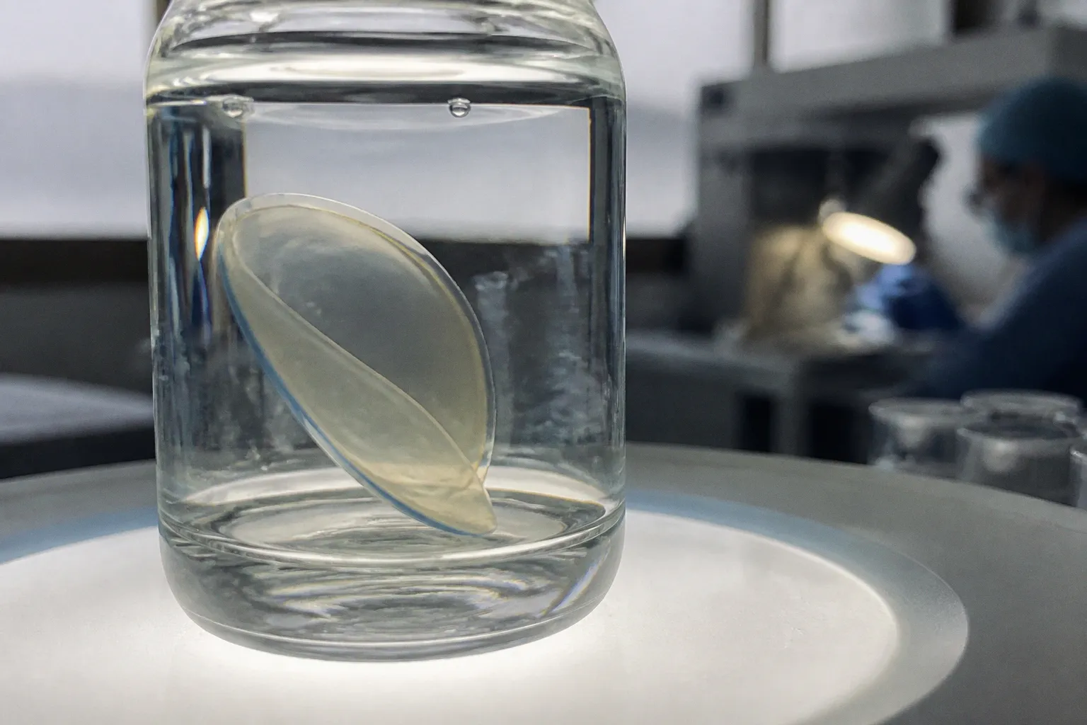
A DMEK graft consists of Descemet membrane with its attached corneal endothelium. The preparation step converts a transparent, layered tissue into a thin, mobile sheet that can later be loaded, delivered, unfolded, and positioned inside the anterior chamber. The useful product is structurally fragile by design. It has minimal stromal content and depends on the integrity of the membrane-endothelium interface.
Preparation failure occurs when that interface does not behave as a uniform plane.
The practical failure modes are familiar:
- A peripheral or central tear propagates while the membrane is being stripped.
- A focal adhesion resists separation and concentrates force at its margin.
- The tissue rolls or folds in a way that prevents controlled peeling.
- The endothelial monolayer becomes discontinuous or otherwise unusable for transplantation.
- A graft that appears intact at the start of preparation loses functional integrity during handling.
These events should not be collapsed into one category. A tear caused by a focal adhesion is not equivalent to a broad separation problem. A difficult peel is not automatically a technician error. The tissue itself can carry altered adhesion mechanics that become visible only when the membrane is manipulated.
DMEK preparation is a mechanical stress test of donor tissue. The failure signal appears at the interface, but the risk may originate in systemic donor biology.
The posterior corneal structure is not a passive substrate. Its handling behavior reflects the condition of the donor tissue, the history of ocular surgery, the preservation interval, and the preparation protocol. The same operator can encounter two corneas with different stripping resistance and different failure probability. A workflow that records only pass or fail loses the intermediate data needed to explain why.
That missing layer matters. Eye banks often have strong data on donor eligibility, endothelial cell density, preservation, and final tissue disposition. They may have less granular information on the actual preparation event: the presence of central adhesions, the location of a tear, the number of manipulation attempts, or the point at which the graft became unusable. Without those fields, a preparation failure is reduced to a binary endpoint rather than treated as a quality signal.
Systemic donor pathology as a predictor
Diabetes mellitus is the clearest risk variable in the available evidence. Across several datasets, diabetic donor tissue shows a higher frequency of DMEK preparation failure. The association is not limited to one processing center or one technique.
A study of 359 corneas found failure in 15.3% of diabetic donor tissues and 1.9% of nondiabetic tissues. A separate analysis of 2,205 DMEK preparations from 1,441 donors reported an overall failure rate of 4.1% and an odds ratio of 4.026 for diabetes, with a 95% confidence interval of 2.567–6.314. The difference between the observed nine-fold increase in one dataset and the lower odds ratio in another does not invalidate either result. It reflects variation in donor composition, processing methods, endpoint definitions, and case selection.
The signal is consistent enough to affect procurement planning.
Diabetes is not an absolute exclusion criterion. Many corneas from diabetic donors can be prepared successfully. The correct interpretation is probabilistic: diabetes shifts the expected failure distribution. It raises the risk that a tissue will consume processing capacity without producing a usable DMEK graft.
Other systemic variables appear in the data. Multivariate analysis at Lions VisionGift identified diabetes mellitus and hyperlipidemia or obesity as independent factors associated with preparation failure. When donors with those conditions were excluded, overall failure declined from 5.2% to 2.2%. The result suggests that donor selection can alter yield before any modification to the bench protocol.
Additional risk factors reported for Descemet membrane splitting include:
- Type 2 diabetes mellitus.
- Heart failure.
- Chronic kidney disease.
- Previous cataract surgery.
- Hyperlipidemia or obesity in the analyzed donor cohort.
The variables do not operate as a universal scoring system. The evidence does not establish a single threshold at which diabetes duration, metabolic status, or renal disease should automatically disqualify a donor cornea. It does establish that a flat donor profile is a weak representation of tissue behavior. Systemic history belongs in the preparation-risk model, not only in the eligibility file.
Why the mechanism remains incomplete
The clinical association is stronger than the biochemical explanation. Diabetes and other vascular or metabolic disorders may alter extracellular matrix properties, tissue hydration, membrane adhesion, endothelial resilience, or the interaction between the posterior stroma and Descemet membrane. The exact mechanism is not resolved across all non-diabetic systemic conditions.
That distinction matters for evidence handling. It is defensible to state that diabetes is associated with higher DMEK preparation failure. It is not defensible to state that a specific molecular change is responsible unless that mechanism has been directly established.
The same restraint applies to chronic kidney disease, heart failure, and prior cataract surgery. Their presence may identify tissue with altered preparation behavior, but the dataset does not convert those variables into a complete causal model. For an eye bank, the operational value is predictive before it is explanatory.
Traditional stripping versus no-touch preparation
Technique has a measurable effect on tissue utilization. In an eye bank evaluation of 1,416 donor corneas, overall graft preparation failure was 3.9%. The traditional technique produced a 7.0% failure rate, while a standardized no-touch technique produced a 2.9% rate.
The absolute difference is 4.1 percentage points. In a high-throughput environment, that difference is material. It affects the number of tissues that must enter the workflow to generate a target number of usable grafts. It also affects rework, technician time, material consumption, and scheduling reliability.
| Processing variable | Traditional technique | Standardized no-touch technique |
|---|---|---|
| Reported preparation failure | 7.0% | 2.9% |
| Difference within the study | Higher failure burden | Lower failure burden |
| Main operational implication | Greater tissue loss and reprocessing exposure | Higher expected tissue utilization |
| Interpretation | More vulnerable to handling-related variability | Reduces direct manipulation during preparation |
The technique comparison should not be read as proof that protocol alone controls the result. Donor pathology remains a major variable. A no-touch method can reduce avoidable manipulation, but it cannot erase structural abnormalities in the donor interface.
The correct systems interpretation is layered:
1. Donor selection defines the baseline risk distribution.
2. Preservation and handling determine how much of that baseline risk is expressed.
3. The preparation technique changes the mechanical exposure applied to the tissue.
4. Operator standardization influences variance between preparations.
5. Documentation determines whether the eye bank can improve the next processing cycle.
This is a process-control problem. The aim is not to attribute every failed graft to the last person who touched it. The aim is to separate tissue-origin risk from procedural variance.
A standardized no-touch approach reduces unnecessary contact and may lower the frequency of preparation damage. It also creates a more reproducible operating environment. That reproducibility is valuable for training, quality audits, and cross-site comparison. If two centers use different definitions of a failed graft or different stripping protocols, their headline failure rates are not directly comparable.
Diabetes, metabolic burden, and graft integrity
The association between diabetes and failure is strongest when the endpoint is defined as a preparation event: tearing, splitting, central adhesion, or inability to produce a usable endothelial graft. This endpoint should remain distinct from postoperative graft survival, endothelial cell loss after transplantation, or visual outcomes.
A tissue can fail during preparation before it reaches the recipient. Conversely, a tissue that survives preparation may later be affected by factors unrelated to the original stripping event. Combining these endpoints creates an unhelpful composite and obscures the location of the actual bottleneck.
The available figures show several different scales of the problem:
- In one retrospective cohort, diabetic tissue failed at 15.3%, versus 1.9% for nondiabetic tissue.
- In a multivariate analysis, diabetes and hyperlipidemia or obesity remained independently associated with failure.
- Removing donors with diabetes, hyperlipidemia, or obesity reduced overall preparation failure from 5.2% to 2.2%.
- In another eye bank study, diabetes was associated with an odds ratio of 4.026 for preparation failure.
- Across 1,416 corneas, the total failure rate was 3.9%, with a substantial difference between traditional and no-touch preparation.
These figures should not be averaged into one universal rate. They come from different cohorts and workflows. The useful conclusion is directional and operational: metabolic and systemic donor variables alter the probability that a cornea will yield a viable DMEK graft, and protocol design can either amplify or reduce the resulting loss.
Central adhesions deserve separate attention. The reported odds ratio for central adhesions in type 2 diabetes donor tissue was 2.2. A central adhesion has a different impact from a peripheral irregularity because it places the manipulation force closer to the functional field of the graft. It may also make the stripping plane less predictable. The location of the defect therefore carries process information that a generic failure code does not.
The data fields that should survive the bench
An eye bank trying to improve DMEK yield requires more than donor age, death-to-preservation interval, and final endothelial cell count. The preparation record should capture the failure geometry and the workflow conditions.
Useful fields include:
- Diabetes status and, where available, type of diabetes.
- Hyperlipidemia, obesity, heart failure, and chronic kidney disease.
- Previous cataract surgery or other relevant anterior-segment procedures.
- Preparation technique, including traditional or no-touch workflow.
- Presence and location of central or peripheral adhesions.
- Tear location and stage of preparation when it occurred.
- Whether the membrane split during initial stripping or later manipulation.
- Final disposition: usable graft, alternate research use, or discarded tissue.
- Operator and processing site, when appropriate for quality analysis.
- Preservation and preparation timestamps for latency analysis.
This is not administrative excess. It is the minimum structure needed to distinguish donor-driven loss from protocol-driven loss.
A binary field marked preparation failed cannot answer whether donor diabetes increased risk, whether central adhesions were overrepresented, or whether one technique reduced the failure rate. A structured event record can.
Donor selection as a yield optimization problem
Donor selection is often framed as a clinical eligibility exercise. For DMEK processing, it is also a yield optimization problem. The relevant output is not the number of corneas accepted into the bank. It is the number of transplantable grafts produced per unit of incoming tissue, labor, and processing time.
That changes the value of donor metadata.
If a center excludes every diabetic donor, it may reduce preparation failures but also shrink the available tissue pool. If it accepts all tissues without risk stratification, it may increase the number of failed preparations and create avoidable throughput volatility. Neither policy is a complete solution. The efficient model is tiered allocation.
A practical system can classify tissue before preparation:
- Lower predicted preparation risk: no recorded diabetes or high-risk systemic condition, no relevant prior anterior-segment surgery, and no known handling concern.
- Intermediate predicted risk: isolated systemic variables or incomplete donor history requiring additional review.
- Higher predicted risk: diabetes combined with hyperlipidemia, obesity, chronic kidney disease, heart failure, or prior cataract surgery.
The classification should not be treated as an automatic discard rule. It should direct the tissue to the appropriate use case. A high-risk cornea may still be suitable for DMEK preparation under a controlled protocol, but it should not be invisible in the production forecast.
Allocation decisions can then incorporate:
- Expected graft demand.
- Available technician capacity.
- Required delivery window.
- Acceptable preparation-loss rate.
- Backup tissue availability.
- Potential value of the tissue for corneal endothelium or Descemet membrane research if transplantation preparation fails.
That final pathway matters. A failed DMEK preparation is not necessarily an information failure. The tissue may still support research on donor endothelial cells, Descemet membrane adhesion, Fuchs dystrophy biology, corneal biomechanics, or anterior-segment histology, provided the collection and consent framework supports that use. The database should preserve the reason for transplant unsuitability rather than reducing the specimen to discarded material.
A failed DMEK preparation is a lost graft, but it can still be a high-value data point if the failure mode is recorded with enough resolution.
Avoiding overcorrection
The strongest risk factor in the data is not a deterministic rule. Diabetes does not guarantee preparation failure. Excluding all diabetic tissue may also conceal the biological range within that group and eliminate potentially usable grafts.
A more defensible approach is to combine donor risk with process controls:
1. Record systemic history before preparation.
2. Use a standardized preparation method.
3. Route higher-risk tissue through operators and workflows with documented performance.
4. Track failure by donor factor and failure mode.
5. Reassess selection thresholds against local yield data.
6. Preserve research pathways for tissue that cannot be used clinically.
This converts donor selection from a static list of exclusions into a monitored decision system. Its performance can be evaluated over time. If the local failure rate remains elevated among low-risk donors, the bottleneck may be protocol or preservation rather than procurement. If failures cluster around diabetic or previously operated donors despite technique standardization, the donor-risk model is capturing a real tissue property.
The procurement and data pipeline
DMEK preparation begins before the cornea reaches the preparation bench. The relevant pipeline includes donor referral, medical-record abstraction, consent, recovery, preservation, transport, tissue inspection, preparation, quality assessment, and final allocation. Each stage can introduce latency or data loss.
The highest-value information is often collected early and used late. Diabetes status may be known during donor screening but omitted from the preparation queue. Previous cataract surgery may be recorded in a recovery note but not transferred to the graft record. The result is a technically clean processing log with an incomplete causal history.
A tissue database designed for anterior-segment research should connect four layers:
Donor layer
This contains systemic conditions, ocular history, age category, cause and timing of death where relevant, and the completeness of the available history. Missingness itself should be visible. An unknown diabetes status is not equivalent to no diabetes.
Tissue layer
This includes corneal identity, preservation medium, storage conditions, endothelial assessment, tissue dimensions, and any pre-existing visible abnormality. The record should retain longitudinal updates rather than only the final status.
Process layer
This records preparation method, operator, timestamps, stripping observations, adhesions, tears, and the point of failure. Process data are what allow a bank to calculate preparation latency and identify high-variance steps.
Outcome layer
This includes clinical usability, alternate allocation, research use, discard reason, and any subsequent quality assessment. The outcome must remain linked to the original donor and process data without exposing unnecessary identifiable information.
The data model should also support denominator discipline. A 4.1% failure rate across 2,205 preparations is not interchangeable with a rate calculated only among tissues selected for a particular technique. A center must know which corneas entered the analysis, which were excluded before preparation, and whether repeated preparations from one donor were treated as independent observations.
This matters because donor-level clustering can distort interpretation. Multiple corneas from one donor may share the same systemic risk factors. If the analysis treats each cornea as fully independent, the apparent sample size can exceed the number of biologically distinct donor profiles. That does not erase the association, but it affects how confidently the result transfers to another tissue bank.
A more precise definition of graft failure
The term DMEK graft preparation failure should identify the point at which the intended endothelial graft cannot be produced. It should not function as a catch-all for every quality concern.
A useful taxonomy separates:
- Preparation failure: the membrane-endothelium sheet is torn, split, or otherwise unusable during stripping.
- Adhesion-limited preparation: a focal attachment prevents controlled separation but may not immediately produce a full-thickness tear.
- Handling artifact: damage associated with manipulation after successful separation.
- Quality failure: the graft is structurally intact but does not meet the bank’s defined clinical or research criteria.
- Post-preparation failure: damage or degradation occurring after the graft has been prepared.
This distinction improves both operations and research. For operations, it shows where tissue is lost. For research, it creates a phenotype for studying Descemet membrane-stromal adhesion. For procurement, it allows the bank to estimate how many incoming corneas are required to meet demand under a specific protocol.
It also prevents a common analytical error: attributing all tissue loss to technician performance. The data show that donor systemic disease and ocular history influence the preparation event. The technician remains part of the process, but not the entire causal system.
Closing assessment
The evidence supports a strict conclusion. DMEK graft preparation failure in donor tissue is a measurable, risk-stratified process outcome. Diabetes is the most reproducible donor-associated signal in the available data, with reported failure rates of 15.3% versus 1.9% in one cohort and an odds ratio of 4.026 in another. Hyperlipidemia or obesity, heart failure, chronic kidney disease, and previous cataract surgery add further risk signals. Preparation technique also changes yield: 7.0% failure with a traditional method compared with 2.9% using a standardized no-touch technique in one eye bank evaluation.
The operational response should not be blanket exclusion. It should be structured intake, standardized stripping, granular failure coding, and continuous linkage between donor history and graft outcome.
The tissue bank that records only whether a graft passed or failed is measuring inventory loss. The tissue bank that records why the membrane tore, where the adhesion formed, which donor variables were present, and which technique was used is building a predictive system. For DMEK, that distinction determines whether procurement remains reactive or becomes evidence-based.