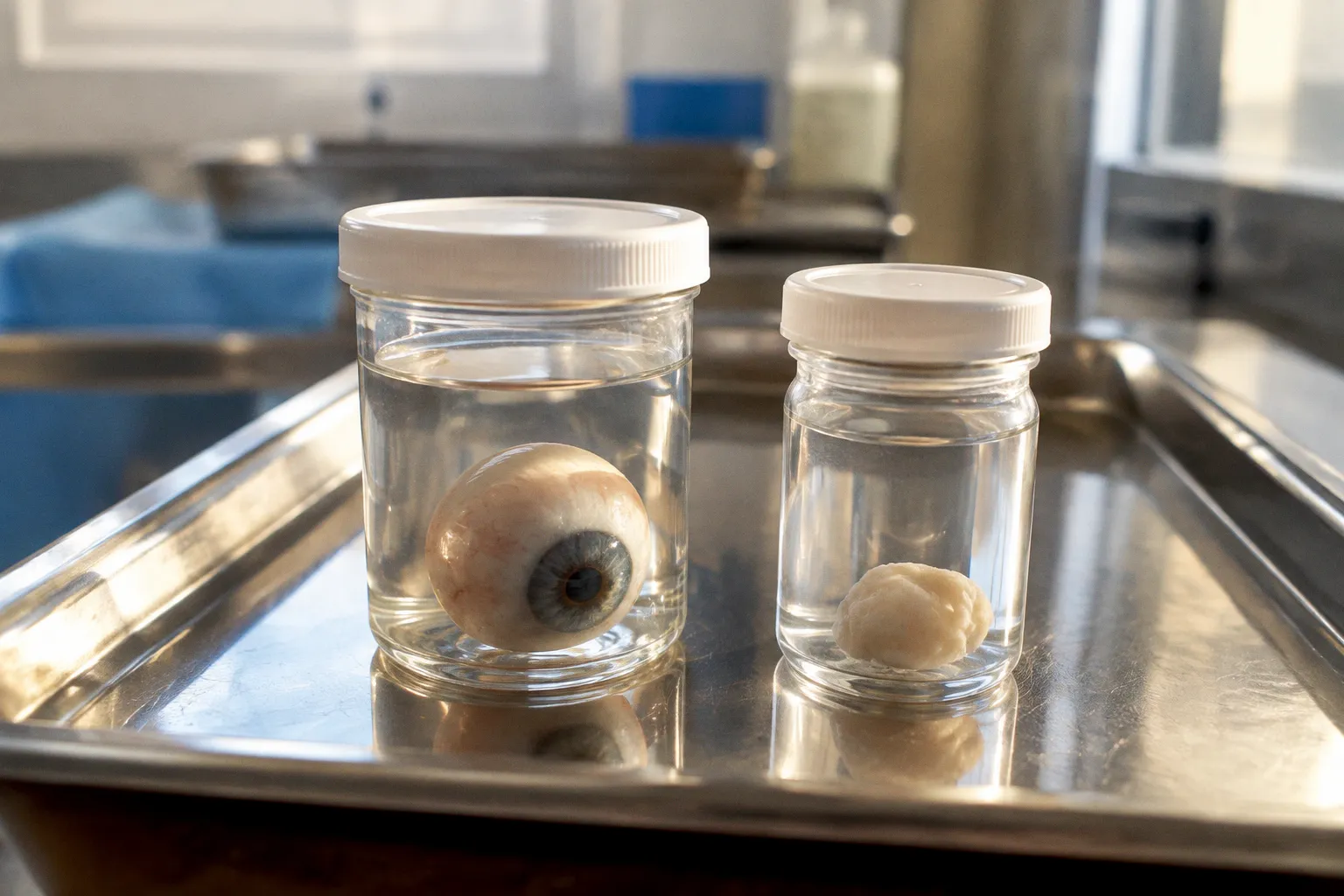
The logic feels airtight — recover everything intact, minimize manipulation, preserve endothelial integrity by leaving the globe undisturbed until excision occurs under controlled laboratory conditions. Eye banks built procurement protocols around this axiom, and for decades, the surgical theatre and the mortuary table both defaulted to the same answer. Yet the data, when you actually look at it, tells a far more inconvenient story than the dogma suggests.
Across thousands of donor cases, in situ corneoscleral excision — the technique where the corneoscleral button is removed directly at the donor site without extracting the whole globe — produces endothelial cell densities and graft failure rates that are statistically indistinguishable from enucleation. The numbers are not ambiguous. They are not preliminary. They are flatly equivalent. And yet the procurement landscape still clings to a hierarchy that the evidence does not support for corneal tissue. The real question is not which method is "better" for corneal transplantation. It is why we continue to treat them as if one were clearly superior, and what that misplaced confidence costs eye biobanks in donor availability and research yield.
Endothelial Integrity: The Numbers That Should Have Ended the Debate
The most robust dataset comes from the Lions Eye Bank of Oregon, comparing 3,618 eyes recovered via whole globe enucleation against 2,048 eyes recovered via in situ corneoscleral disc excision. Mean donor endothelial cell density (ECD) measured 2,726 ± 419 cells/mm² in the enucleation cohort and 2,645 ± 395 cells/mm² in the in situ cohort. That is an 81-cell differential — clinically negligible when you consider the variability inherent in donor age, death-to-preservation interval, and storage conditions.
Primary graft failure rates tell the same story from a different angle: 0.72% for enucleation versus 0.68% for in situ excision. If anything, the in situ group edges fractionally ahead, though the difference is so small it falls well within the noise of surgical variability.
Three months post-transplantation, endothelial cell density in the whole globe group measured 1,708 ± 104.8 cells/mm² versus 1,674 ± 117.4 cells/mm² for in situ. Again, functionally equivalent. The surgical community's attachment to enucleation as the endothelial-superior method has no empirical foundation in these outcomes.
The paradox is uncomfortable: a procurement technique that was supposed to protect the endothelium better produces the same endothelial results. The assumption was never tested; it was inherited.
Death-to-Preservation and Contamination: A Closer Look
Where the data offers some nuance — and where a skeptic should push harder — is on contamination rates and preservation timing. Death-to-preservation intervals in the Oregon dataset were 9.81 ± 3.56 hours for whole globe enucleation and 8.90 ± 3.65 hours for in situ excision. The in situ group moves faster, which is expected: a field excision eliminates the logistics of whole globe transport, container packing, and arrival at the eye bank processing facility before the cornea is even isolated.
Post-harvest contamination rates, measured by positive corneoscleral rim cultures, were 20% in the enucleation group and 24% in the in situ group (P = 0.62). Not statistically significant. But the direction of that four-percentage-point gap deserves attention, particularly for biobank coordinators who need to justify procurement method selection to institutional review boards. In situ excision occurs in a non-sterile field — a mortuary, a hospital room, wherever the donor is located — while enucleation specimens are typically excised under more controlled laboratory conditions at the eye bank. The fact that the contamination differential is clinically meaningless should reassure those who worry about field sterility, but it does not eliminate the question entirely.
| Parameter | Whole Globe Enucleation | In Situ Excision |
|---|---|---|
| Donor ECD (cells/mm²) | 2,726 ± 419 | 2,645 ± 395 |
| Primary graft failure rate | 0.72% | 0.68% |
| Death-to-preservation (hours) | 9.81 ± 3.56 | 8.90 ± 3.65 |
| Rim culture positive rate | 20% | 24% (P = 0.62) |
| 3-month post-op ECD | 1,708 ± 104.8 | 1,674 ± 117.4 |
| Remote site stromal haze incidence | Higher | Lower |
Donor Family Consent: The Hidden Variable That Reshapes the Supply Chain
This is where the procurement debate shifts from a clinical question to a logistics and bioethics one — and where the practical case for in situ excision becomes difficult to dismiss.
Eye banks that transitioned from whole globe enucleation to in situ corneoscleral disc excision reported measurable increases in donor numbers. The reason is straightforward: donor families are more willing to consent to a procedure that does not involve removing their loved one's eyes from the body. The cosmetically less invasive nature of in situ excision — a disc excision that leaves the eye structure largely intact externally — translates directly into higher acceptance rates during an already emotionally charged consent conversation.
This is not an abstract benefit. For ocular biobanks, every declined consent is a lost tissue specimen, a gap in the repository, a research project that cannot be completed. The procurement method that families are more likely to accept is, by definition, the method that builds a larger and more diverse tissue inventory. Yet many procurement protocols still default to whole globe enucleation because the institutional assumption — inherited, again, not tested — holds that it is the "complete" recovery.
Stromal Quality and the Remote Location Problem
A separate study evaluating 468 donor corneas harvested from remote locations found that in situ procurement resulted in a lower incidence of moderate or severe stromal haze and Descemet membrane folds compared to whole globe enucleation. For eye banks operating across dispersed geographies — and this describes the majority of national and regional eye bank networks — this finding matters considerably.
The likely mechanism is mechanical: whole globes are subject to more handling, transport agitation, and pressure fluctuation during transit than a corneoscleral disc resting in a viewing chamber. The intact globe may look like the more "protected" specimen, but the fluid dynamics inside a sealed container being driven across a county or shipped by courier are not benign. The endothelium and stroma experience stress differently when the entire anterior chamber is intact versus when a disc has been excised and immediately placed in storage media.
The assumption that whole globe recovery is gentler on the tissue inverts the actual mechanical reality of specimen transport — and the histopathological evidence bears that out.
What You Actually Lose Without the Globe
Here is where intellectual honesty demands that the case for in situ excision acknowledge its genuine limitation. Whole globe enucleation preserves non-corneal ocular structures: the retina, the sclera, the optic nerve, and the vitreous humor. For research biobanking specifically, this distinction is not trivial. Retinal tissue is central to studies of age-related macular degeneration, diabetic retinopathy, glaucoma neurodegeneration, and a broadening field of retinal gene therapy research. Sclera is sought for biomechanical studies of myopia progression. The optic nerve head is indispensable for understanding glaucomatous cupping at the tissue level.
If an eye bank's mission is purely corneal transplantation, in situ excision is the more efficient and clinically equivalent procurement method. But if the biobank serves a multi-tissue research mandate — which is increasingly the standard expectation for modern ocular biorepositories — then whole globe recovery remains the only method that delivers the full complement of ocular structures in a single retrieval.
This is not a reason to default to enucleation for every donor. It is a reason to stratify: reserve whole globe recovery for donors meeting specific research tissue needs, and deploy in situ excision as the routine procurement standard for transplant-eligible and cornea-only research donors.
Toward a Procurement Protocol That Follows the Evidence
The paradigm deficit in ocular tissue procurement is not a gap in knowledge — the data has existed for over a decade. It is a gap in institutional willingness to update protocols that were designed in an era when in situ excision was untested and donor consent logistics were less scrutinised.
Modern eye biobanks need procurement workflows that are responsive to both clinical outcome data and research inventory requirements. The binary thinking that once characterised this debate — enucleation or in situ, one must be better — should give way to a stratified model where the donor's eligibility, the family's consent threshold, and the biobank's research tissue needs all determine which technique is deployed.
| Scenario | Recommended Method | Rationale |
|---|---|---|
| Transplant-eligible donor, cornea research only | In situ excision | Equivalent ECD and graft outcomes; higher family consent rate |
| Multi-tissue research donor (retina, sclera, optic nerve) | Whole globe enucleation | Only method preserving full posterior segment |
| Remote or delayed procurement location | In situ excision | Lower stromal haze; faster death-to-preservation interval |
| Uncertain research mandate at time of recovery | Whole globe enucleation | Maximum tissue optionality; cornea still usable |
The data is unambiguous on one point: for corneal outcomes, the procurement method does not matter. Endothelial cell density, graft clarity, and primary failure rates are equivalent whether you excise the disc in the field or in the lab. Every year that procurement protocols default to whole globe enucleation without considering the consent and logistics benefits of in situ excision is a year of avoidable donor attrition.
The challenge to the field is not to abandon whole globe recovery — it has irreplaceable utility for posterior segment research — but to stop pretending that one method is generically superior. Stratify by purpose. Follow the data. And update the protocols before the next generation of biobankers inherits the same unexamined assumptions we are still labouring under now.