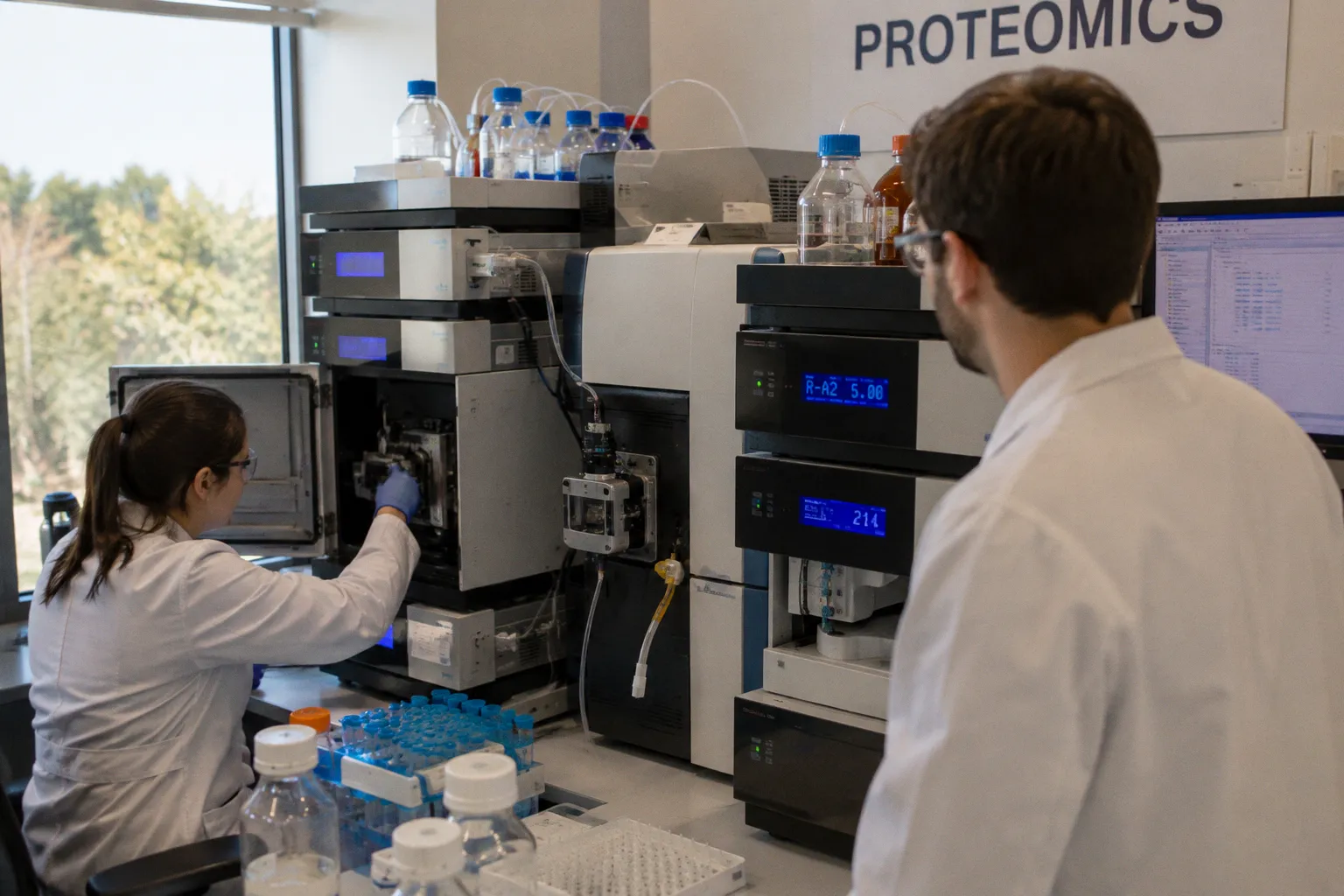
In a six-plex iTRAQ LC-MS/MS workflow, investigators identified 526 proteins at a 95% confidence limit in stretched LC cells; a parallel study of cultured human optic nerve head astrocytes under the same biomechanical regimen returned 573 proteins at the same confidence threshold. These are not theoretical counts. They are the molecular fingerprints of what the optic nerve head experiences when intraocular pressure translates into mechanical load — and they form the substrate on which glaucomatous remodeling is now being redrawn, one named protein at a time.
The clinical observation has been familiar for decades: in primary open-angle glaucoma, the cup deepens, the neuroretinal rim thins, the retinal ganglion cell axons withdraw. What proteomics is now doing is converting that familiar topography into a list of molecules — and, more importantly, into a sequence of events. The optic nerve head is no longer a black box between the intraocular pressure measurement and the visual field defect. It is a worked surface, with strain-sensitive signaling hubs, an extracellular matrix that is being dismantled and rebuilt, and astrocytes that are increasingly populating the very pores the axons once occupied.
Biomechanical Strain and the Proteomic Landscape of the Lamina Cribrosa
The lamina cribrosa is a meshwork of collagenous beams through which retinal ganglion cell axons exit the eye. It is also a structure under continuous mechanical negotiation. Even at physiological intraocular pressures, the beams flex; in glaucoma, the cyclical load increases, and the cells embedded in the beams — primarily LC fibroblasts and astrocytes — register the change at the protein level long before any structural deformation is visible on imaging.
To capture that registration, investigators applied 3% and 12% cyclic equiaxial stretch to primary human LC cells for durations of two and twenty-four hours, then subjected the lysates to nano LC-MS/MS with iTRAQ labeling. The 526 proteins identified at 95% confidence are not a closed list of suspects — they are a survey of which intracellular and extracellular compartments respond to strain within a defined experimental time window. The choice of 3% and 12% was intended to bracket a range of mechanical conditions — a modest stretch magnitude at the lower end and a more severe deformation at the upper — without mapping directly to a specific intraocular pressure threshold or a particular stage of clinical cupping.
The proteome of a stretched lamina cribrosa cell is not a snapshot of damage — it is a ledger of which pathways the cell is willing to pay to keep its architecture standing.
What the proteome shows, even at this resolution, is that strain does not trigger a single response. It triggers layered responses. Some proteins shift within two hours; others require the twenty-four-hour window. Some peak at 3% stretch; others only resolve at 12%. The two-hour time point captures the acute signaling cascade — kinases, heat-shock proteins, immediate early gene products — while the twenty-four-hour window begins to reveal the downstream structural and metabolic commitments the cell makes once the mechanical insult persists. This is not the behavior of a passive scaffold being squeezed. It is the behavior of a living tissue making decisions, allocating resources, and routing signals along pathways that no single time point can describe.
Among the 526 proteins, the distribution across cellular compartments is itself informative. Cytoskeletal proteins, endoplasmic reticulum chaperones, mitochondrial enzymes, and extracellular matrix components all appear, indicating that mechanical strain does not confine itself to the cytoskeleton or the cell membrane. The cell-wide response suggests that even modest stretch initiates a coordinated metabolic reprogramming — one that proteomics is only beginning to resolve into individual pathway threads.
Molecular Signaling Hubs: TGFβ1, TNF, and p53 in Glaucomatous Stress
Among the 526 proteins identified in strained LC cells, pathway analysis has repeatedly surfaced a small, recurrent set of hubs: TGFβ1, TNF, CASP3, and p53. None of these are strangers to glaucoma literature, but their co-occurrence in a single strained proteome reframes them as parts of a coordinated circuit rather than independent findings.
TGFβ1 is the most familiar architect of extracellular matrix remodeling. In the LC, it drives the synthesis of collagens, fibronectin, and the crosslinking enzymes that stiffen the beams. Its appearance in the strain proteome suggests that mechanical load is being translated, biochemically, into a fibrosis program — one that progressively reduces the compliance of the lamina cribrosa and may create a self-reinforcing loop in which stiffer beams transmit more strain to the cells within them. TNF, the inflammatory hub, points in a different direction — toward microglial activation, astrocyte reactivity, and the chronic low-grade neuroinflammation that increasingly characterizes the glaucomatous optic nerve head. CASP3 and p53 mark the apoptotic axis: the cellular decision to withdraw rather than remodel. Together, these hubs describe a tissue that is simultaneously rebuilding its matrix, recruiting immune surveillance, and pruning its own resident cells.
A subset of less prominent proteins — BAG5, NO66, eIF-5A — has drawn attention because of their regulatory character. BAG5 sits at the interface of Bcl-2 family signaling and CHIP E3 ligase activity, modulating how LC astrocytes handle misfolded protein load under conditions of mechanical and oxidative stress. Its presence in the strain proteome suggests that the ubiquitin-proteasome system is engaged early in the response, not merely as a cleanup mechanism but as a regulatory switch determining which misfolded client proteins are triaged for degradation versus refolding. NO66, a Jumonji-domain histone demethylase, hints at the possibility that biomechanical strain reaches into chromatin-level regulation, not just cytoplasmic signaling — a finding that, if confirmed, would place mechanical stress in the same epigenetic category as metabolic and hypoxic insults to the optic nerve head. eIF-5A, the hypusine-modified translation factor, links cellular stress to the production of specific proteins required for survival under hypoxic and mechanical insult. Its hypusination is an oxygen- and polyamine-dependent modification, meaning the cell's translational response to stretch may be gated by the same metabolic conditions that worsen in glaucomatous tissue. Each is a foothold for further study, and each has begun to appear in adjacent ocular multiomics atlases being built from donor tissue.
Astrocyte Accumulation and Basement Membrane Replacement in Donor Tissue
Proteomics tells one story in cultured cells; histology of donor tissue tells another from the intact organ, and the convergence is striking. In a quantitative study of 27 glaucoma and 19 age-matched postmortem donor eyes, the proportionate area of GFAP and F-actin processes within lamina cribrosa pores was significantly lower in glaucoma eyes (P = 0.01). That number is not marginal: it describes a measurable withdrawal of the astrocytic cytoskeleton from the pores — the very channels through which axons have historically transited.
What occupies the space the axons left behind is the question that histology has begun to answer with unusual precision. SOX9 labeling of postmortem glaucoma eyes demonstrated that 92.3% of the increased cell count in LC pores was astrocytic. The axons were not merely being lost — the cellular composition of the pore was being rewritten. Anti-collagen IV labeling, meanwhile, identified basement membrane material occupying the space where axons had once traveled. The co-occurrence of astrocytic cells and collagen-IV-rich matrix in the same remodeled pores raises a compelling question about causality — whether the astrocytes are the source of that new basement membrane or merely the resident cells that remain after axonal withdrawal has opened space for matrix deposition by other mechanisms.
In glaucomatous LC pores, the predominant cell identified by SOX9 labeling is the astrocyte, and the matrix occupying vacated axonal channels is predominantly collagen IV — the causal link between the two remains to be established.
The implication is that what the clinician sees as "cupping" is, at the molecular scale, an active substitution. The remodeled pore is not simply empty space; it is reoccupied by cells and matrix, and the resulting tissue is qualitatively different from what preceded it. A porous, axon-permissive meshwork converts into a denser, more cellular, basement-membrane-rich tissue. This is remodeling in the strict sense: the demolition of one architecture and the construction of another, executed by the same resident cell population that once supported the axons — even if the precise cellular choreography, who deposits what and when, has yet to be fully resolved.
Translating In Vitro Strain Data to Clinical Glaucomatous Neuropathy
The temptation in glaucoma proteomics is to read the in vitro strain data as a direct portrait of the in vivo optic nerve head. The available evidence does not support that shortcut. Cultured LC cells and ONH astrocytes, even when subjected to physiologically calibrated cyclic stretch, exist outside the context of vascular perfusion, of true translaminar pressure gradients, and of the cellular diversity that the intact optic nerve head contains. A 3% stretch on a fibronectin-coated silicone well is not the same biomechanical event as a 3% elongation within a perfused, microglia-infiltrated lamina cribrosa beam. The in vitro system isolates one variable — mechanical strain — and removes nearly everything else: the oxygen tension gradients, the paracrine signaling from axons and microglia, the basement membrane interactions, and the blood supply that determines whether stressed cells can meet the metabolic demand of their own survival pathways.
What the in vitro studies do offer is a controlled dissection of which pathways a single cell type activates when exposed to mechanical strain alone. The rat ocular hypertension model complements this picture from the other side: in vivo proteomic changes in the optic nerve head at early time points have implicated Rho family GTPase signaling, NRF2-mediated oxidative stress response, ERK/MAPK signaling, and Sirtuin signaling. These are not the same hubs the human LC cell proteome foregrounds, and the divergence matters. The rat model captures an integrated tissue response under a sustained intraocular pressure elevation; the human in vitro model captures a purified strain response in a single cell type. Reading one through the other requires acknowledging that both are partial views of the same disease process, and that neither alone defines the molecular trajectory a human optic nerve head follows as pressure-related damage accumulates.
The donor eye histology — 27 glaucoma and 19 control — sits in between these two extremes. It is human, it is in vivo, but it is postmortem: a snapshot of the tissue at the time of procurement, in which remodeling has already reached a particular structural configuration. The temporal gap between the two-hour strain proteome and the postmortem histology is the central unresolved geography of the field.
| Evidence stream | System | Time window | What it captures | What it misses |
|---|---|---|---|---|
| Human LC cell strain proteomics (526 proteins) | In vitro, monoculture | 2 h and 24 h | Purified strain-induced signaling in a single cell type | Vascular context, multi-cell interactions, in vivo pressure gradients |
| Human ONH astrocyte strain proteomics (573 proteins) | In vitro, monoculture | 2 h and 24 h | Astrocyte-specific strain response | Microglial and axonal contribution, basement membrane dynamics |
| Rat ocular hypertension proteomics | In vivo, intact tissue | Early time points | Integrated tissue response, Rho/NRF2/ERK/Sirtuin axes | Human-specific protein isoforms, long-term human remodeling |
| Postmortem donor eye histology | In vivo, human, postmortem | At procurement | Cellular and matrix composition of remodeled pores | The molecular events that produced that composition |
The table is not a hierarchy — it is a triangulation. Each stream answers a question the others cannot, and the disease sits at the convergence point that none of them fully occupy.
Future Directions in Mapping the Temporal Proteome of the Optic Nerve Head
What the field does not yet have is a continuous proteomic record of the optic nerve head as it moves from compensated intraocular pressure elevation through early structural change to the clinical phenotype that presents with visual field loss. The in vitro studies cover the early hours; the donor tissue studies describe a postmortem endpoint. The intervening years — the period in which a clinician might intervene — remain molecularly dark, and several specific gaps define the next phase of work:
1. Single-cell proteomics applied to donor optic nerve heads at multiple disease stages, to identify when TGFβ1, TNF, CASP3, and p53 hubs are activated relative to one another — and whether late-stage tissue is still listening to them at all. Current bulk proteomics averages the signal across every cell in the lysate; single-cell resolution would reveal whether the apoptotic and fibrotic programs coexist within the same cell or are segregated into distinct populations occupying different regions of the lamina.
2. Longitudinal sampling of aqueous or vitreous humor in patients, where proteins shed from the optic nerve head might be detected in the biofluid and correlated with structural and functional progression on OCT and perimetry. The anterior chamber is not a direct readout of posterior segment proteomics, but the molecular spillover from a remodeling lamina cribrosa — collagen fragments, TGFβ pathway products, apoptotic markers — may eventually serve as a biofluid signature of active optic nerve head remodeling.
3. Phosphoproteomic and acetylomic profiling of freshly recovered donor tissue, to capture post-translational states that standard proteomics workflows discard during tissue processing. The phosphorylation status of p53, the acetylation of histones near fibrotic gene loci, and the hypusination of eIF-5A are all modifications that degrade rapidly postmortem and require procurement pipelines designed for molecular preservation.
4. Integration of proteomic data with transcriptomic atlases from the same donor eyes, to test whether the mRNA changes that single-cell RNA-seq is beginning to catalogue are reflected at the protein level in the same cells. The correlation between transcript abundance and protein abundance in post-transcriptionally regulated pathways — particularly those involving stress granules and translational control — is often weaker than assumed, and proteomic confirmation is essential before a transcriptomic finding is treated as a therapeutic target.
Each of these directions depends on procurement pipelines that can deliver matched human specimens — intact optic nerve heads with documented intraocular pressure history, donor eyes recovered under protocols that preserve labile post-translational modifications, and biobanks that catalogue not only the tissue but the molecular context from which it was drawn. The infrastructure for such procurement is growing, but it remains the rate-limiting step for every approach that aspires to move beyond the dish and the animal model.
The proteome of a single stretched cell and the proteome of a postmortem glaucomatous pore are both real — and neither, alone, is the disease.
The unresolved question that sits at the bottom of this stack of evidence is whether the molecular hubs identified in early strain — TGFβ1, TNF, CASP3, p53, and the lesser-studied BAG5, NO66, eIF-5A — are the same hubs that sustain remodeling across the course of a human disease, or whether the tissue has reorganized around a different set of regulators by the time it reaches the configuration observed at procurement. Until that question is answered with a continuous, time-resolved proteomic record drawn from human optic nerve heads, every therapeutic target identified in the dish will arrive at the clinic with the same caveat: we know what the cell does when it is stretched, but we do not yet know whether the disease is still listening to the stretch when the patient arrives.