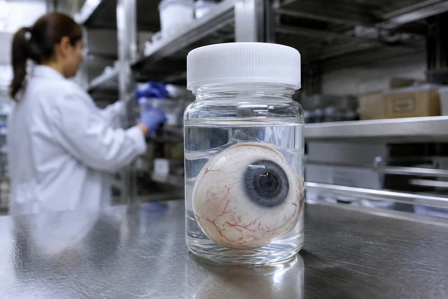
Human donor retina is not automatically molecularly invalid because procurement takes several hours. The harder fact is more inconvenient: DNA methylation can remain sufficiently stable for high-throughput profiling across common eye-bank retrieval windows, while other molecular layers may already be drifting in ways that are less visible, less predictable, and much more consequential.
This is the central problem with discussing retinal epigenetic alterations post mortem interval as though PMI were a single switch between valid and unusable tissue. It is not. A 4-hour specimen, a 12-hour specimen, and a 17-hour specimen do not carry the same logistical history. Nor do DNA methylation, hydroxymethylation, histone modifications, RNA abundance, and chromatin accessibility respond to post-mortem ischemia in the same way.
The real question is therefore not whether post-mortem delay distorts data. It is which data it distorts, by how much, and whether the study design is capable of detecting the distortion.
The 12-hour window is a logistical compromise, not a biological guarantee
Researchers requesting human donor retinal tissue typically specify a post-mortem interval between 4 and 12 hours before preservation. That range sounds precise until it is compared with the actual workflow of ocular procurement.
In eye-banking operations, the average time from donor death to authorization is 7.0 hours, with a median of 5.5 hours and a standard deviation of 4.7 hours. The average death-to-recovery interval is longer: 12.6 hours, with a median of 11.5 hours and a standard deviation of 6.1 hours. By the time recovered ocular tissue is checked back into the eye bank, the average death-to-check-in interval reaches 17.0 hours, with a median of 15.8 hours and a standard deviation of 8.2 hours.
Those numbers create an uncomfortable mismatch between what researchers request and what procurement systems routinely deliver. A study may specify a 4–12-hour PMI, yet the operational chain includes authorization, transport, recovery, processing, and check-in. Each step adds time, and each step may introduce different thermal and ischemic conditions.
The familiar response is to treat the delay as a nuisance variable. Record it, adjust for it, and continue. That is better than ignoring it, but it does not solve the underlying biological question. PMI is not merely a number attached to a specimen. It is a composite of:
- the duration of ischemia before cooling;
- the temperature during transport and temporary storage;
- the interval between recovery and tissue dissection;
- the preservation method used for the retina, RPE, choroid, or optic nerve;
- the cellular composition of the sampled region;
- and the molecular assay applied afterward.
A donor retina that spent several hours under controlled cooling is not equivalent to one exposed to prolonged uncooled delay, even if both are labeled with the same nominal PMI. The label compresses a process into one number. Biology, predictably, refuses to cooperate.
A post-mortem interval is not a biological verdict. It is a compressed summary of several different stresses that should not be treated as interchangeable.
The practical implication is not that human ocular tissue becomes unusable after 12 hours. The available evidence does not support that claim. It supports a more demanding conclusion: researchers need to distinguish operational delay from molecular damage rather than assuming that one perfectly predicts the other.
DNA methylation is more resilient than the standard warning suggests
For many retinal epigenomics studies, the most reassuring evidence concerns DNA methylation. Human donor RPE and choroid tissue has been profiled using DNA methylation microarrays and bisulfite pyrosequencing across post-mortem intervals of up to 9 hours. These approaches have been used to examine epigenetic changes without treating the entire dataset as biologically compromised simply because the tissue was obtained after death.
That does not mean methylation is frozen at death. It means the degree of stability can be sufficient for a defined analytical purpose.
Global DNA methylation and hydroxymethylation may show mild alterations under extreme delay or ischemic conditions, but short PMIs—particularly within the 4–12-hour range commonly requested for donor retina research—can preserve molecular integrity suitable for high-throughput epigenetic and transcriptomic profiling when cooling and storage protocols are properly applied.
The distinction between methylation and hydroxymethylation matters. In retinal and neural tissue, 5-methylcytosine and 5-hydroxymethylcytosine are not interchangeable signals. A study that reports only a broad methylation measure may conceal a more complex redistribution between these marks. Even when the total signal appears relatively stable, the biological interpretation can change if one mark declines while another rises.
There is also a difference between measuring a stable chemical modification and inferring an active regulatory state. Bisulfite-based methods are capable of generating useful methylation measurements, but they do not automatically reveal whether a regulatory element remained functionally active after death. A methylated CpG profile can survive long enough for measurement while the transcriptional machinery that gave the mark its biological meaning has already become unstable.
That is where the usual confidence in methylation data becomes too broad. A robust DNA methylation array does not validate every other layer of the same specimen. It validates the measurement of a particular molecular feature under particular conditions.
What the evidence can support
The current evidence supports several restrained conclusions:
1. Short-to-moderate PMI does not automatically invalidate retinal or RPE/choroid methylation studies. Tissue retrieved within common eye-bank windows can retain sufficient molecular integrity for high-throughput analysis.
2. Cooling and storage are not procedural footnotes. They are part of the biological exposure history and should be incorporated into sample metadata, not left as vague handling notes.
3. Methylation stability is not equivalent to total epigenomic stability. DNA CpG methylation may be comparatively resilient while RNA, chromatin accessibility, histone modifications, or cell-state markers behave differently.
4. The longer the delay, the more important degradation-aware interpretation becomes. Even when the assay remains technically measurable, increasing DNA degradation can correlate with greater methylation variance.
A bisulfite sequencing evaluation found reliable measurement quality up to a PMI threshold of 72 hours, but methylation variance still correlated with advancing DNA degradation. The reported relationship was statistically measurable, with an R² of 0.4311 and p = 0.0392. This is not a permission slip to treat 72 hours as a routine target. It is evidence that analytical measurability and biological comparability are separate questions.
A sample can produce a technically valid readout and still be a poor match for another sample collected under different conditions. The sequencing instrument does not know whether the donor tissue experienced a controlled four-hour delay or a poorly documented period of ischemia. The resulting file may look equally polished. The confounding does not.
The 5mC and 5hmC problem: small shifts, different meanings
The most useful warning comes from controlled observations of uncooled post-mortem tissue. After a prolonged interval of up to 540 minutes—nine hours—global 5-methylcytosine content declined from 3.7% to 2.9%, while 5-hydroxymethylcytosine increased from 0.19% to 0.29%. The same conditions were associated with elevated reactive oxygen species.
These are not trivial details. They show that post-mortem conditions can produce directional molecular changes rather than merely causing a vague loss of quality. The decrease in 5mC and increase in 5hmC suggest that oxidative and ischemic processes may reshape the apparent epigenetic landscape even before a specimen reaches the most extreme PMI ranges.
But the context is decisive: the reported changes occurred under uncooled conditions. They should not be transplanted wholesale onto every eye-bank specimen collected within a standard retrieval window. That would be an equally careless form of overinterpretation. Controlled cooling, faster recovery, and appropriate preservation can alter the exposure substantially.
The point is not that every nine-hour specimen has undergone the same 5mC-to-5hmC shift. The point is that the molecular direction of change depends on the environment surrounding the delay. A nominal PMI without thermal and handling information is therefore a weak proxy for molecular state.
Why oxidative stress complicates the interpretation
Reactive oxygen species matter because post-mortem tissue is not simply waiting in suspended animation. Oxygen delivery stops, energy metabolism fails, membranes destabilize, and cellular compartments begin to lose their normal regulatory boundaries. The epigenome is embedded in that system.
For DNA methylation studies, this creates at least three interpretive risks:
- Apparent disease-associated changes may partly reflect PMI-associated drift. If diseased and control donors have systematically different retrieval times, the epigenetic contrast may absorb a logistics effect.
- Global averages may conceal cell-type-specific changes. A stable overall methylation percentage can coexist with altered marks in retinal ganglion cells, photoreceptors, Müller glia, RPE, or vascular-associated populations.
- Oxidative and ischemic effects may not be uniform across genomic regions. A promoter, enhancer, repetitive element, or heterochromatic region may respond differently to post-mortem stress.
This is precisely where claims about ocular epigenetic alterations in donor tissue need to become narrower. A global shift is not the same as a locus-specific regulatory change. A locus-specific change is not automatically disease-specific. And a technically reproducible signal is not necessarily a signal preserved in vivo.
The assay can be reproducible and the biological comparison still be unfair. Technical precision does not erase unequal post-mortem exposure.
Methylation stability should not be used to certify chromatin accessibility
The most dangerous extrapolation in current ocular multiomics is the assumption that if DNA methylation survives, chromatin accessibility must have survived too. There is no adequate basis for treating those layers as interchangeable.
DNA methylation is a chemical modification on relatively stable DNA. Chromatin accessibility depends on the organization and activity of nucleosomes, transcription factors, histone modifications, ATP-dependent remodeling, and cellular energy state. Those systems are more directly entangled with post-mortem physiology.
For researchers planning donor eye chromatin accessibility assays such as ATAC-seq, the evidence is notably less complete. The exact quantitative PMI threshold at which cell-type-specific chromatin accessibility in human retinal cells experiences irreversible decay remains unresolved. That unknown should not be filled with optimism simply because bisulfite sequencing remains measurable at longer intervals.
The distinction is not academic. An ATAC-seq profile is often interpreted as a map of regulatory potential: open chromatin near a promoter or enhancer is treated as evidence that a cell is prepared to express a gene or respond to a stimulus. Post-mortem ischemia may alter that accessibility landscape before DNA methylation becomes visibly unreliable. If so, an investigator could obtain a clean-looking library from a molecular state that no longer represents the living retina.
The problem becomes sharper in single-cell experiments. Bulk tissue can average away cell-type-specific degradation. Single-cell data can reveal the damage—but it can also expose the fact that some cell populations are disproportionately lost, stressed, or technically underrepresented after delay. A change in cluster abundance may reflect biology, tissue preservation, dissociation efficiency, or some combination of the three.
For that reason, a methylation result should not be used as a general certificate of tissue quality. The most defensible interpretation is narrower:
- DNA methylation can remain suitable for analysis within common, well-controlled PMI windows.
- The stability of hydroxymethylation requires separate consideration.
- Chromatin accessibility requires its own validation strategy.
- Histone post-translational modifications remain especially uncertain in human post-mortem retina.
- Transcriptomic and proteomic measurements should be interpreted with their own degradation and ischemia controls.
The last point is critical for ophthalmic multiomics. A donor retina may support a valuable methylome and a compromised transcriptome. Or it may produce adequate RNA sequencing data while failing to preserve the regulatory state required for a meaningful ATAC-seq experiment. Multiomic integration does not remove these differences. It can amplify them if each layer is treated as equally trustworthy.
A practical quality-control framework for human ocular epigenomics
The right response to PMI is not to discard every specimen outside an ideal window. That would waste scarce human tissue and confuse logistical imperfection with biological failure. The better approach is to make the exposure history visible and design the analysis around it.
A rigorous study should capture, at minimum:
- time of death, authorization, recovery, preservation, and laboratory check-in;
- whether the tissue was cooled, when cooling began, and how temperature was controlled;
- the anatomical region and cell populations included in the sample;
- the interval between recovery and dissection;
- the preservation chemistry and storage conditions;
- DNA integrity and, where relevant, RNA integrity;
- batch, extraction, library-preparation, and sequencing variables;
- disease status, age-related variables, and other donor metadata that could correlate with procurement time.
The temptation is to collapse all of this into a single PMI covariate. That is convenient but often biologically crude. Death-to-authorization, death-to-recovery, and death-to-check-in describe different stages. A specimen with a long authorization delay but rapid cooled recovery is not necessarily equivalent to one with a shorter administrative interval and prolonged uncooled transport.
How to compare specimens without manufacturing certainty
A useful analysis can proceed through several layers:
1. Stratify by PMI rather than assuming a linear effect. The difference between 4 and 8 hours may not have the same biological meaning as the difference between 12 and 16 hours.
2. Separate cooled from uncooled exposure. The 5mC and 5hmC changes observed under prolonged uncooled conditions demonstrate why temperature history cannot be treated as decorative metadata.
3. Test molecular layers independently. A methylation-based quality assessment should not stand in for RNA quality, chromatin accessibility, or histone-state validation.
4. Look for variance, not only mean shifts. Increasing degradation may widen methylation variance without producing a dramatic global average change.
5. Match cases and controls operationally. If diseased donors consistently have longer retrieval times, statistical correction may not fully recover the lost comparability.
6. Report exclusions transparently. Removing high-PMI specimens after seeing the molecular results can create a different kind of bias unless the decision rule is defined in advance.
A compact comparison helps clarify the logic:
| Molecular layer | What short-to-moderate PMI evidence supports | Main interpretive risk |
|---|---|---|
| DNA CpG methylation | Often measurable across common 4–12-hour research windows when tissue is properly cooled and stored | Degradation-associated variance and confounding by unequal donor handling |
| DNA hydroxymethylation | Can show directional change under prolonged or uncooled conditions | Treating 5hmC as interchangeable with 5mC or assuming stability from one mark |
| Transcriptomics | May remain suitable under controlled workflows, but requires independent quality assessment | RNA degradation and ischemia-related expression shifts |
| Chromatin accessibility | No established quantitative threshold for irreversible cell-type-specific decay in human retina | Using methylation stability as a substitute for ATAC-seq validation |
| Histone modifications | Exact post-mortem behavior in human retina remains insufficiently resolved | Inferring active regulatory states from marks that may be highly labile |
| Proteomics | Potentially informative with appropriate tissue and assay controls | Post-mortem proteolysis, compartment changes, and unequal preservation |
This table is not an argument for a universal cutoff. It is an argument against one. The relevant threshold depends on the question, the tissue compartment, the preservation protocol, and the molecular layer being measured.
The 72-hour threshold is a measurement boundary, not a biological permission slip
The reported reliability of bisulfite sequencing up to 72 hours PMI is easy to misuse. In a field accustomed to binary quality labels, the number appears to offer a clean answer: below 72 hours, proceed; above 72 hours, stop. That is not what the evidence establishes.
It establishes that measurement quality can remain reliable to a defined threshold, while methylation variance correlates with advancing DNA degradation. The distinction is fundamental. A sequencing workflow may still generate interpretable data after a considerable delay, but the biological comparability of those data may be weaker than the technical readout suggests.
The same error appears in reverse when researchers assume that a 4–12-hour specimen is automatically pristine. Standard retrieval windows are reassuring, not magical. Even within them, tissue can experience different temperatures, transport conditions, anatomical sampling choices, and delays before preservation. A narrow PMI range reduces one source of variation; it does not eliminate the rest.
The phrase post mortem delay retinal DNA methylation therefore needs a qualifier every time it is used in a serious study: under what preservation conditions, in which tissue compartment, measured by which assay, and compared with what control structure?
Without those qualifiers, the field is left with a misleading hierarchy:
- short PMI equals good tissue;
- long PMI equals bad tissue;
- methylation equals stable epigenome;
- usable assay equals biologically faithful sample.
Every line in that hierarchy is too simple.
What remains unresolved beyond DNA methylation
The next phase of human ocular epigenomics will not be defined by another broad claim that post-mortem tissue is either reliable or unreliable. It will be defined by resolving which regulatory marks persist, which marks drift, and which cell populations carry the distortion.
Two uncertainties are particularly important.
First, the field does not yet have an exact quantitative threshold for irreversible decay of cell-type-specific chromatin accessibility in human retinal cells after death. This gap matters for single-cell and single-nucleus ATAC-seq, where a technically successful library may still represent a selectively altered cellular landscape.
Second, the behavior of active histone modifications under post-mortem ischemic hypoxia remains insufficiently defined in human retina. Marks such as H3K27ac and H3K4me3 are often interpreted as indicators of active regulatory regions, but it is not yet clear how selectively and how quickly these states change after death compared with relatively stable DNA CpG methylation.
These are not minor technical details. They determine whether an epigenomic atlas describes living retinal biology or the molecular afterimage of procurement.
The field should also resist a familiar form of paradigm deficit: treating multiomics as automatically self-correcting. Combining methylation, RNA, chromatin, and proteomic data can reveal concordant biology, but it can also produce a polished consensus among measurements that share the same post-mortem exposure. Agreement between layers is informative only when their failure modes are understood.
The answer: delays can distort retinal epigenetics, but not uniformly
So, do post-mortem delays distort retinal epigenetic data?
Yes—but not in the indiscriminate way often implied.
Within properly controlled short-to-moderate PMI windows, especially the 4–12-hour range commonly requested for donor retinal research, DNA methylation data can remain suitable for high-throughput profiling. Human RPE/choroid methylation has been measured across intervals up to 9 hours, and bisulfite sequencing can retain reliable measurement quality at much longer intervals under defined conditions.
At the same time, prolonged or uncooled delay can alter global 5mC and 5hmC levels and increase oxidative stress. Methylation variance can rise with DNA degradation. None of this licenses researchers to assume that transcriptomic states, chromatin accessibility, histone modifications, and methylation marks share one stability curve.
The responsible position is therefore neither dismissal nor reassurance. PMI should be treated as a structured biological exposure, not a ceremonial timestamp. Studies using human donor eyes need to report the procurement chain, separate cooled from uncooled delay, analyze each molecular layer on its own terms, and avoid calling a technically measurable specimen biologically pristine.
The traditional paradigm says the post-mortem window determines whether tissue is trustworthy. The emerging evidence is less tidy and more useful: the window matters, but the molecular question matters just as much. Until chromatin accessibility and active histone states are mapped across real-world ocular procurement conditions, any claim that a stable methylome represents a stable retinal epigenome is not rigor. It is simply the old assumption wearing multiomic terminology.