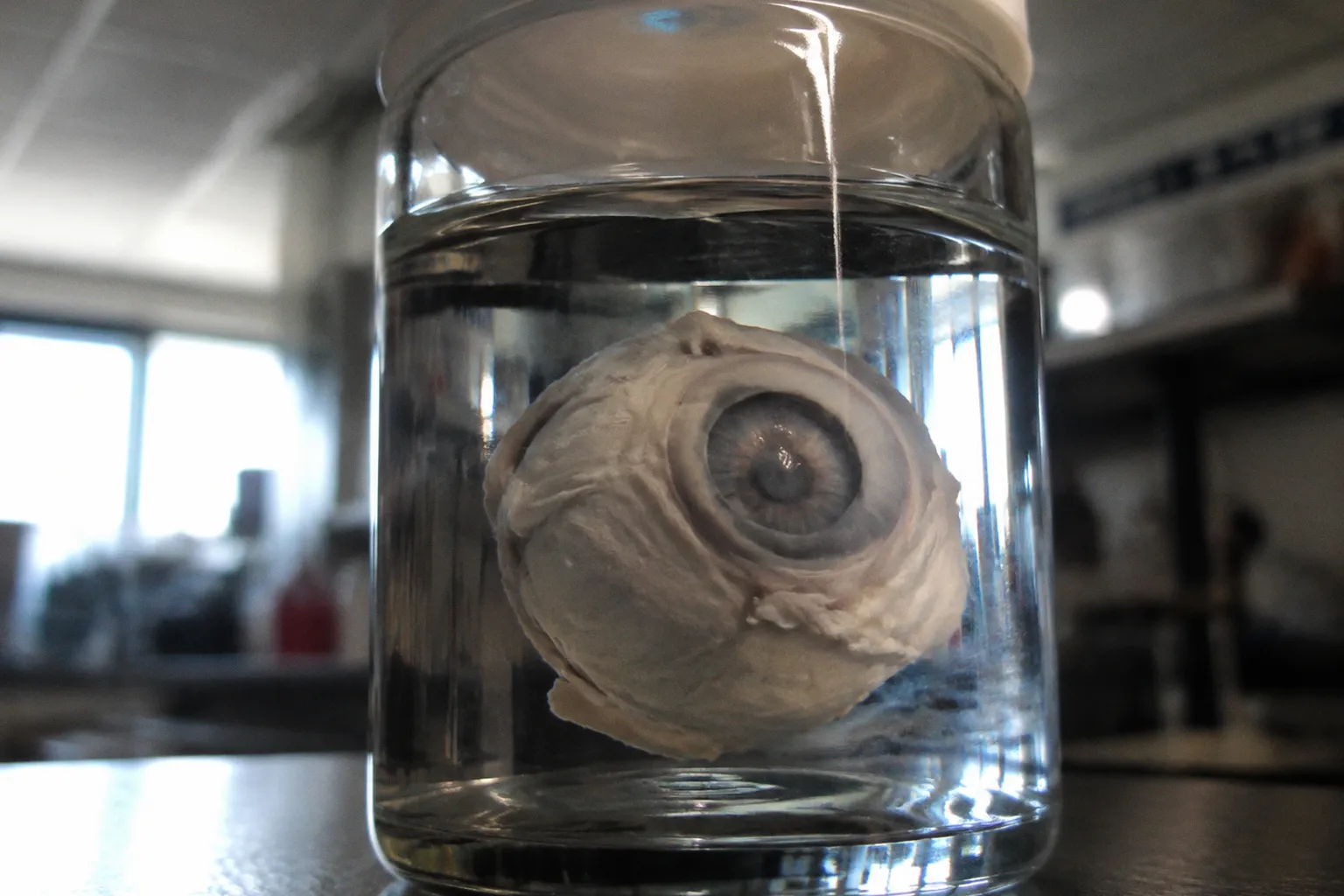
Every ocular pathologist has, at some point, encountered the assumption that donor retinal tissue is “too autolyzed to trust.” The usual shorthand is that anything beyond a few hours post-mortem is little more than cellular mush: useful at best for orientation, and at worst a setup for misdiagnosis. Yet the histology tells a more inconvenient story. Rod photoreceptor outer segments can retain disc membrane architecture long after the inner retinal circuits have lost functional integrity. At the same time, metabolic activity and synaptic transmission decline on much shorter timescales. The dogma, it turns out, is not simply too strict. It is built around a single idea of viability that donor tissue does not support.
The question is no longer whether donor eyes carry artifacts. They do. The question is whether the field has the discipline to separate genuine photoreceptor loss from the predictable consequences of ischemia, cooling, delayed enucleation, and fixation. The tools for making that distinction exist. The willingness to use the procurement record as part of the interpretation still lags behind.
Histological Signatures of Post-Mortem Retinal Degradation
Post-mortem artifacts in donor eyes are not subtle, and they are not random. They tend to follow a recognizable sequence: retinal detachment, separation within the photoreceptor layers, vacuolation of the nerve fiber and outer plexiform layers, cleavage of the Henle fiber layer, and detachment of the RPE from Bruch’s membrane. In a living patient, any one of these findings could prompt clinical concern. In a paraffin block marked with a prolonged post-mortem interval, the same findings may be consequences of tissue handling and ischemic decay rather than evidence of a primary retinal disorder.
That distinction is not semantic. A detached retina can reflect mechanical separation during recovery, processing, or fixation. A cleft in the outer retina can arise from post-mortem swelling or tissue fragility. RPE separation may say more about the condition of the globe at fixation than about the state of the RPE in life. The slide records the final arrangement of the tissue; it does not, by itself, identify when or why that arrangement developed.
A histological survey of 24 donor eyes graded artifact severity without reference to donor age or cause of death: 8 specimens were classified as mild, 7 as moderate, and 9 as severe. The practical takeaway is not that donor demographics are irrelevant in every context. It is that artifact severity cannot be safely inferred from age or cause of death alone. A younger donor with a long death-to-enucleation interval may show more severe disruption than an older donor recovered promptly. Once the cohort is examined without demographic assumptions, the procurement timeline becomes impossible to ignore.
The vacuolation pattern deserves particular attention. Vacuoles in the nerve fiber layer may appear early, sometimes within the first few hours, and their distribution is not necessarily uniform. When they are concentrated along arcuate bundles, they can invite comparison with the topography of glaucomatous damage. A pathologist working without procurement metadata could mistake artifactual spaces for axonal loss, particularly when the surrounding tissue is poorly preserved.
The safer interpretation is comparative rather than categorical. Post-mortem vacuolation may be patchy and asymmetric between the two eyes, and it may occur alongside small vacuolar changes elsewhere in the inner retina. Those findings can help identify a preservation-related pattern, but they should not be treated as a universal signature or as proof that a lesion cannot occur in disease. The important point is narrower: vacuoles should not be read in isolation. Their distribution, the condition of adjacent layers, the degree of retinal separation, and the recorded processing interval all matter.
The same caution applies to the Henle fiber layer. The oblique course of photoreceptor axons through the foveal slope makes this region vulnerable to mechanical distortion, edema, and autolytic change. In donor tissue, the layer can separate and form a cleft that resembles a schisis-like space on cursory review. That appearance is not enough to diagnose foveal schisis or a lamellar macular defect. Nor is the absence of additional clinical features enough to establish that the cleft is purely artifactual. A more defensible description is morphological: a separation within the Henle fiber layer in tissue with a documented post-mortem interval and associated preservation changes.
This is the difference between recognizing an artifact pattern and claiming a disease mimicry that the specimen cannot establish. Histology can show what is present. The procurement record helps determine how much biological meaning can be assigned to it.
A detached retina on a histology slide is not a diagnosis. It is a reason to read the procurement timestamp before reading the lesion.
The pathologist therefore cannot outsource interpretation to the donor chart, but neither can the pathologist ignore it. Every layer separation, every vacuole, and every apparently “abnormal” finding should be considered against the cold-chain record and the order of processing steps. The tissue retains evidence of what happened to it. The slide does not lie, but it does not volunteer context either.
Metabolic Decay and Functional Viability Windows
The metabolic clock starts before the body cools. Between 2 and 4.5 hours post-mortem, human donor retinas show declining 3H-taurine uptake, rhodopsin synthesis, and 32P-phosphate incorporation into rhodopsin, with reported losses of roughly 16% to 19% per hour in the relevant experiments. These are not peripheral readouts. Taurine handling and rhodopsin turnover are closely tied to photoreceptor maintenance. A tissue can therefore look anatomically recognizable while its metabolic performance has already fallen substantially.
That is the first separation researchers need to make: visible structure is not the same thing as intact physiology.
The same body of work that documents metabolic decline also indicates that rod outer segments can retain recognizable disc membrane architecture in donor specimens harvested up to 48 hours after death. By the fourth or fifth hour, metabolic activity may be markedly reduced, while outer segment morphology remains sufficiently preserved for structural analysis. Under ex vivo conditions, photoreceptor cell bodies can also retain recoverable light-evoked electrical responses within an appropriately short recovery window. These observations are not contradictory. They describe different compartments and different definitions of preservation.
A preserved disc membrane does not prove that the photoreceptor is metabolically normal. A recoverable electrical response does not prove that the entire retinal circuit remains functional. Conversely, a decline in metabolic assays does not mean that every relevant structural feature has disappeared. Donor tissue has several clocks running at once.
The useful model is therefore a gradient rather than a binary label:
| Post-mortem window | What may remain interpretable | What becomes increasingly unreliable |
|---|---|---|
| Under 20 minutes | Photoreceptor-to-inner-retina signaling may remain close to the recovery conditions required for physiological study | Delay-sensitive synaptic measurements begin to lose their margin of confidence |
| Up to approximately 5 hours | Photoreceptor cell bodies and some light-evoked responses may remain recoverable under ex vivo conditions | Circuit-level transmission and metabolic performance are no longer equivalent to those of living tissue |
| Approximately 4–4.5 hours | Outer segment architecture and measurable rhodopsin-related activity may still be assessed, with substantial decline | Quantitative comparisons with fresh tissue require careful normalization and metadata |
| Up to 48 hours | Rod outer segment disc membranes may remain morphologically recognizable | Functional viability and normal metabolic activity cannot be inferred from morphology alone |
The table is not a comfort. It is a warning about the danger of using a single word—“viable”—for several incompatible endpoints. A sample can be suitable for examining outer segment organization and unsuitable for studying synaptic transmission. It can retain cell bodies while losing the physiological relationship between photoreceptors and bipolar cells. It can produce interpretable histology while carrying a transcriptional stress state that would distort a disease comparison.
This is why donor eye post-mortem interval should be treated as an experimental variable, not as a footnote in the methods section. The interval determines which questions remain answerable and which conclusions have already passed beyond the tissue’s evidentiary range.
What Structural Preservation Can and Cannot Establish
Outer segment preservation is particularly easy to overinterpret because it is visually persuasive. The discs remain organized; the cell outline is recognizable; the retina may look sufficiently intact under light microscopy. But morphology is a record of architecture, not a direct assay of energy balance, ion homeostasis, or synaptic release.
For structural studies, this distinction may be entirely acceptable. A project focused on disc membrane arrangement, outer segment length, or the spatial relationship between photoreceptors and the RPE can still extract meaningful information from tissue with a longer post-mortem interval, provided the preservation pattern is reported and matched across groups. The same tissue may be inappropriate for claims about phototransduction kinetics, neurotransmitter release, or intact retinal circuitry.
The most reliable practice is to state the endpoint in concrete terms. “Outer segment morphology was preserved” is a defensible observation. “Photoreceptors remained viable” is a broader claim that requires additional evidence. If the study does not measure function or metabolism, it should not imply that morphology has established either.
Transcriptional Shifts in Post-Mortem vs. Living Tissue
If histology is the surface and metabolism is the engine, transcriptomics is the record of the tissue’s response to what happened. Single-cell RNA sequencing comparing living surgical tissue preserved within a short interval to post-mortem donor eyes harvested later can reveal distinct transcriptional states. Cell populations may be represented differently, stress programs may be elevated, and RNA quality may vary across retinal compartments. A post-mortem expression profile is not simply a living profile with a little technical noise added.
This is where Müller glia activation becomes important. With longer death-to-enucleation intervals, Müller cells can show transcriptional changes consistent with ischemic stress, ionic dysregulation, and loss of perfusion. These changes may include increased expression of GFAP, vimentin, nestin, and genes involved in inflammatory signaling and cytoskeletal remodeling. Such a profile can overlap with reactive gliosis, but overlap is not identity. Without clinical and procurement context, the dataset may encourage an interpretation in which post-mortem stress is mistaken for disease-associated gliosis.
The appropriate language is therefore conditional. Müller glia activation in a donor retina may reflect a response to the post-mortem environment; it may also coexist with pre-existing disease. The transcriptomic signal alone cannot decide between those explanations. The death-to-enucleation interval, enucleation-to-processing interval, tissue temperature, fixation or dissociation protocol, and clinical history all contribute to the interpretation.
This problem becomes more serious when datasets are compared across laboratories. One group’s six-hour donor retina may be treated as equivalent to another group’s much later specimen, even though the tissue has passed through different metabolic and transcriptional states. Without standardized annotation, the biological variability introduced by procurement logistics can contaminate every cluster in a dimensionality-reduction plot. Genes that appear differentially expressed between “diseased” and “control” donor retinas may instead distinguish faster and slower procurement cohorts.
The confound remains invisible unless the metadata is present. In a research biologics database, “human donor retina” is not a sufficient description. The record should distinguish at least:
- death-to-enucleation interval;
- enucleation-to-fixation, dissociation, or freezing interval;
- storage and transport temperature;
- preservation medium and fixation method;
- retinal region and dissection strategy;
- tissue quality grading and visible detachment;
- whether the sample was collected from a living surgical procedure or after death.
These fields do not eliminate variability. They make it possible to model it.
There is also the problem of RNA degradation kinetics. Total RNA integrity, including measures such as RIN where available, declines with post-mortem delay, but the decline is not uniform across transcript classes. Some transcripts may persist longer than others, and normalization panels can themselves become unstable if their degradation behavior differs from that of the genes under study. A housekeeping gene is not automatically a neutral reference in post-mortem tissue.
The technical literature on post-mortem RNA degradation in brain tissue is extensive. The corresponding evidence specific to human retina is thinner, and brain-derived correction factors should not be transferred without validation. The retina has its own cellular composition, metabolic demands, and handling constraints. A correction model borrowed from another tissue may be useful as a hypothesis, but it is not a substitute for ocular validation.
The lesson is not that transcriptomics is unreliable. It is that there is no single biological entity called “post-mortem retina.” There are retinas harvested after different intervals, processed through different workflows, and carrying different degrees of ischemic stress. Each is a distinct experimental state. Pooling them without annotation is not integration. It is averaging away the variable most likely to explain the result.
The Threshold of Synaptic Integrity in Donor Photoreceptors
This is where the assumption of universal post-mortem decay breaks down most clearly. Photoreceptor cell bodies collected from human maculae within several hours after death can, under ex vivo conditions, retain light-evoked electrical responses. The photoreceptor itself may still respond when supplied with an appropriate perfusing environment and recovery protocol. The catch is that synaptic communication with downstream ON-bipolar cells is far more delay-sensitive than the gross morphology of the photoreceptor.
Evidence from rapid-recovery preparations indicates that ON-bipolar transmission is best preserved when tissue is recovered within a very short post-mortem interval, on the order of minutes rather than hours. Once that interval is exceeded, the ability to infer normal photoreceptor-to-bipolar signaling falls rapidly. The exact boundary depends on the preparation, temperature, perfusion, and readout, so a rigid universal cutoff should be avoided. The practical conclusion is nevertheless clear: a donor retina that is acceptable for examining photoreceptor structure may already be unsuitable for studying intact synaptic transmission.
The outer segment is a durable record of architecture. The ribbon synapse is a much less forgiving record of function.
The differential vulnerability has a straightforward physiological explanation. The ribbon synapse is an energy-intensive structure. Vesicle replenishment, calcium cycling, neurotransmitter release, and maintenance of ionic gradients depend on continuous metabolic support. When perfusion stops, those processes are exposed to rapid disruption. Functional transmission can therefore fail before the photoreceptor cell body develops obvious morphological signs of destruction.
That statement should not be expanded into a more specific ultrastructural claim than the available evidence supports. It is reasonable to say that synaptic function is lost or severely compromised rapidly after death and that photoreceptor cell bodies may remain morphologically recognizable for longer. It is less defensible to assign a universal minute-by-minute schedule to the disappearance of every ultrastructural component of the synapse. Fixation delay, tissue temperature, recovery method, and the definition of “synaptic integrity” all affect the result.
For the same reason, the persistence of photoreceptor RNA should not be treated as proof of a fully functioning cell. Transcriptional activity can outlast physiological competence. A cell may retain detectable transcripts while its membrane gradients, outer segment renewal, or synaptic release machinery have already deteriorated. Morphology, transcription, metabolism, and circuit function must be reported as separate endpoints.
For researchers studying photoreceptor degeneration biology, this distinction is not academic. A study aiming to characterize synaptic pathology in age-related macular degeneration cannot rely on a standard delayed donor interval if its principal readout is bipolar-cell coupling. The tissue may still support carefully framed observations about outer segment morphology, RPE relationships, or intrinsic photoreceptor structure. It may also support molecular studies, provided the post-mortem stress state is modeled rather than ignored. The recovery window defines the question.
Photoreceptor Loss or Photoreceptor Distortion?
The central diagnostic error is to equate a missing or displaced structure on a donor-eye section with cell death. Post-mortem separation can make photoreceptor layers appear thinned, interrupted, or uneven. Swelling can distort the relationship between the outer nuclear layer, inner and outer segments, and RPE. Folding and tearing during processing can create local gaps that look more severe in a two-dimensional section than they were in the intact globe.
A convincing claim of photoreceptor loss therefore needs more than a single altered plane. It should be supported by the distribution of nuclei, the continuity of neighboring layers, the condition of the RPE, the presence or absence of corresponding changes in adjacent sections, and the procurement history. If immunolabeling is used, loss of a marker should not automatically be equated with loss of the cell: antigen preservation and epitope accessibility are also affected by fixation and delay.
The same principle applies to outer segment shortening. A shortened or detached outer segment may indicate genuine degeneration, but it may also reflect post-mortem collapse, mechanical separation, or processing. The question is not whether an artifact can resemble disease. Many artifacts can. The question is whether the specimen contains enough independent evidence to distinguish the possibilities.
Navigating Artifact Variability in Donor Eye Histology
What, then, is the working pathologist or vision scientist to do? The honest answer is to refuse the binary. Tissue is neither “viable” nor “non-viable” in the abstract. It is a continuous experimental variable defined by time, temperature, preservation method, and the specific question being asked.
Several operational principles follow from the evidence.
- Match the assay to the recovery window. Outer segment morphology tolerates longer intervals than bipolar-cell physiology. Choose the assay to fit the procurement reality, not the other way around. A laboratory studying disc membrane organization may work with tissue that would be unacceptable for synaptic physiology. Those are different experiments, even when the source material is described with the same phrase: “donor retinal tissue.”
- Document the procurement clock at every step. Death-to-enucleation time, enucleation-to-fixation or freezing time, transport conditions, and fixation protocol all influence the final specimen. A slide without provenance is a hypothesis, not a complete finding. Reporting only the interval between death and preservation is insufficient if the tissue then remained unfixed during transport or dissection.
- Treat Müller glia activation as a context-dependent signal. GFAP or related stress signatures may reflect ischemic injury after death rather than the disease process under study. They may also coexist with genuine reactive gliosis. The correct response is not to discard the signal, but to interpret it against the post-mortem interval and include procurement variables in the analysis.
- Do not infer artifact severity from age or cause of death alone. Demographic information can be relevant, but it cannot replace the recovery timeline. A young eye is not automatically a well-preserved eye, and an older eye is not automatically unusable.
- Separate structural, metabolic, transcriptional, and functional endpoints. Outer segment architecture, rhodopsin-related activity, RNA profiles, photoreceptor electrical responses, and ON-bipolar transmission answer different questions. A result in one category should not be used as a proxy for all the others.
- Use matched controls whenever possible. Disease and control tissue should be balanced not only for age and region but also for death-to-enucleation interval, enucleation-to-processing interval, temperature exposure, and tissue handling. If matching is impossible, the imbalance should be modeled and stated rather than buried in a supplementary table.
- Describe the artifact field, not only the target lesion. Retinal detachment, layer clefts, vacuolation, RPE separation, folds, tears, and uneven staining are not background annoyances. They are part of the specimen’s biological and technical history. Recording them allows later readers to judge whether a proposed photoreceptor loss pattern is convincing.
There is no universal biochemical marker that cleanly separates post-mortem outer segment detachment from pathological in vivo detachment in paraffin-embedded tissue. Anyone promising a single decisive readout is selling certainty the field does not have. The honest path forward is to publish the procurement parameters alongside the data and to state which conclusions the tissue can support.
Building a More Useful Donor-Tissue Record
For an ocular tissue procurement and research biologics database, quality assessment should be designed around use rather than around a single pass–fail label. A sample can be valuable for one project and unsuitable for another. A database that records only “good” or “poor” tissue throws away precisely the information that makes the specimen useful.
A more informative entry would connect the sample to its intended analytical range:
| Database field | Why it matters |
|---|---|
| Death-to-enucleation interval | Helps estimate the degree of early ischemic stress and likely preservation of delay-sensitive function |
| Enucleation-to-processing interval | Separates biological delay from later handling effects |
| Fixation or freezing method | Influences morphology, antigen preservation, RNA quality, and downstream assay choice |
| Temperature and transport history | Adds context to metabolic decline and tissue distortion |
| Retinal region | Macula, periphery, and specific layers may not show identical artifact patterns |
| Histological quality grade | Records detachment, vacuolation, layer cleavage, folds, and other visible changes |
| Intended assay | Prevents a structural-quality grade from being mistaken for a functional-quality grade |
| Clinical and donor metadata | Helps distinguish pre-existing pathology from preservation-related changes without treating either source as definitive |
This is not administrative excess. It is the minimum context required to avoid turning a procurement variable into a false biological discovery. If a study finds increased Müller glial stress genes in a disease cohort, the first question should be whether the cohort also had longer post-mortem intervals. If photoreceptor density appears lower, the next question should be whether the sections show detachment or mechanical loss. If synaptic markers are absent, the interpretation should acknowledge that synaptic structures are especially vulnerable to delay.
The database should also preserve uncertainty. “Suitable for outer retinal morphology; unsuitable for circuit physiology” is more useful than “moderate quality.” “RNA profile affected by prolonged post-mortem interval” is more useful than a generic warning that the sample was “degraded.” Precision does not always mean adding another number. Sometimes it means naming the limitation correctly.
Closing Position
The retinal research community has spent decades treating donor tissue as a degraded proxy for the living eye. The histology is messier. The transcriptome is shifted. Synaptic transmission is lost far earlier than the outer segment architecture that remains visible on a slide. And yet the same tissue that fails as a reliable synaptic preparation may preserve enough structural information for serious work on photoreceptor organization, RPE relationships, and post-mortem retinal biology.
The reasonable position is not blanket skepticism toward donor tissue. It is skepticism toward conclusions that do not name the procurement window. A donor eye is not one experiment. It is a collection of compartments, each with its own tolerance for delay and its own definition of preservation.
Photoreceptor loss post mortem artifact donor retina is therefore not a problem solved by choosing one preferred morphology. It is a problem of calibration. The researcher must ask what survived, what failed first, which changes reflect ischemia, and whether the available metadata can support the distinction. Once those questions become routine, donor eyes stop looking like compromised versions of living tissue and start being treated as what they are: time-sensitive biological specimens whose value depends on matching the right question to the right window.