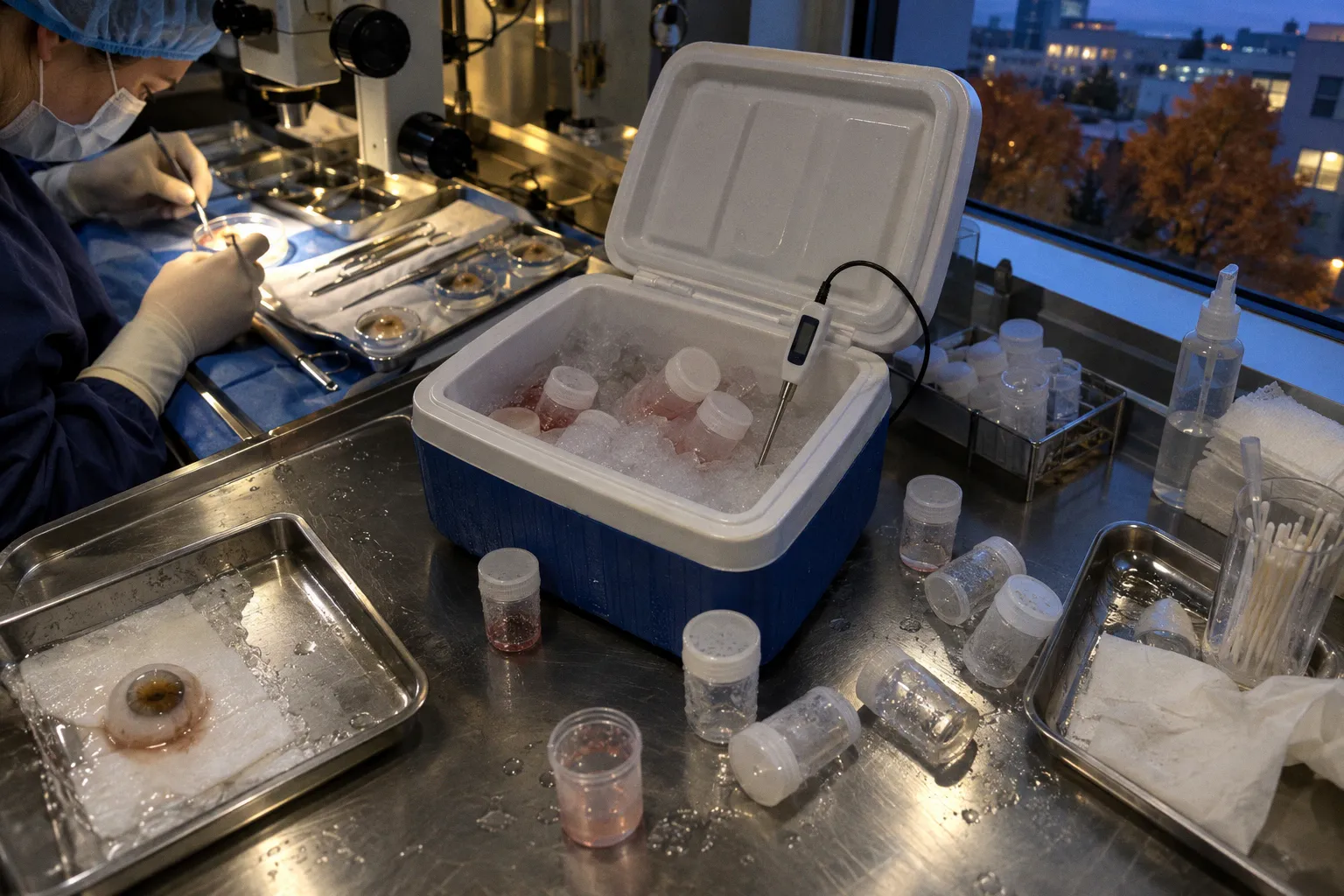
The molecular record is less reassuring. Endothelial cell loss in donor corneal storage is not a marginal footnote in eye-banking logistics. It is a measurable biological event, and its severity depends not only on the nominal storage method but also on the thermal history between procurement, processing, shipment, and surgery.
That distinction matters because a storage label describes a target temperature, not necessarily the temperature the tissue experienced. A cornea may be assigned to hypothermic storage at approximately 4°C and still encounter repeated excursions during packing, transport, temporary holding, or preparation for implantation. In organ culture, the problem is reversed: the tissue is deliberately maintained in a metabolically active state, making stability around the intended incubation range especially important. In both systems, the recorded endpoint can conceal the path taken to reach it.
The central question is not whether every temperature fluctuation causes clinically meaningful damage. The available evidence does not support that claim. The more precise question is how specific deviations, sustained for specific durations and occurring in specific preservation systems, alter endothelial density, cellular stress, and the tissue’s margin for recovery. That is where the evidence becomes useful—and where the certainty ends.
Thermal Sensitivity in Hypothermic and Organ Culture Preservation
Two preservation regimes dominate contemporary eye banking. Hypothermic storage holds donor corneas at 2°C to 8°C, commonly around 4°C, in media such as Eusol-C or Optisol-GS, with a maximum preservation window of 14 days. Organ culture maintains tissue at approximately 30°C to 37°C and can extend the preservation period to as long as 34 days. These are not simply two versions of the same intervention. They place the endothelium in different metabolic states and create different failure modes when temperature control is lost.
Hypothermia suppresses metabolism without eliminating it. Endothelial cells remain viable, continue to exchange ions, and retain the machinery needed to preserve corneal deturgescence, but their activity is slowed. The reduction in metabolic demand is part of the protective effect. It is not biological silence. Oxygen consumption, membrane stress, and the capacity for oxidative injury do not disappear merely because the tissue is cold.
Organ culture takes the opposite approach. The tissue remains metabolically active under conditions closer to physiological temperature, with the potential advantages of longer preservation and ongoing assessment. The cost is that the system depends on stable incubation. A downward deviation is not merely a small change in a number on a monitor; it shifts the tissue toward a state for which the preservation protocol was not designed.
A comparative dataset illustrates the asymmetry. Porcine corneas maintained in organ culture and then exposed to 21°C for 48 hours showed a statistically significant reduction in endothelial cell density compared with controls, with p = 0.025. In a parallel arm, exposure of the same tissue to 4°C for 12 hours did not produce a clinically relevant difference. The result should not be converted into a universal rule that all warming is safe or all cold exposure is harmful. It describes a defined experimental comparison. Within those conditions, however, the important signal is clear: organ-cultured tissue can be vulnerable to cooling below the intended incubation range, and the response may depend on both the depth and duration of the excursion.
That is a different problem from ordinary hypothermic storage. A temperature that is expected in one preservation regime may be an adverse deviation in another. The label “cold” has little value without the protocol around it.
Nominal range versus experienced range
For tissue-quality review, the nominal storage temperature answers only one question: what environment was intended? A useful thermal record should also address:
- the temperature at procurement and initial placement into preservation medium;
- the time required to reach the target range;
- the temperature profile during packaging and transit;
- any period of temporary holding outside the eye bank or surgical facility;
- the duration and conditions of pre-surgical preparation;
- whether the tissue was returned to the intended range after an excursion.
This is not administrative overreach. It is the minimum information needed to distinguish a brief, recoverable deviation from a prolonged exposure that may have altered endothelial function. Yet many records remain better at documenting release and arrival than the interval between them.
The preservation label describes the destination. Endothelial injury may be written in the journey.
The difference between the two systems also explains why comparisons between hypothermic and organ-culture data require restraint. An observation made in porcine organ culture at 21°C for 48 hours cannot be used to define a universal threshold for human corneas in hypothermic media. Conversely, the absence of a clinically relevant difference after 12 hours at 4°C in that experimental arm does not establish that every cold excursion is harmless. It establishes what happened under the stated conditions.
Quantifying Endothelial Cell Density Decline During Storage
Cell density remains the most familiar operational measure of corneal endothelial quality, and it is still indispensable. It is also incomplete. A density count describes how many cells are visible at a given assessment point; it does not fully describe cellular morphology, pump reserve, junctional integrity, or the capacity of the monolayer to tolerate rewarming and surgical stress.
In a cohort of donor corneas stored at 4°C in Eusol-C, mean endothelial cell density fell from 2,195 ± 383 cells/mm² at procurement to 1,658 cells/mm² by the eighth day. The reported mean loss was 24.5% over the first week of cold storage. For corneas held longer, between day 9 and day 24 in the same medium, the decline proceeded at approximately 3.1% per day. The curve was front-loaded: the most pronounced reduction occurred early, as the tissue adapted to the storage environment.
Those figures are useful as a description of that storage experience. They are not a universal decay law for every donor cornea, medium, eye bank, or transport history. Initial endothelial density varies. Donor characteristics vary. Assessment methods vary. So do the conditions under which tissue reaches the laboratory or operating room. A percentage derived from one cohort should be treated as a benchmark for interpretation, not a guaranteed forecast for an individual graft.
The practical value of the curve lies in what it makes visible. The early storage interval deserves particular scrutiny because the first days may carry a disproportionate share of the observed decline. A stable later rate does not mean the tissue has stopped changing; it means that the pattern in this dataset became less steep after the initial adjustment. If a temperature excursion occurs during that early period, the effect may be difficult to separate from the baseline loss associated with storage itself unless the thermal history has been recorded.
A side-by-side view helps clarify what each method is designed to do:
| Parameter | Hypothermic storage, 2–8°C | Organ culture, approximately 30–37°C |
|---|---|---|
| Common operating point | Approximately 4°C | Approximately 31–37°C |
| Maximum preservation window cited here | Up to 14 days | Up to 34 days |
| Metabolic state | Suppressed but not absent | Metabolically active |
| Documented endothelial pattern | In one Eusol-C cohort, 24.5% mean loss by day 8, followed by approximately 3.1% per day from days 9–24 | Sensitive to defined downward deviations below the intended incubation range |
| Principal thermal concern | Duration and history of cold exposure | Cooling below the organ-culture set point |
| What the available evidence does not establish | A single universal damage threshold for all media and corneas | A universal human threshold derived from the porcine comparison |
The table is not a verdict on which method is superior. It is a reminder that the same temperature cannot be interpreted outside its preservation context. The relevant question is always whether the tissue remained within the range for which the protocol, medium, and evidence base were designed.
Molecular Mechanisms of Stress Response to Thermal Instability
Density counts capture the visible residue of injury. They do not show how the tissue arrived there. Molecular measurements add another layer, but they must be read with the same discipline as cell counts.
In one study, donor corneas held under standard hypothermic conditions showed substantial changes in gene expression relative to fresh controls: 1,264 genes were upregulated and 2,058 were downregulated. The total of 3,322 altered genes belongs to that specific comparison. It should not be presented as a fixed molecular signature of every cold-stored cornea, nor as a per-cornea count that applies regardless of donor, medium, storage duration, or analytical method.
The study’s direction of change is more informative than the headline total. The downregulated group was enriched for processes associated with stress response, cell–cell junction integrity, and oxidative-stress signaling. That pattern is compatible with an endothelium that remains alive under hypothermic conditions but is functioning under constraint. It does not, by itself, prove that a particular graft will fail after transplantation or that a particular temperature excursion caused the observed expression profile.
Three mechanisms help organize the findings.
Junctional destabilization
The endothelial monolayer depends on coordinated cell–cell contacts to maintain the barrier and pump functions that keep the cornea relatively dehydrated and optically clear. Changes affecting tight and adherens junctions may weaken that coordination even before a dramatic fall in cell density becomes apparent.
This is one reason a cell count should not be treated as a complete quality certificate. Two tissues can show similar density while differing in cell shape, junctional organization, pump reserve, or susceptibility to edema after rewarming. A transcriptomic signal associated with junctional stress raises a biologically plausible concern, but it does not replace functional assessment or establish an outcome for an individual graft.
Oxidative stress
Hypothermic storage reduces metabolic activity; it does not make the tissue oxygen-free or chemically inert. Residual mitochondrial activity and the transition between storage and rewarming can generate oxidative pressure. If antioxidant responses are altered, the tissue may return to physiological temperature with less reserve than its density count suggests.
The critical point is not that cold storage inevitably produces irreversible oxidative injury. The available evidence does not justify that universal statement. It is that temperature changes can interact with an already stressed biological system. The effect may depend on the starting condition of the tissue, the length of storage, the medium, and the sequence of cooling and warming.
Apoptotic priming and recovery limits
Stress-response signaling may leave cells more vulnerable without killing them immediately. A tissue can therefore appear acceptable at one assessment point while having a reduced capacity to withstand rewarming, manipulation, or implantation. This is a mechanistic possibility supported by the broader stress-response picture, not a direct prediction that every affected cornea will undergo post-storage cell death.
That distinction matters clinically. The phrase “survived storage” can mean that cells remained countable at release. It does not necessarily mean that the endothelial monolayer retained its full functional reserve. Conversely, a molecular change does not automatically mean that the graft has crossed an irreversible threshold. The two forms of evidence should be interpreted together rather than made to compete.
A transcriptomic signal is evidence of altered state, not a verdict on a single graft.
Clinical Implications of Transit Fluctuations and Pre-Surgical Warming
The shipping manifest often records the intended route more reliably than the tissue’s complete thermal history. Refrigerated containers drift. Coolant performance changes over time. Shipments may wait during loading, transfer, or receipt. A final arrival temperature can therefore conceal an excursion that occurred earlier.
That uncertainty creates a practical problem for tissue release. If the only recorded value is the temperature at arrival, the reviewer may be unable to distinguish a stable shipment from one that returned to the target range after a period of deviation. In the absence of a continuous record, the tissue is not automatically unusable—but the confidence attached to the release decision is different.
Pre-surgical warming complicates the story in a useful way. Donor corneas exposed to room temperature for up to approximately seven hours immediately before surgery did not show a statistically significant difference in endothelial cell density, with p = 0.353, or corneal thickness, with p = 0.415, measured immediately after warming. Those findings support a limited conclusion: under the conditions studied, that period of pre-surgical warming did not produce a detectable short-term difference in the measured endpoints.
They do not show that every warming protocol is safe, that warming has no molecular effect, or that the same exposure would have no consequence after prolonged storage or prior temperature instability. Nor do they establish anything about long-term graft survival. Immediate post-warming measurements and later clinical outcomes are different endpoints.
The asymmetry is still operationally important. A brief, controlled period of warming before surgery may be tolerated in a way that a prolonged, undocumented deviation during preservation is not. The distinction should guide review rather than produce a new absolute rule.
What a temperature excursion should trigger
When a tissue experiences a documented deviation, the response should be proportionate to what is known:
1. Reconstruct the event. Record the highest and lowest temperatures, duration, point in the chain of custody, and whether the tissue later returned to the intended range.
2. Identify the preservation system. A deviation below 30°C has a different meaning in organ culture than a reading within the expected range for hypothermic storage.
3. Review the biological context. Consider storage day, initial endothelial density, medium, tissue appearance, and any available assessment of cell morphology or corneal thickness.
4. Separate evidence from inference. A documented excursion may justify additional review; it does not, by itself, prove irreversible endothelial loss or predict graft failure.
5. Record the uncertainty. If the thermal history is incomplete, that limitation belongs in the quality assessment rather than being silently converted into reassurance.
A tissue that has experienced a substantial or prolonged deviation may need to be flagged, re-evaluated, or excluded according to the eye bank’s validated procedures. The decision should not be based on a universal threshold that the current evidence has not established.
Defining the Thresholds for Irreversible Endothelial Degradation
This is where precision tends to outrun the data. The exact combination of temperature, duration, medium, donor condition, and tissue history that produces irreversible endothelial cell death across preservation systems is not established. Nor is the long-term graft-survival effect specifically attributable to minor transit temperature fluctuations established.
That uncertainty is not a reason to ignore temperature. It is a reason to describe the evidence accurately. The available findings support concern about measurable endothelial decline and molecular stress under defined conditions. They do not support the claim that a particular thermal history determines whether a graft will remain viable three years after keratoplasty.
Several boundaries can be discussed, but they should not be confused with universal biological thresholds.
Hypothermic storage
The cited Eusol-C cohort provides a reference pattern: a fall from 2,195 ± 383 cells/mm² at procurement to 1,658 cells/mm² by day eight, corresponding to a reported mean loss of 24.5%, followed by approximately 3.1% per day between days 9 and 24. That pattern describes endothelial density decline during the specified storage experience. It does not identify the point at which cells become irreversibly damaged, and it does not isolate the contribution of transit excursions from ordinary storage-related change.
The 2°C to 8°C range is therefore best treated as the intended hypothermic operating range in the context of the relevant protocol, not as proof that every value inside it has identical biological consequences. A short deviation near a boundary and a prolonged excursion well outside the range should not be treated as equivalent, but the evidence supplied here does not provide a single cutoff that resolves every case.
Organ culture
The porcine comparison offers a more specific warning about a defined deviation: 21°C for 48 hours after organ culture was associated with a statistically significant reduction in endothelial cell density, whereas 4°C for 12 hours did not produce a clinically relevant difference in the parallel arm. This supports heightened concern about cooling organ-cultured corneas below the intended range, especially when the exposure is prolonged.
It does not establish a human threshold, a universal 30°C damage line, or the proposition that human endothelium must respond identically to porcine tissue. The result is valuable precisely because its conditions are clear. Extending it beyond those conditions requires validation, not confidence.
Duration limits
The cited maximum windows—up to 14 days for hypothermic storage and up to 34 days for organ culture—are operational and regulatory boundaries within the described systems. They should not be recast as biological guarantees. Remaining within a maximum duration does not erase an adverse temperature history; exceeding it does not reveal, by itself, the exact degree of injury. Duration and temperature interact.
For research and quality systems, the unresolved variables are therefore straightforward:
- continuous rather than point-in-time temperature recording;
- agreed definitions for the depth and duration of an excursion;
- separate validation for hypothermic and organ-culture systems;
- correlation of thermal history with density, morphology, thickness, and functional recovery;
- follow-up that distinguishes immediate tissue quality from long-term graft outcomes.
The field needs those links before it can responsibly claim that a particular thermal event predicts a particular clinical result.
What the Evidence Can—and Cannot—Say
The strongest conclusion is narrower than the most dramatic version of the argument. Storage temperature matters. Endothelial density can decline substantially during cold storage. Organ-cultured tissue can respond adversely to a defined period of cooling below its intended range. Cold-storage conditions can be associated with broad transcriptomic changes relative to fresh controls, including 1,264 upregulated and 2,058 downregulated genes in one reported comparison.
But none of those findings establishes that every cold-stored cornea carries the same molecular signature. The 3,322 altered genes are the total reported for a specific study comparison, not a per-cornea universal count. Nor do the findings establish that a thermal excursion determines graft survival at three years, or that release density alone predicts that outcome. Long-term survival effects specifically attributable to storage-temperature history remain insufficiently defined.
The clinical implication is not to discard the tools already in use. Specular microscopy, endothelial density, morphology, corneal thickness, and release criteria remain necessary. The implication is to stop treating them as a complete record of preservation. A release measurement is a snapshot. Temperature logging is the history that gives the snapshot context.
A graft is not only what the microscope sees at release. It is also the thermal history that the release record may fail to show.
For ocular biobanking and research biologics databases, that means temperature should be captured as a structured biological variable rather than a single compliance field. A record that preserves storage medium, nominal range, continuous or interval temperature data, duration of excursions, and timing relative to procurement and surgery can support better retrospective analysis. It can also prevent a common analytical mistake: assigning a clinical outcome to “storage” while leaving the actual storage history undefined.
The evidence does not yet justify a universal irreversible-degradation threshold. It does justify more exact records, more cautious language, and studies designed to connect thermal exposure with both short-term endothelial function and long-term graft performance. Until those links are established, the responsible position is neither that temperature drift is harmless nor that every excursion condemns a graft.
The biology is more specific than that. A defined temperature, in a defined preservation system, for a defined duration, can produce a defined change. Everything beyond those conditions remains a question for validation rather than a fact to be filled in by assumption.