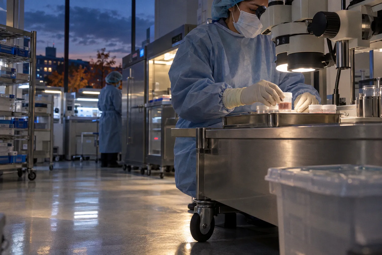
Hypothermic corneal preservation typically requires continuous exposure to 2–8°C and supports viability for up to 14 days in media such as Optisol-GS or McCarey–Kaufman medium. European organ-culture systems use a different thermal regime entirely: approximately 28–37°C, with storage extending to about four weeks under controlled incubation.
These are not interchangeable preservation models. They use different metabolic assumptions, release criteria, microbiological controls, and transport architectures. A shipment designed around refrigerated corneal media cannot be evaluated by organ-culture parameters. Conversely, a warm organ-culture workflow is not a substitute for cold-chain control during recovery and transfer.
The central logistics problem is therefore alignment. Death-to-recovery time, post-mortem refrigeration, preservation medium, container performance, temperature monitoring, and receiving-site processing must form one traceable chain. A failure at any interface can reduce transplant suitability even when the final storage temperature appears compliant.
Hypothermic storage and organ culture are separate operating systems
The phrase “cold chain” is technically accurate for hypothermic corneal preservation but incomplete for ocular biobanking as a whole. Eye banks operate across at least two major preservation architectures.
Hypothermic storage suppresses metabolic activity. The tissue is placed in a validated preservation medium and maintained between 2°C and 8°C. The objective is to slow cellular degradation while retaining sufficient endothelial function for later use. Under this model, the storage interval may extend up to 14 days, depending on the medium, tissue condition, release criteria, and local protocol.
Organ culture works differently. Corneal tissue is maintained at approximately 28–37°C in a controlled culture environment. The higher temperature does not represent a failure of refrigeration. It is part of the preservation method. European eye banks frequently use this approach to extend storage to approximately four weeks and to support microbiological screening before tissue release.
The operational variables are therefore distributed differently:
| Parameter | Hypothermic preservation | Organ culture |
|---|---|---|
| Primary thermal range | 2–8°C | 28–37°C |
| Typical preservation medium or environment | Refrigerated media such as Optisol-GS or McCarey–Kaufman medium | Controlled culture medium and incubator |
| Approximate storage horizon | Up to 14 days | Up to 4 weeks |
| Main logistics dependency | Continuous refrigerated transport and temperature control | Validated incubation, culture monitoring, and controlled transfer |
| Main release concern | Viability within the storage window and preservation history | Culture integrity, microbiological screening, and tissue quality |
| Typical failure mode | Temperature excursion, delayed recovery, or inadequate refrigeration | Incubator deviation, contamination, or culture-process interruption |
This distinction matters for research biorepositories. A dataset that records only the final storage temperature cannot reconstruct the preservation state of the specimen. The same cornea may pass through room-temperature handling, refrigerated media, insulated transport, inspection, and further storage. Each phase contributes to degradation kinetics.
A useful record must therefore capture at least:
- time of death or the operational timestamp used by the recovery organization;
- time of body refrigeration;
- time of ocular recovery;
- time of placement into preservation medium or culture;
- temperature range during transport;
- container type and coolant configuration;
- arrival time at the receiving eye bank or research facility;
- storage method after receipt;
- deviations, corrective actions, and final disposition.
The data model is not administrative overhead. It is the minimum structure required to interpret tissue quality. Without event timestamps, a later failure in endothelial viability or transcriptomic yield cannot be assigned to a specific stage in the workflow.
A compliant endpoint does not prove a compliant journey. Ocular tissue quality is a function of the entire thermal and temporal sequence.
Why the preservation medium does not solve the logistics problem
Refrigerated media slow tissue deterioration. They do not neutralize an uncontrolled transport profile. A vial or corneal chamber can remain inside a nominally suitable medium while the surrounding package undergoes a temperature excursion. The resulting risk depends on duration, peak temperature, tissue condition, and the time required to return the specimen to the target range.
The reverse problem also occurs. Direct contact with hard, super-cooled ice can expose living tissue to local freezing conditions even when the average package temperature appears acceptable. Freezing living corneal tissue without specialized cryoprotectants can generate intracellular ice crystals, rupture cell membranes, and produce irreversible cell death.
Packaging must therefore control both extremes:
1. It must maintain the required temperature range for the validated transit interval.
2. It must prevent localized freezing at the tissue container.
3. It must preserve physical separation between coolant and specimen.
4. It must limit mechanical shock, leakage, and orientation errors.
5. It must generate a record that can be reviewed after receipt.
A box filled with ice is not a validated shipping system. The relevant unit is the complete package: insulation, coolant mass, specimen placement, closure, ambient exposure, route duration, and monitoring method.
Death-to-preservation time is an upstream quality variable
Temperature control begins before the specimen enters a shipping container. The pre-recovery interval establishes the initial condition of the tissue. Early post-mortem body refrigeration is associated with a lower rate of donor tissue unsuitability for transplantation. The documented adjusted odds ratio is 0.78, corresponding to a 22% reduction in the odds of unsuitability during the death-to-preservation interval.
That finding should be interpreted precisely. It supports early refrigeration as a quality-protection measure. It does not establish that refrigeration increases corneal endothelial cell density. The reported effect concerns overall suitability, while endothelial cell count is a separate parameter with its own biological and measurement variability.
Operationally, many recovery workflows target ocular recovery within approximately 6–8 hours after death. The target is not a universal guarantee of suitability. It is a control point that limits uncontrolled post-mortem exposure before preservation begins.
The interval can be represented as a sequence of separate delays:
- notification latency between death and the eye bank;
- authorization and eligibility-screening latency;
- body refrigeration latency;
- dispatch latency for the recovery team;
- recovery procedure time;
- transfer latency to preservation media;
- transport latency to the processing or research facility.
These delays should not be collapsed into one field labelled “recovery time.” They carry different interventions and different failure causes. A delay before refrigeration is not equivalent to a delay after placement in validated refrigerated medium. A shipment delay in a qualified container is not equivalent to a delay in an uncontrolled vehicle.
Eligibility and recovery data are part of the specimen record
For transplantation, donor eligibility criteria and tissue examination determine whether a cornea can be released. For research, the tissue may remain valuable even when it is unsuitable for transplantation. This creates a branching disposition model rather than a simple pass/fail outcome.
A cornea that does not meet surgical release criteria may still be relevant for:
- endothelial morphology studies;
- epithelial wound-healing models;
- infectious disease research;
- molecular profiling;
- biomarker development;
- method validation;
- training and instrument calibration.
The research value, however, depends on the intended assay. A specimen that is unsuitable for grafting may still carry usable morphology but degraded RNA. Another may support structural analysis while failing to provide adequate transcriptomic yield. A repository that records only transplant suitability loses the information needed by investigators working with non-transplant tissue.
The intake record should distinguish at least three outcomes:
1. Surgical release: tissue meets the applicable transplantation criteria.
2. Research allocation: tissue is unsuitable or not allocated for surgery but remains fit for a defined research use.
3. Discard or restricted use: tissue condition, contamination risk, missing documentation, or uncontrolled exposure prevents approved use.
This is a systems distinction. “Unsuitable” is not synonymous with “without scientific value.” But research allocation cannot be used to obscure missing chain-of-custody data or unexplained temperature excursions.
Shipping containers require thermal validation, not nominal specifications
Transport is the most visible portion of donor eye tissue cold chain logistics, but it is often evaluated too narrowly. Package performance depends on coolant mass, insulation geometry, ambient temperature, opening frequency, specimen load, and transit duration.
Validation testing of insulated containers packed with wet ice provides concrete reference points. Under the tested ambient conditions, a small container with 685 grams of ice maintained temperatures at or below 8°C for 25.1 hours. A medium container with 1,929 grams of ice maintained the same threshold for 58.9 hours. A large container with 4,439 grams of ice maintained it for 85.7 hours.
These figures are not universal transit guarantees. They describe validated configurations under defined conditions. Changing the coolant mass, insulation, packing density, external temperature, or container dimensions changes the thermal profile.
The relationship is operationally useful:
| Container configuration | Wet ice load | Validated duration at or below 8°C |
|---|---|---|
| Small | 685 g | 25.1 hours |
| Medium | 1,929 g | 58.9 hours |
| Large | 4,439 g | 85.7 hours |
The numbers show why package selection should be based on route risk rather than specimen count alone. A small package may be adequate for a short, controlled regional transfer. It provides little margin for a missed pickup, weekend hold, airline disruption, customs delay, or receiving-site closure.
A medium or large container increases thermal margin, but it does not remove other risks. More ice can create localized freezing if the specimen is not physically separated from the coolant. A larger package can also increase dead volume, handling complexity, and the number of surfaces that require inspection after receipt.
The route is a thermal experiment
Every transport lane has a distinct risk profile. The same container can behave differently on a short ground route in winter and a long air route through warm terminals. A validated packaging configuration should therefore be matched to the expected route envelope.
The relevant variables include:
- forecast ambient temperature along the route;
- maximum planned transit duration;
- terminal dwell time;
- probability of overnight storage;
- carrier handling restrictions;
- opening or inspection points;
- handoff count;
- receiving-site operating hours;
- availability of immediate refrigerated storage;
- temperature logger placement and activation time.
The last variable is frequently underestimated. A temperature logger placed outside the specimen chamber can describe the package environment without describing the tissue container. A logger placed directly against the coolant can report a freezing event that does not represent the specimen. Monitoring design must match the question being asked.
For donor cornea shipping protocols, the receiving site should be able to determine:
- whether the package remained within the validated range;
- whether the tissue was exposed to a freezing risk;
- when the thermal excursion began;
- how long the excursion lasted;
- whether the specimen was released, quarantined, reclassified, or rejected;
- who made the disposition decision.
A temperature trace without a disposition rule is only a graph. It becomes a quality-control instrument when linked to predefined actions.
The package is not the unit of validation. The unit is the package, the route, the logger, the receiver, and the decision rule operating together.
Temperature excursions need classification, not improvisation
A temperature excursion is not automatically equivalent to tissue failure. The effect depends on its magnitude, duration, location, and interaction with the preservation method. At the same time, the absence of an established universal threshold means that facilities should not invent a false precision for every specimen type.
For hypothermically stored corneal tissue, a deviation above 8°C may indicate loss of validated conditions. A deviation below the safe lower boundary may indicate freezing risk, especially when the tissue is in direct proximity to ice or a super-cooled surface. The correct response depends on the recorded profile and the applicable eye bank or study protocol.
A practical deviation workflow has five stages:
1. Quarantine the tissue. Prevent automatic release while the record is under review.
2. Preserve the evidence. Retain the temperature file, package images, shipping label, coolant configuration, and receipt timestamp.
3. Reconstruct the event. Identify the start, peak, duration, and recovery of the excursion.
4. Relate the event to tissue state. Review preservation medium, time since recovery, tissue examination, and intended use.
5. Document disposition. Release, restrict, reclassify for research, or reject according to a defined authority.
The workflow should not rely on the receiving technician’s visual impression. A clear media container does not demonstrate preserved endothelial viability. A slightly softened ice pack does not establish that tissue was exposed to an unacceptable temperature. The record must be assessed against the validated package and the tissue-specific protocol.
Data quality determines whether the excursion is interpretable
Biorepository cold chain integrity is partly a sensor problem and partly a data-governance problem. A missing timestamp can be as damaging as a missing temperature measurement. If the logger starts after dispatch, the first segment of the route is unknown. If the receiving time is entered manually days later, the interval between arrival and refrigeration may be irrecoverable.
A minimum monitoring record should include:
- logger identification;
- calibration or verification status;
- activation time;
- sampling interval;
- sensor location;
- recorded minimum and maximum temperature;
- duration outside the target range;
- package opening or inspection events;
- receipt time;
- time of transfer to final storage;
- reviewer and disposition.
For research specimens, metadata should also include the intended assay category. A temperature excursion may have different implications for endothelial transplantation, RNA sequencing, proteomic analysis, histology, or cell culture. The same tissue should not be assigned one universal quality label when different downstream assays impose different stability requirements.
The known evidence is stronger for corneal preservation than for many non-corneal ocular biologics. Long-term structural stability benchmarks for isolated retinal pigment epithelium, scleral tissue, and other research materials under −80°C or vapor-phase nitrogen storage are not universal across repositories. Those materials require assay-specific validation rather than automatic transfer of corneal benchmarks.
Accreditation converts local practice into auditable process
Eye bank logistics are not standardized by equipment alone. They are standardized through documented procedures, competency records, inspections, and traceable corrective actions.
Eye Bank Association of America accreditation requires eye banks to document the handling of at least 25 surgical corneas for each function and to undergo on-site inspections at least once every three years. The requirement addresses process proficiency. It does not mean that every facility has identical shipping conditions or that accreditation eliminates transport risk.
Accreditation functions as a control layer over several operational domains:
- donor screening and eligibility review;
- recovery and aseptic technique;
- tissue examination;
- preservation and storage;
- labeling and chain of custody;
- temperature monitoring;
- release and disposition;
- staff competency;
- deviation management;
- record retention.
For a research-facing repository, the same architecture should extend beyond surgical release. A specimen database should connect the donor record to each physical aliquot, chamber, or tissue segment. The record should preserve lineage when the original globe is divided between transplantation, research, pathology, and discard.
This is where procurement systems and laboratory information systems often fail to connect. Procurement teams may track donor availability and recovery scheduling. Eye banks may track tissue suitability and release. Research laboratories may track sample receipt and assay performance. If these systems use different identifiers, the final dataset cannot reconstruct the full tissue history.
A robust ocular biorepository data model needs a common specimen identifier and event-level timestamps. The identifier should persist across:
- donor referral;
- eligibility review;
- recovery;
- preservation;
- transport;
- receipt;
- examination;
- allocation;
- subdivision;
- assay;
- storage;
- final disposition.
The objective is not maximal data collection. It is causal traceability. Every field should help answer a specific question: where was the tissue, under what condition, for how long, and what happened next?
The current benchmark is a controlled range, not a single number
The current evidence supports several practical conclusions.
Hypothermic corneal preservation is generally organized around 2–8°C, with preservation media supporting viability for up to 14 days under the applicable protocol. Organ culture uses a warmer 28–37°C environment and can extend storage to approximately four weeks in European workflows. These systems should remain analytically separate.
Early post-mortem refrigeration reduces the odds of tissue unsuitability, with an adjusted odds ratio of 0.78 in the cited evidence. The effect should not be restated as an improvement in endothelial cell density. It is a suitability outcome, not a universal cellular-performance guarantee.
Transport validation must include the full package configuration. Tested wet-ice systems maintained temperatures at or below 8°C for 25.1, 58.9, and 85.7 hours across small, medium, and large container configurations. Those durations establish reference performance under defined conditions, not a universal safe limit for every route.
Direct freezing is a separate hazard. Living corneal tissue should not contact hard, super-cooled ice, and freezing without specialized cryoprotectants can produce intracellular ice formation and membrane rupture. A low average package temperature is not automatically a quality signal.
Finally, the strongest control remains the quality of the data pipeline. Recovery time, refrigeration time, preservation time, transport temperature, logger status, receiving time, and disposition must be connected. Without that continuity, tissue quality becomes an endpoint without a reconstructable cause.
The donor eye tissue cold chain is therefore best understood as a temporal system. Temperature is one variable. The other variables are delay, medium, route, monitoring, and decision authority. Current benchmarks provide workable boundaries, but they do not replace validation. A tissue repository that cannot reconstruct the path from death to preservation to receipt cannot reliably distinguish biological degradation from logistical failure.