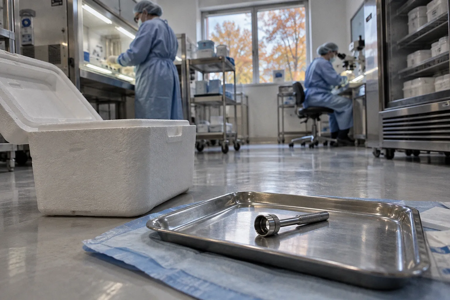
That conclusion sounds plausible—until endothelial viability is measured rather than inferred.
Hypothermic storage preserves corneoscleral tissue at 2–8°C, commonly around 2–6°C, for a maximum recommended period of 14 days. Organ culture keeps the tissue in cell culture medium at warm temperatures, typically around 31°C and within a broader 30–37°C range, extending the permissible storage window to roughly four or five weeks. The difference is not simply a matter of refrigerator versus incubator. It changes when quality can be assessed, how microbial testing is integrated, how stromal swelling is managed, and whether the tissue remains useful for research beyond immediate graft preparation.
The central question in this corneal endothelial cell preservation methods comparison is therefore not which technique is universally superior. There is no credible basis for that claim. The sharper question is this: what kind of uncertainty does each method leave the eye bank, surgeon, or researcher to manage?
Temperature determines more than storage duration
Hypothermic storage suppresses cellular metabolism by lowering temperature. The operational appeal is obvious. The tissue can be placed in a validated cold-storage system, transported within a defined time window, and prepared without maintaining a prolonged living-tissue culture workflow. For programs built around predictable turnover and established transport routes, that simplicity is not trivial.
But simplicity is not the same as transparency. At low temperature, the tissue is preserved in a metabolically quiet state; the method does not provide the same opportunity for extended observation of endothelial behavior during storage. The cornea may arrive within the accepted time window, yet the window itself says little about the quality of every individual endothelial mosaic.
Organ culture takes the opposite approach. Rather than placing the tissue into metabolic suspension, it maintains the cornea in a warm medium for several weeks. That longer interval permits intermediate endothelial cell quality assessment and microbiological testing before transplantation or further processing. The tissue is not merely stored; it is kept in a controlled biological environment in which its condition can be reviewed over time.
This sounds like an uncomplicated advantage. It is not. Warm preservation demands a more involved workflow, a functioning culture infrastructure, validated media handling, environmental control, and a team able to interpret tissue changes rather than simply record a dispatch date. A longer storage window creates options, but it also creates more points at which the process can fail.
The basic contrast is clear:
| Parameter | Hypothermic storage | Organ culture |
|---|---|---|
| Typical temperature | 2–8°C, commonly 2–6°C | Approximately 30–37°C, typically around 31°C |
| Maximum recommended storage window | Up to 14 days | Approximately 4–5 weeks |
| Operational profile | Relatively simple and transport-friendly | More demanding, culture-based workflow |
| Intermediate assessment | More limited during storage | Allows endothelial and microbiological assessment during the storage period |
| Stromal condition | Does not produce the same culture-associated swelling pattern | Stromal swelling is expected and requires later deswelling |
| Research utility | Useful when rapid processing is the priority | More time for selection, testing, and coordinated tissue allocation |
| Main operational liability | Shorter scheduling window and less prolonged observation | Culture complexity and mandatory deswelling before use |
The table does not identify a winner. It identifies where each method places the burden: cold storage places more pressure on timing; organ culture places more pressure on infrastructure and interpretation.
A longer preservation window is not automatically a better preservation system. It is a longer period in which quality can be examined—or in which poor process control can be exposed.
Hypothermic storage versus organ culture: the endothelial question
For corneal transplantation, the endothelium is not a decorative layer attached to an otherwise interchangeable scaffold. It is the functional barrier and pump system that controls stromal hydration and therefore optical clarity. A graft can have an acceptable gross appearance while carrying a compromised endothelial population. That is precisely why endothelial cell density, morphology, and cell-death measures matter more than the storage label alone.
Hypothermic storage has earned its position because it is practical, familiar, and clinically serviceable. It avoids the prolonged warm-culture workflow and fits the logistics of many North American eye banks, where cold preservation is widely favored. Yet the standard operational argument often slides into a biological argument: because cold storage is widely used, it is treated as though it must be the more reliable method for preserving endothelial quality. That leap is not supported by the available comparison.
A clinical study from the Rome Eye Bank compared 540 organ-cultured corneas with 92 corneas preserved under hypothermic conditions. The average endothelial cell density and morphology outcomes were clinically comparable. This is the inconvenient middle ground that dogmatic comparisons tend to avoid. Organ culture did not produce a demonstrated universal clinical victory, but neither did cold storage establish a decisive advantage.
For researchers attempting endothelial cell isolation and expansion, however, the comparison becomes more discriminating. In one study context, donor tissues preserved under organ culture conditions showed fewer than 1% trypan-blue-positive cells before cell isolation. The corresponding figure for cold-storage corneas was 9.3% ± 4.0%. The finding does not justify claiming that organ culture guarantees better graft survival. It does suggest that preservation conditions can materially affect the starting population available for cell-based work.
That distinction matters. A graft assessment asks whether tissue is clinically acceptable for a surgical purpose. A cell-isolation project asks how many cells remain viable, recoverable, and suitable for manipulation. The same cornea can be adequate for one use and disappointing for another.
Why average endothelial density is not enough
Endothelial cell density is a useful metric, but it is not a complete description of tissue quality. Density can remain within an acceptable range while cell morphology, size variation, or regional distribution raises questions about functional reserve. Likewise, a single viability measurement does not explain how the cells will respond to isolation, enzymatic handling, expansion attempts, or preparation for a specific anterior segment application.
This is where a corneal endothelial viability assessment becomes more than a release criterion. It is a reconstruction of what happened to the tissue during procurement and preservation.
A practical assessment may need to consider:
- Endothelial cell density: a basic indicator of the remaining cellular population, but not a standalone prediction of downstream performance.
- Cell morphology: variation in cell size and shape can reveal stress that a density count alone may conceal.
- Trypan-blue exclusion or related viability measures: particularly relevant when tissue is intended for endothelial cell isolation rather than routine grafting.
- Time from procurement to preservation: the storage method cannot erase the biological consequences of earlier delays.
- Tissue purpose: a DMEK graft, a full-thickness graft, an experimental cell isolation, and a histological study do not impose identical quality requirements.
- Condition of the stroma and Descemet membrane: especially when the tissue will be dissected, stripped, or mechanically tested.
The familiar question—how long has this cornea been stored?—is therefore too blunt. The more useful question is whether the preservation method has generated the kind of evidence needed for the intended use.
The complication organ culture cannot hide: stromal swelling
Organ culture has a biological and logistical consequence that cannot be edited out of the protocol: stromal swelling. Corneal tissue maintained in warm culture becomes edematous. Before transplantation or graft preparation, it must be transferred to a macromolecular deswelling medium, commonly containing agents such as dextran, to reduce stromal hydration.
This is not a minor finishing step. It is part of the preservation method.
A cold-stored cornea arrives with a workflow designed around low-temperature preservation and relatively direct preparation. An organ-cultured cornea arrives from a system in which the tissue has been metabolically maintained but structurally swollen. The laboratory must then manage deswelling before the graft can be prepared or used. If that step is treated as an administrative afterthought, the comparison becomes clinically meaningless.
The deswelling phase also changes how organ-cultured tissue should be evaluated. A swollen cornea may not offer the same immediate visual or handling characteristics as a deswelled graft. Thickness, tissue manipulation, and interface preparation need to be interpreted within the context of the protocol rather than compared as though both methods deliver identical physical material at the end of storage.
That difference becomes particularly relevant for posterior lamellar procedures. Descemet membrane endothelial keratoplasty tissue is thin, delicate, and highly dependent on controlled handling. The preservation method does not determine every aspect of graft behavior, but it shapes the condition in which the tissue reaches preparation. A protocol that is entirely acceptable for one graft format may require refinement for another.
The same logic applies to experimental tissue. If a laboratory is studying donor corneal endothelial cells, the fact that organ culture requires deswelling does not erase its potential value. It simply means that the process must be documented as a sequence: procurement, culture, quality assessment, deswelling, dissection or isolation, and downstream analysis. The tissue is not a static specimen. It is a biological system moving through several controlled states.
Regional preference is operational, not proof of biological superiority
Organ culture is used by approximately 65% of European eye banks. Hypothermic storage is widely favored in North America. These regional patterns are often presented as if they settle the scientific argument. They do not.
Adoption reflects more than comparative endothelial biology. It incorporates local regulation, transport geography, eye-bank infrastructure, staffing, media supply, laboratory validation, surgeon expectations, and the historical development of each preservation network. A method becomes dominant partly because an entire system has been built around it.
This is the paradigm deficit in many preservation discussions: infrastructure is mistaken for evidence.
If an eye bank has robust incubator capacity, trained personnel, validated culture media, and a distribution model that benefits from several weeks of scheduling flexibility, organ culture may be a rational default. If a program prioritizes straightforward processing, rapid dispatch, and a cold-chain workflow already integrated into procurement and transplantation, hypothermic storage may be the more coherent choice.
Neither conclusion requires pretending that one technique has solved endothelial attrition. Both methods remain vulnerable to the quality of the donor tissue at procurement, the interval before preservation, handling stress, and the intended use of the cornea.
The clinical comparison from the Rome Eye Bank is useful precisely because it resists the usual binary. With 540 organ-cultured corneas and 92 hypothermic corneas, average endothelial density and morphology were clinically comparable. That result does not mean every preservation protocol is equivalent. It means the storage category alone is a poor substitute for actual quality data.
A regional preference can tell us what a system has optimized for. It cannot, by itself, tell us which method preserves every biologically relevant property of every donor cornea.
The geography of eye-bank practice reveals where workflows are mature. It does not reveal where endothelial biology stops mattering.
Storage duration creates flexibility—and a different kind of risk
The fourteen-day maximum recommended window for hypothermic preservation imposes a firm scheduling discipline. That can be a limitation when a graft must be matched, transported, prepared, and allocated across a complex network. It can also be a form of control. A shorter window reduces the temptation to treat time as an unlimited resource.
Organ culture extends storage to approximately four or five weeks. For a biobank or research coordinator, that additional time can be transformative. It may allow more deliberate donor-recipient matching, staged assessment, microbiological testing, and allocation of tissue to projects that cannot accept a narrow dispatch window. For studies involving human corneal tissue, where procurement opportunities are irregular and experimental schedules rarely align neatly with collection dates, the longer window has obvious practical value.
But extended storage is not a free biological extension. The relevant question is not whether tissue can remain in the system for four or five weeks. It is whether the quality data collected during that period are sufficient to justify the intended use.
A prolonged storage interval may affect different components of the cornea differently. The endothelium, stroma, Descemet membrane, limbal tissue, and trabecular meshwork are not interchangeable biological materials. A protocol optimized for preserving a transplantable central cornea may not preserve the features needed for anterior chamber drainage histology or trabecular meshwork biology. The same donor may be highly valuable for one research endpoint and unsuitable for another after processing.
This is why donor cornea storage techniques should be selected with the downstream assay already in view. A project focused on endothelial cell expansion will prioritize viable, recoverable endothelial cells and standardized isolation conditions. A biomechanics study may care more about stromal architecture, hydration history, and mechanical handling. A limbal stem cell project requires attention to the peripheral cornea and limbal region rather than treating the central button as the entire specimen.
Storage time is therefore not merely a countdown. It is an argument about which biological properties the workflow is trying to preserve.
What the methods mean for research tissue
Clinical transplantation has a relatively clear endpoint: prepare a graft that meets the program’s release requirements. Research procurement is more fragmented. One donor cornea may be divided among several questions, each with its own definition of acceptable tissue.
For endothelial cell isolation and expansion, the contrast between fewer than 1% trypan-blue-positive cells in organ-cultured tissue and 9.3% ± 4.0% in cold-storage tissue is difficult to dismiss. It points toward a meaningful difference in the starting material under that study condition. Yet even here, the result must be interpreted narrowly. It supports an advantage for cell-isolation utility in the reported setting; it does not prove superior long-term clinical graft performance.
For human corneal stroma research, preservation introduces a different concern. Organ culture-associated swelling and subsequent deswelling may affect how tissue thickness and hydration are interpreted. A laboratory measuring corneal biomechanics cannot treat preservation history as background metadata. The sequence of storage and re-equilibration belongs in the experimental design.
For anterior chamber drainage studies, the trabecular meshwork and adjacent structures demand still greater specificity. Tissue procurement, dissection orientation, time to fixation, and preservation medium can influence the quality of histological interpretation. A cornea preserved for transplantation is not automatically an ideal anterior segment research specimen. The procurement request must state what structures are required and what handling history is acceptable.
A research-grade comparison should therefore record more than preservation category. At minimum, the dataset should distinguish:
1. The purpose of the tissue. Clinical grafting, endothelial cell isolation, stromal biomechanics, limbal biology, or histology each place different demands on the specimen.
2. The timing of preservation. Time from death and procurement to placement in the preservation system may be as consequential as the nominal storage method.
3. The condition at release. Endothelial density, morphology, and viability should be linked to the specific tissue rather than inferred from the bank’s general protocol.
4. The processing path. Deswelling, stripping, dissection, enzymatic isolation, and fixation can all alter the material available for analysis.
5. The remaining tissue architecture. A central endothelial preparation may have little value for a project requiring limbal or trabecular meshwork tissue.
6. The endpoint used to judge success. Clinical suitability, cell recovery, histological preservation, and mechanical integrity are not interchangeable outcomes.
This may sound less elegant than ranking two storage methods. It is also more honest. Preservation is not a single outcome, and the cornea is not a single research reagent.
Why cell expansion exposes the difference
Cell expansion is especially unforgiving because it tests the tissue after preservation rather than merely inspecting it before use. A cornea can pass a clinical quality screen and still yield a less productive cell-isolation experiment. The reasons may include nonviable cells, altered adhesion, regional endothelial damage, or handling effects that become visible only once the cells are removed from their native environment.
The lower proportion of trypan-blue-positive cells reported in organ-cultured tissue before isolation suggests that organ culture may offer a more favorable starting point for certain expansion workflows. That finding is biologically plausible and practically relevant. It is not a universal permission slip.
Expansion outcomes depend on the isolation protocol, donor characteristics, culture medium, substrate, passage conditions, and definition of a successful culture. The preservation method contributes to the starting condition; it does not control the entire experiment. Treating organ culture as a guarantee would simply replace one dogma with another.
The sensible conclusion is narrower: when the primary value of a donor cornea lies in recovering viable endothelial cells, preservation protocols should be judged by cell-level evidence, not by transplant history or regional popularity.
The false comfort of a single preservation label
Biobanks often need concise categories. A database field may say cold storage or organ culture, and the label is useful for first-pass coordination. It is not enough for serious tissue selection.
A useful record should connect preservation history to the specific research question. For example, the fact that tissue was held under organ culture for several weeks tells a coordinator that a deswelling phase may be required and that intermediate assessment may be available. It does not, by itself, establish that the endothelium is suitable for expansion. Likewise, a cold-storage label identifies a shorter preservation pathway but does not prove that the specimen has suffered unacceptable endothelial loss.
The phrase endothelial cell loss in storage is also more complicated than a simple before-and-after number. Loss may occur during the interval before preservation, during storage, during warming, during dissection, or during isolation. If the stages are not separated, the final count becomes an accusation without a location.
This is where careful procurement records matter. The useful database is not the one with the most fields; it is the one that preserves the causal chain. Temperature range, storage duration, assessment results, deswelling status, tissue region, and intended allocation should remain connected. Once those details are detached, researchers are left comparing labels instead of specimens.
For translational vision science, that distinction is decisive. A donor cornea is not merely available or unavailable. It is available for a defined purpose under a defined handling history, with a defined level of uncertainty.
A choice between workflows, not ideologies
The comparison between hypothermic storage and organ culture is often framed as a contest between a traditional technique and an emerging biological alternative. That framing is too crude. Hypothermic corneoscleral storage was introduced in 1974, while organ culture preservation was introduced in 1976; neither method belongs neatly to a simple old-versus-new narrative. The real contrast is between two operational philosophies.
Hypothermic storage prioritizes procedural simplicity, cold-chain control, and a shorter allocation cycle. It is well suited to systems that can move tissue efficiently and make decisions within approximately two weeks.
Organ culture prioritizes an extended preservation interval and the opportunity for intermediate assessment. It may be especially useful where donor tissue must be coordinated across multiple research or clinical demands, or where endothelial viability before cell isolation is a central concern. But it requires a culture workflow and a deswelling phase; there is no credible shortcut around those requirements.
The available evidence supports several restrained conclusions:
- Organ culture extends the storage window from up to 14 days under hypothermic conditions to approximately four or five weeks.
- Organ culture provides opportunities for endothelial quality assessment and microbiological testing during preservation.
- Stromal swelling is an inherent consequence of organ culture and must be addressed with a macromolecular deswelling medium before transplantation or graft preparation.
- Clinical endothelial density and morphology can be comparable between the two approaches, as shown in the cited Rome Eye Bank comparison.
- Organ-cultured tissue may offer a lower proportion of nonviable endothelial cells for cell-isolation work in the reported study context.
- No established basis supports claiming that organ culture universally improves long-term clinical graft survival over hypothermic storage.
The practical decision is therefore conditional. Choose hypothermic storage when the program’s strength is rapid, controlled movement through a short workflow. Choose organ culture when the program can support warm preservation and needs time for assessment, coordination, or research allocation. In both cases, treat the tissue’s measured condition—not the reputation of the method—as the final argument.
The unresolved issue is not whether one storage technique will defeat the other. It is whether corneal research will continue to compare preservation systems using broad clinical averages while under-documenting the cell-level outcomes that determine research utility. Until that paradigm deficit is corrected, the most defensible method is not the one with the strongest tradition or the longest storage window. It is the one that produces the clearest evidence for the exact biological question the tissue must answer.