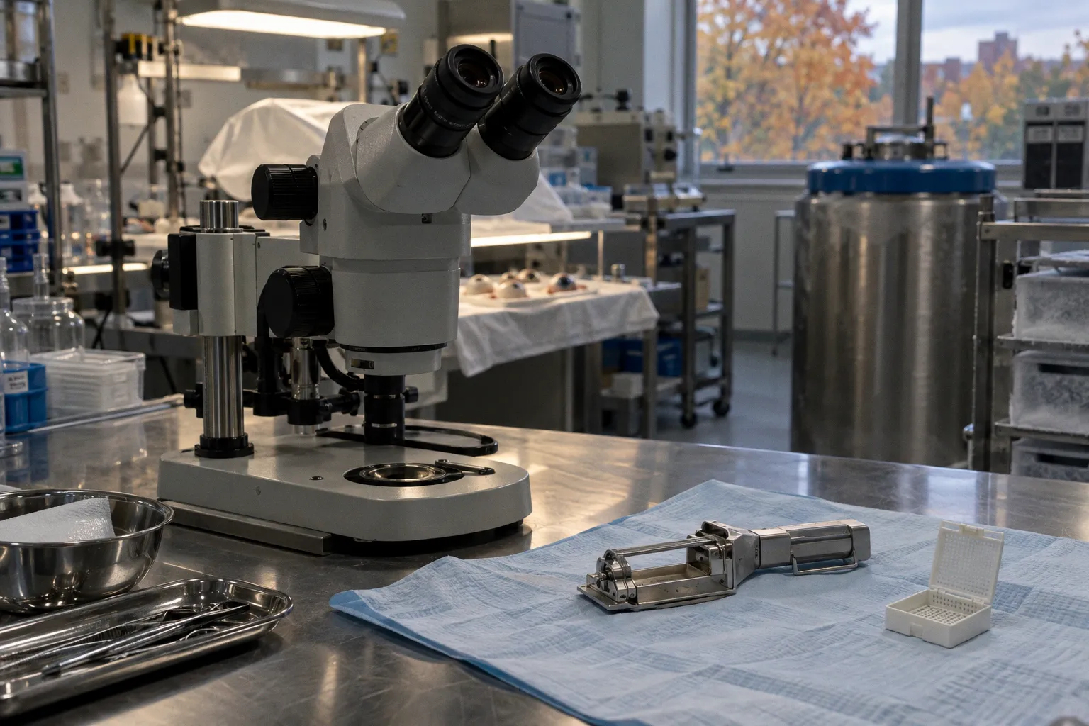
For a research eye bank, the operational problem is not simply whether tissue is “fresh.” It is whether the procurement timeline and preservation history are compatible with the intended assay.
Post-mortem interval (PMI) is therefore a routing variable as much as a timestamp. Retinal morphology, corneal clarity and RNA integrity each provide different information about tissue condition. None can stand in for the others. A specimen may retain useful structural features while showing molecular degradation, or meet a molecular quality target despite a PMI that exceeds a preferred threshold for another study.
Histological signatures of early retinal autolysis
After death, oxygen delivery stops and cellular homeostasis fails. Autolytic changes then emerge in tissue morphology. In the retina, early findings can include separation and folding of the retinal layers, pyknosis of nuclei in the inner nuclear layer, and swelling of ganglion-cell cytoplasm. With progression, ganglion cells may shrink and become more basophilic.
These findings matter because the same slide can contain both biological signal and procurement-related artifact. Retinal detachment, for example, is not by itself evidence of a pre-mortem retinal disease. It can occur as an early post-mortem change. Treating it as a diagnostic feature without considering PMI and handling history risks assigning a false biological interpretation to a logistics effect.
The practical reading is comparative. Histological assessment should be interpreted alongside the recorded interval from death to recovery, the condition of the globe, and the preservation pathway. If the study depends on layer relationships or cellular morphology, the tissue request should specify the structural features that must remain interpretable. A broad label such as “retina suitable for research” leaves too much unresolved: suitability for which region, which endpoint, and which tolerance for separation or cellular change?
A useful procurement record separates at least three times: estimated time of death, time of recovery, and time the tissue entered the specified preservation condition. These timestamps answer different questions. A reported PMI without a clear endpoint can conceal whether the clock stops at enucleation, dissection, stabilization or freezing. That ambiguity makes comparisons between samples and repositories less reliable.
Retinal morphology is a time-sensitive readout, not a self-interpreting record of disease.
For retinal disease research, requests commonly set a PMI below 12 hours because nucleic acids and proteins may degrade with time. That threshold expresses a conservative study requirement; it does not establish a universal point beyond which all ocular tissue becomes unusable. Histology, molecular profiling and other applications can have different acceptance criteria. The threshold should be tied to the assay, not treated as a general verdict on the specimen.
Corneal opacification depends on exposure
Corneal clouding is an observable marker, but its onset depends on whether the eye remains open. If the eye is open after death, corneal haziness can develop within 2 to 3 hours. In closed eyes, opacification typically begins later, between 12 and 24 hours post-mortem.
This difference makes exposure history part of the tissue record. A cloudy cornea does not provide a direct, universal measure of elapsed time. It may reflect the post-mortem interval, the state of the eyelids, or both. Conversely, apparent clarity should not be used as proof that the retina or molecular material remains intact. The cornea and retina are distinct compartments with different observable changes and different research uses.
For corneal studies, the relevant endpoint also needs definition. Optical clarity, endothelial assessment and histological structure are not interchangeable measures. The available facts establish the timing of visible haze under open- and closed-eye conditions; they do not establish a universal rate of endothelial cell loss at particular storage temperatures or in specific preservation media. Where endothelial viability is central, a clouding observation alone cannot answer the quality question.
A tissue intake workflow can capture the exposure variable without turning it into a false precision score:
- Record whether the eye was open or closed when observed, if that information is available.
- Document the time of the observation and distinguish it from the time of recovery.
- Describe corneal appearance using a consistent local terminology, rather than collapsing all findings into “clear” or “poor.”
- Keep the corneal assessment separate from retinal morphology and molecular quality fields.
These fields improve interpretation, but they do not reconstruct missing history. If exposure status is unknown, it should remain unknown in the dataset. Assigning a presumed state creates an apparently complete record at the cost of making the underlying evidence less accurate.
Biochemical indicators and the post-mortem interval
Vitreous humor potassium rises predictably after death and can serve as a biochemical indicator when estimating PMI. Its value is contextual: it can contribute information about the interval, but it does not directly measure the integrity of every ocular tissue compartment.
That distinction is important in repository data. A biochemical estimate of elapsed time and a tissue-quality assessment answer related but separate questions. Potassium concentration may help characterize the post-mortem timeline. Retinal histology records structural changes. RNA integrity reflects a different aspect of molecular preservation. Combining them can improve the description of a specimen, but no single marker should be made to certify all downstream uses.
The basic data model should preserve raw observations and their provenance. For vitreous analysis, that means retaining the measured value, the collection context and the method used to interpret it. For tissue morphology, it means recording the sampled region and the observation method. For molecular work, it means reporting the quality metric alongside the relevant timing and handling data. Without these distinctions, a repository may accumulate records that look comparable but were produced by different workflows.
| Marker | What it can indicate | What it cannot establish by itself |
|---|---|---|
| Retinal folding or separation | Post-mortem morphological change may be present | That the finding represents pre-mortem disease |
| Corneal haze | A visible change associated with time after death and exposure conditions | A precise PMI or retinal quality |
| Vitreous potassium | A biochemical clue for PMI estimation | Molecular or histological fitness of every tissue |
| RNA Integrity Number | RNA quality in the tested sample | Overall suitability of the whole globe for every assay |
| Anterior-segment OCT | In situ structural features in accessible anterior tissues | Reliable retinal morphology when lens opacity blocks imaging |
A robust tissue record is not necessarily a longer record. It is one that keeps measurements, estimates and interpretations in separate fields. A measured potassium concentration is not the same thing as an estimated PMI; an estimated PMI is not the same thing as a conclusion about assay suitability. Preserving that separation allows later users to apply their own study-specific criteria.
Molecular stability and RNA integrity
RNA degradation is one of the principal constraints on transcriptomic use of donor neural retina. The timing of procurement matters, but the available evidence also shows why a single hard cutoff is not an adequate description of molecular stability. High-quality gene-expression profiling has been achieved with an average RNA Integrity Number (RIN) of 8.5 when neural retina procurement occurred within a controlled PMI, averaging 20.5 hours and extending to a maximum of 26 hours.
That result does not erase the reason many retinal disease studies request tissue within 12 hours. A conservative threshold narrows variability and reduces the risk that degradation will confound a sensitive assay. The RIN finding instead demonstrates that controlled procurement can preserve usable RNA beyond that request window in some workflows. It should not be generalized into a guarantee for every donor, tissue region, preservation method or transcriptomic protocol.
For biobanking operations, the implication is to report distributions and specimen-level quality rather than convert one successful range into a universal promise. If a repository supplies neural retina for gene-expression work, the record should make the PMI definition explicit and attach the RIN result to the tested material. A quality value from one aliquot should not silently become a whole-globe label.
Several variables need to remain visible in the data pipeline:
1. The timing convention. State what event ends the reported PMI. “Twenty hours” is difficult to compare across repositories if one measures to recovery and another to stabilization.
2. The sampled tissue. Neural retina is not a generic proxy for all ocular material. The region and preparation used for the assay should be identifiable.
3. The measured endpoint. RIN is a quality measure for the RNA assessed. It is not a complete account of protein stability, histological preservation or cellular viability.
4. The handling record. Procurement and preservation conditions should be linked to the specimen record, not left in free-text notes that are difficult to compare or retrieve.
The phrase molecular stability of ocular tissue can suggest a single property. In practice, stability is endpoint-specific. RNA integrity, protein preservation and morphological interpretability are separate outputs of the same procurement system. A sample can perform well on one and less well on another.
Non-invasive monitoring with spectral-domain OCT
Portable spectral-domain OCT can visualize post-mortem morphological changes in the cornea, sclera and anterior chamber in situ for up to 72 hours. This creates a useful observation window for the anterior segment, where imaging can document structural change without first reducing the globe to a set of isolated tissue sections.
The window is not equivalent across the eye. Retinal imaging is hindered by lens opacification that can occur within the first 24 hours. The instrument may therefore continue to provide useful anterior-segment information after the optical path to the retina has become compromised. This is an access limitation, not evidence that retinal tissue has remained unchanged.
OCT should consequently be treated as one layer of quality documentation. It can add spatially resolved observations to the specimen record and may help distinguish visible anterior changes over time. It does not replace histology, biochemical assessment or molecular quality testing. Nor does a successful anterior-segment scan establish that the neural retina is suitable for transcriptomics.
The repository workflow needs to preserve both the image and its acquisition context. At minimum, later interpretation depends on when imaging occurred relative to death and recovery, which structures were assessable, and whether lens opacity restricted the view. A scan that lacks those fields may be technically legible but operationally weak: users cannot tell whether a missing retinal feature was absent or simply not observable.
Quality is a linked set of endpoints
Post-mortem ocular tissue autolysis markers are most useful when treated as a coordinated set of observations, not as a ranking from “good” to “bad.” PMI is a key routing variable, but it does not replace tissue-specific assessment. Retinal folds, corneal haze, vitreous potassium, RNA integrity and OCT findings describe different components of the specimen’s state.
The procurement system should therefore match donor tissue to the assay through explicit endpoint requirements. A retinal morphology study may prioritize short PMI and preserved layer relationships. A transcriptomic study may specify RNA quality and a defined timing convention. An anterior-segment study may make use of OCT observations while accounting for the separate limits imposed by lens opacity. Those are not competing definitions of quality. They are different specifications applied to the same biological supply chain.
The evidence supports a strict but non-binary assessment. Earlier recovery generally reduces exposure to autolytic change, and sensitive retinal research may reasonably set a PMI below 12 hours. Yet tissue beyond that interval is not automatically invalid: controlled procurement has supported high-quality neural-retina RNA profiling at longer intervals, while anterior-segment OCT can remain informative for up to 72 hours. The defensible conclusion is not that one cutoff governs all ocular research. It is that each specimen needs a traceable timeline, tissue-specific observations and a quality decision tied to the intended assay.