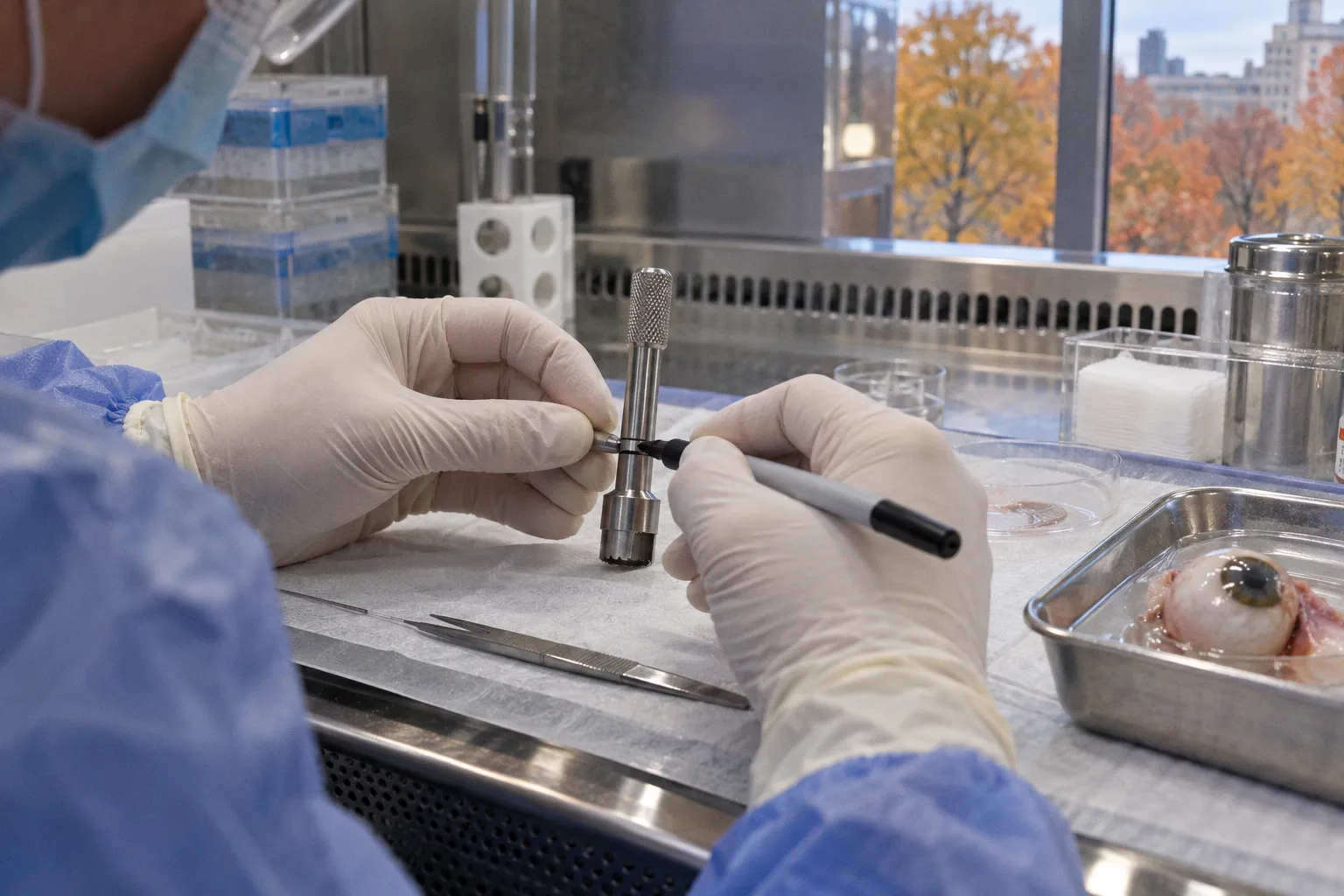
In donor retina and retinal pigment epithelium, the interval between death, cooling, recovery, dissection, and freezing directly influences whether open-chromatin signatures remain strong enough for reproducible interpretation.
ATAC-seq and related assays can recover meaningful regulatory information from post-mortem human eyes. They do not erase the procurement clock. Fresh donor tissue used for retinal and RPE chromatin profiling is typically processed within 14 hours after death. Within that window, the data remain usable for profiling major cellular and regulatory features, but longer intervals are associated with weaker accessibility signals and smaller peaks. The practical problem is therefore not whether a donor eye is usable in binary terms. It is whether the tissue still supports the resolution required by the study design.
The temporal window for epigenomic integrity in donor retina
Chromatin accessibility is a molecular readout of genome regulation. ATAC-seq measures regions of relatively open chromatin, where regulatory elements are more accessible to transcriptional machinery and associated factors. In the retina, these regions may help resolve cell-type-specific regulatory programs across photoreceptors, retinal neurons, Müller glia, vascular-associated cells, and RPE.
Post-mortem tissue introduces a second variable: the biological state of the sample is changing while the procurement chain is still in progress. The tissue is no longer under physiological regulation, but enzymatic activity, structural deterioration, temperature effects, and loss of nuclear integrity do not stop at the moment of death. They continue through the pre-cooling and recovery intervals.
This makes the procurement timeline part of the experimental design. A donor record that contains age, disease status, medication history, and cause of death but lacks reliable timing fields is incomplete for epigenomic interpretation. The time metadata are not administrative decoration. They describe the conditions under which the molecular signal was retained or degraded.
Standardized donor eye protocols for high-resolution molecular analysis target a death-to-cooling interval of approximately 2.75 ± 1.5 hours and a death-to-recovery interval of approximately 6.0 ± 1.5 hours. Recovered globes are generally maintained at 2–8°C until dissection. These values define a controlled operating range rather than a universal biological cutoff.
The distinction matters. There is no supported basis for treating the end of that range as an abrupt failure point. The available evidence instead indicates a gradient: as procurement is delayed, chromatin accessibility signals decline. The exact hour at which a specific cell type becomes unsuitable for single-cell detection remains unresolved.
The procurement clock is not a background variable. In donor-eye epigenomics, it is one of the primary determinants of signal strength.
Why longer procurement intervals reduce ATAC-seq signal quality
In retinal ATAC-seq assays, longer post-mortem procurement intervals correlate with reduced signal quality and smaller chromatin accessibility peaks. That relationship affects more than visual peak height. It changes the information density of the dataset.
A strong accessibility peak provides a clearer distinction between an open regulatory region and background signal. As peak intensity decreases, several downstream effects become more likely:
- low-amplitude regulatory elements become harder to separate from technical noise;
- cell-type-specific peaks become less robust across replicates;
- differential accessibility analyses lose statistical power;
- subtle disease-associated changes can be obscured by procurement-related variation;
- cross-donor comparisons become more dependent on normalization and covariate modeling.
The impact is not necessarily uniform across the genome or across ocular cell types. Regulatory regions with strong baseline accessibility may remain detectable longer than weak or narrowly restricted elements. A bulk retinal sample may therefore appear technically acceptable while still losing the fine-grained information required for a cell-resolved analysis.
This is a recurring issue in multiomic procurement. A sample can pass a broad quality screen and fail the intended analytical question. If the objective is to describe broad epigenetic architecture in the human retina, moderate degradation may be tolerable. If the objective is to identify weak enhancer activity in a rare retinal cell population, the same tissue may be insufficient.
The correct unit of assessment is therefore not simply tissue usability. It is tissue suitability for a defined assay resolution.
Procurement interval as a modeling variable
Post-mortem interval should be carried into the analysis as a structured covariate. At minimum, the dataset should distinguish:
1. time of death, where available;
2. time of initial cooling;
3. time of eye recovery;
4. time of dissection;
5. time of nuclear isolation or assay initiation;
6. time of freezing for retained tissue.
The practical objective is to separate biological variation from logistics-induced variation. Without those fields, a reduction in accessibility at a genomic locus may be attributed to disease, age, or cell composition when the more immediate cause is a longer procurement delay.
This is particularly important for studies comparing healthy and diseased donor eyes. If one cohort has systematically longer death-to-cooling or death-to-recovery intervals, the resulting chromatin state donor ocular cells may reflect the procurement distribution rather than the disease biology. The confounding can remain invisible in a conventional metadata table if timing is reduced to a single vague field such as “post-mortem interval.”
A better representation treats the chain as multiple intervals, each with a different operational meaning.
| Interval | Operational relevance | Likely analytical consequence |
|---|---|---|
| Death to cooling | Duration before temperature control begins | Early exposure to uncontrolled post-mortem change |
| Death to recovery | Total time before the globe enters the eye-bank recovery workflow | Major determinant of overall molecular preservation |
| Recovery to dissection | Holding time before tissue separation | Additional exposure before region-specific processing |
| Dissection to freezing | Time during which isolated tissue remains unpreserved | Risk of further loss of nuclear and epigenomic integrity |
| Cooling conditions | Temperature environment during holding and transport | Affects the rate of post-mortem molecular change |
The table is not a substitute for empirical quality metrics. It is the minimum infrastructure required to interpret them.
Death-to-cooling and recovery protocols
Rapid cooling is one of the most consequential controllable steps in donor-eye procurement. Standard protocols commonly aim to cool the eyes within roughly three hours of death, with recovery taking place within approximately six hours on average. Recovered globes are held at 2–8°C until dissection, after which selected tissue may be flash-frozen in liquid nitrogen for downstream molecular work.
These steps are often described as a linear protocol. In practice, they form a chain with multiple failure points. A nominally acceptable death-to-recovery interval does not guarantee that the preceding cooling interval was controlled. Conversely, a longer total interval may have a different molecular impact depending on whether the eye was cooled early and maintained within the intended temperature range.
For data systems, the difference is fundamental. One aggregated post-mortem interval cannot represent the full cold-chain history. Procurement records should preserve the sequence of events rather than only the final duration.
The first bottleneck: death-to-cooling latency
The period before cooling is the least forgiving part of the workflow because it occurs before the sample enters temperature control. It is also often the least precisely recorded. The target around 2.75 ± 1.5 hours indicates the operational range used in standardized protocols, not a guarantee that all samples within that range are molecularly equivalent.
A registry should store the measured or estimated value, the source of the timestamp, and the uncertainty of that timestamp. An exact-looking time derived from an imprecise report can create false confidence in downstream modeling. In procurement databases, timestamp provenance is part of sample quality.
The second bottleneck: recovery and transport
Recovery at approximately 2–8°C creates a controlled holding environment, but it does not freeze the molecular state. Cooling slows degradation kinetics; it does not remove the effect of elapsed time. The distinction is especially relevant when tissue is intended for high-resolution ocular tissue epigenomic profiling.
Transport records should therefore include more than dispatch and receipt. They should capture the cooling start, temperature range where available, recovery time, and any transition between facilities. If the eye bank and research laboratory operate on different systems, timestamp reconciliation becomes a data-engineering problem rather than a clerical task.
The third bottleneck: dissection latency
A recovered globe can remain within a nominal temperature range while still accumulating additional delay before region-specific dissection. Retina, RPE, macular tissue, and peripheral retina may not enter the same downstream workflow at the same time. The assay-ready state is determined by the interval relevant to the specific tissue aliquot, not merely by the time the globe arrived at the facility.
This is where sample-level identifiers become essential. The procurement record must remain linked to each dissected region, aliquot, and assay batch. A single donor eye may generate multiple data products with different effective preservation histories.
Implications for single-cell chromatin profiling
Single-cell ATAC-seq and joint scRNA/scATAC-seq workflows have demonstrated that major cell types can be isolated and mapped across macular and peripheral human retinal and RPE tissue. This is a significant technical capability, but it should not be confused with unlimited tolerance to post-mortem delay.
Single-cell assays distribute the available signal across individual nuclei. That creates a different sensitivity profile from bulk ATAC-seq. A bulk sample can accumulate enough material to produce a coherent aggregate signal even when some subpopulations are poorly represented. In a single-cell experiment, low-quality nuclei and weak accessibility profiles can reduce the usable fraction of the library and narrow the range of detectable regulatory elements.
The analytical consequences appear at several levels:
- Nuclei recovery: delayed processing may reduce the number of intact nuclei available for loading.
- Library complexity: damaged or low-integrity nuclei can contribute fewer informative fragments.
- Cell-type representation: fragile or less abundant populations may be under-recovered.
- Peak calling: weaker aggregate signal can produce a less complete accessibility map.
- Joint-assay balance: in scRNA/scATAC workflows, RNA and chromatin measurements may degrade at different rates, complicating cross-modality integration.
The final point is operationally important. Multiomic integration assumes that measurements from the same biological material remain sufficiently informative across modalities. If the chromatin component is disproportionately affected by procurement delay, the joint dataset may still generate clusters while losing the regulatory resolution that justified the experiment.
Macular and peripheral tissue are not interchangeable analytical units
Human donor-eye studies can profile open chromatin across both macular and peripheral retinal and RPE tissues. That spatial range expands the biological value of the material, but it also increases the number of variables that must be tracked.
A macular sample and a peripheral sample from the same eye may share procurement history but differ in cellular composition and baseline regulatory architecture. The comparison must therefore distinguish spatial biology from preservation state. If tissue regions are dissected at different times, the effective processing interval can also diverge within the same globe.
For database design, region should be represented as a first-class field. It should not be buried in free-text notes attached to the donor record. The same applies to retinal layer, RPE inclusion, dissection method, and assay modality. These fields determine whether a signal difference is biological, technical, or logistical.
Single-cell resolution increases the value of a well-controlled donor eye. It also increases the cost of incomplete timing metadata.
Building a usable donor-eye multiomics dataset
The central procurement problem is not the absence of information. It is the fragmentation of information across eye-bank records, transport logs, laboratory intake forms, dissection worksheets, and sequencing metadata.
A usable system must connect these layers without flattening them into one quality label. “Good tissue” is not a sufficient data object. The database should preserve the variables that allow researchers to define their own inclusion thresholds for ATAC-seq, scATAC-seq, joint RNA/chromatin assays, proteomics, or transcriptomic profiling.
A practical donor-eye record should include:
- donor-level clinical and demographic metadata;
- death-time provenance and uncertainty;
- time of initial cooling;
- time of recovery and facility location;
- recorded temperature conditions during holding;
- arrival and dissection timestamps;
- anatomical region and tissue composition;
- aliquot-level freezing or preservation status;
- assay type and library preparation workflow;
- sequencing or profiling batch;
- quality metrics linked back to each procurement interval.
The purpose is not to create administrative volume. It is to support causal separation. If a sample shows reduced peak intensity, the researcher should be able to ask whether the change tracks with post-mortem interval, cooling latency, tissue region, cell composition, or assay batch.
Quality control should be assay-specific
There is no single quality threshold that applies equally to bulk ATAC-seq, single-cell ATAC-seq, joint scRNA/scATAC-seq, and other molecular assays. The required signal depends on the intended output.
For broad regulatory mapping, a dataset may remain useful with lower peak intensity than a study designed to detect subtle changes in a rare population. For disease studies, the relevant question is often comparative: are the procurement differences between disease and control groups smaller than the biological effect being measured?
This is why a categorical sample label can be misleading. A continuous record of procurement intervals is more useful than a binary designation such as pass or fail. It allows analysts to model degradation as a source of variance and to perform sensitivity analyses around different inclusion windows.
The known evidence supports a clear directional relationship: longer procurement intervals are associated with weaker ATAC-seq accessibility signals. It does not establish a universal molecular half-life or a single cutoff hour below which single-cell detection becomes irreproducible. Those thresholds remain dependent on tissue type, assay chemistry, cell population, preservation conditions, and study objective.
That uncertainty should be represented explicitly rather than converted into a fabricated precision.
Optimizing tissue preservation for high-resolution multiomics
Preservation optimization begins before the eye reaches the laboratory. The most effective intervention is usually latency reduction, supported by reliable cooling and complete event logging. Later computational correction cannot fully reconstruct regulatory signal that was never captured.
The workflow can be improved through several linked controls:
1. Reduce death-to-cooling latency. The earliest interval should be treated as a primary operational metric, not an incidental timestamp.
2. Maintain controlled holding conditions. Recovered globes should remain within the intended 2–8°C range until dissection, with deviations recorded rather than inferred.
3. Shorten recovery-to-dissection time. A controlled cold chain still permits continued molecular change.
4. Track aliquots independently. Retina, RPE, macular, and peripheral samples may have different processing times and should not inherit one undifferentiated donor-level status.
5. Record timestamps with provenance. Estimated, reported, and instrument-generated times should be distinguishable.
6. Model procurement variables during analysis. Post-mortem interval and cooling latency should be available as covariates in differential accessibility and multiomic integration workflows.
7. Match tissue to analytical ambition. Samples with stronger preservation are the appropriate substrate for rare-cell, enhancer-level, and high-resolution regulatory questions.
These controls do not eliminate post-mortem biology. They make its effects measurable and comparable.
The 2018 foundational work on post-mortem human retinal chromatin accessibility established that donor eyes can support ATAC-seq-based regulatory profiling. More recent work, including evaluations of temporal molecular stability in post-mortem retina, reinforces the operational conclusion: the assay remains feasible, but signal quality is coupled to procurement timing. The field is moving from proof of feasibility toward calibration of the procurement-to-signal relationship.
That transition requires better longitudinal metadata and more explicit reporting. A study that reports only that tissue was obtained from an eye bank leaves out the variable most relevant to epigenomic reproducibility: how long the tissue remained in the post-mortem workflow before the assay began.
The evidence-based assessment
Human donor retina and RPE can retain usable chromatin accessibility information after death. ATAC-seq, scATAC-seq, and joint scRNA/scATAC-seq workflows can recover meaningful regulatory profiles, including from macular and peripheral tissue. The feasible processing window is commonly framed around recovery within 6 to 14 hours post-mortem, with rapid cooling and storage at 2–8°C before dissection.
But post-mortem stability is not equivalence. Longer procurement intervals reduce ATAC-seq peak intensity, and the loss is most consequential when the study depends on weak, rare, or cell-type-restricted regulatory signals. The exact threshold for failure is not established as a universal number.
The strict conclusion is therefore operational. Donor-eye epigenomics should be designed around timestamped logistics, not only around tissue identity. Death-to-cooling time, death-to-recovery time, holding temperature, dissection latency, and aliquot history belong in the analytical model. Without them, ocular tissue chromatin accessibility post-mortem stability remains only partially observable—and the dataset carries an avoidable source of uncertainty from the procurement chain into every downstream result.