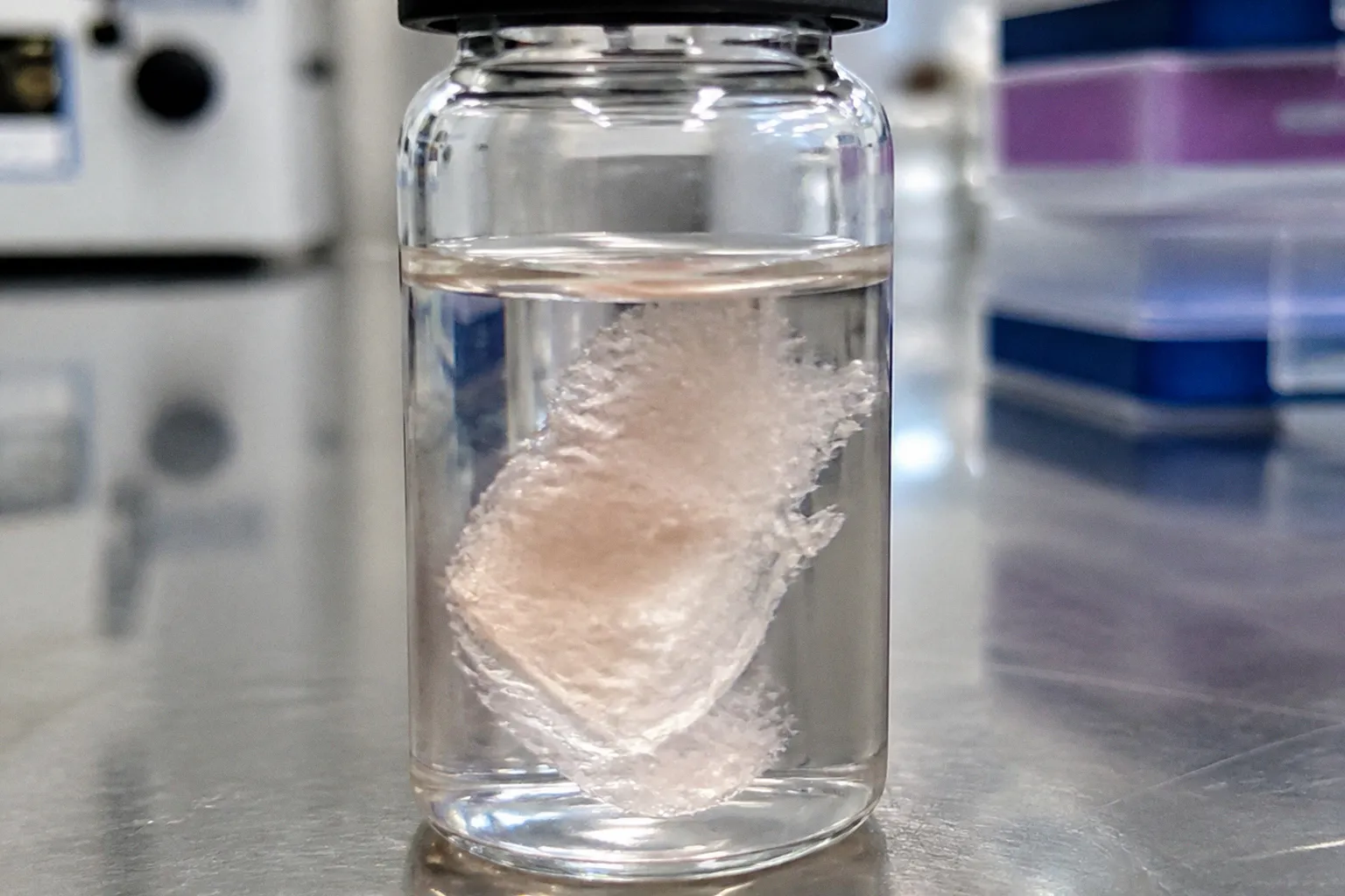
The cells, in theory, settle near the outer nuclear layer, integrate with host circuitry, and either replace lost photoreceptors or secrete trophic factors that slow further loss.
The reality at the operating microscope is less cooperative. Donor cells in a single-cell bolus can clump, reflux through the retinotomy, and die in substantial numbers during the first days after transplantation. The recurring lesson in ocular cell-therapy manufacturing is that suspension injection is not a neutral delivery format. It exposes fragile cells to dissociation stress, fluid shear, pressure changes, and an unfamiliar extracellular environment all at once. A protocol that looks clean at the end of the culture step can therefore produce a very different graft once the cells have passed through the needle and entered the subretinal space.
The central problem is not simply whether the cells are alive before injection. It is whether they remain sufficiently dispersed, viable, and spatially organized to interact with the host retina after injection. That is why retinal progenitor cell grafting protocol mistakes often appear to be surgical failures when the real defect began earlier, during dissociation, filtration, formulation, or time spent waiting in the syringe.
A single-cell suspension is the default, but clumping is the default outcome. The protocol decides which one the surgeon actually gets.
The Mechanics of Suspension Failure: Shear Stress and Extracellular DNA
The first failure mode is mechanical. Dissociating a donor retina or a cultured progenitor sheet into individual cells forces those cells through narrow fluid paths and, eventually, through a fine cannula for subretinal delivery. The passage may be brief, but the cells experience pressure and shear conditions that are far removed from those in the tissue niche. The effect is not limited to immediately ruptured cells. Cells that survive the passage can still carry sublethal damage that compromises membrane integrity, mitochondrial function, motility, or their ability to extend processes after deposition.
This is one reason a basic viability measurement taken before loading the syringe can be misleading. A suspension may meet its release specification immediately after digestion and still lose quality during the interval between final preparation and injection. The relevant question is not only how many cells are viable at the starting point, but how many remain viable and functionally competent after the complete handling sequence.
A second problem appears as damaged cells release intracellular material. Fragmented genomic DNA and other cellular debris increase the viscosity and adhesive character of the suspension. Free DNA can act as a physical bridge between nearby cells, while membrane fragments and proteins provide additional material around which aggregates form. The result is not always a dramatic, visible clump at the outset. Aggregation can develop gradually as the suspension sits, particularly when the cell concentration is high, the medium is not mixed carefully, or the preparation contains a large fraction of damaged cells.
The timing matters. The longer the suspension remains in the syringe, the more opportunity there is for non-uniformity to develop. Cells settle, debris accumulates in local concentrations, and the first part of the dose may no longer resemble the last. A syringe that contains a visually homogeneous suspension when loaded can deliver a more concentrated or aggregated fraction toward the end of the injection. That makes dwell time a manufacturing variable, not merely a matter of operating-room convenience.
DNase treatment is one experimental way to address extracellular DNA. By degrading free DNA, it may reduce the adhesive material that helps link cells into aggregates. It does not, however, repair membrane damage, restore dead cells, or solve every cause of clumping. If the upstream digestion is too harsh or the suspension contains excessive debris, DNase alone will not turn a compromised preparation into a reliable product. Its value depends on how it fits into the broader process: tissue dissociation, enzyme neutralization, washing, filtration, formulation, and the time between preparation and delivery.
The clumping itself is not cosmetic. A cluster of cells has a different relationship with the host retina than a dispersed population. The cells on the outside of an aggregate may contact the host tissue, while cells in the interior remain physically isolated from it. They may receive fewer survival signals, have less access to nutrients, and be less able to extend processes into the surrounding tissue. As the interior cells die, they release more debris and DNA, which can enlarge or stabilize the aggregate. A manageable dissociation artifact can therefore become a self-reinforcing failure.
There is also a scale problem. A small cluster may separate during handling or after deposition. A dense mass is less likely to do so. The larger the aggregate, the more likely it is to behave as a foreign body within the subretinal space rather than as a population of progenitors capable of interacting with the host. This is why an aggregate-size distribution is more informative than a simple statement that the suspension contains viable cells.
Mitigating Dissociation-Induced Apoptosis with ROCK Inhibitors
Once the team accepts that dissociation itself can trigger cell death, the next question is how to preserve the cells during the transition from tissue or culture to a delivery-ready suspension. Rho-associated kinase inhibitors, most commonly Y-27632 in experimental cell-processing workflows, are one tool used to reduce dissociation-associated apoptosis and anoikis. Their role is supportive rather than magical: they can improve survival during a vulnerable handling window, but they cannot compensate for excessive shear, prolonged holding, poor filtration, or an unsuitable formulation.
Anoikis is the form of programmed cell death associated with the loss of normal adhesive contacts. Progenitor cells that are suddenly separated from neighboring cells and extracellular matrix can undergo cytoskeletal contraction, membrane blebbing, and downstream apoptotic signaling. Retinal progenitors are not simply inert particles in a bottle; their behavior is shaped by the niche from which they came and by the contacts that are removed during harvesting. Dissociation strips away much of that context in a matter of minutes.
Y-27632 inhibits ROCK signaling downstream of RhoA. In practical terms, ROCK inhibition can reduce actomyosin contractility and help cells tolerate the transition into a single-cell or small-cluster suspension. The benefit is usually most relevant when the compound is present during or immediately after dissociation, before the full apoptotic program has progressed. Adding it late, after cells have spent an extended period in a stressed suspension, is unlikely to reverse damage that has already become irreversible.
The timing and exposure window therefore need to be treated as process parameters. A protocol should specify when the inhibitor is introduced, how long it remains in contact with the cells, and whether it is removed before the final formulation or carried into the delivery medium. Those decisions affect not only cell survival but also downstream assays, residual-reagent controls, and the interpretation of quality data.
Y-27632 should be described accurately in this context: it is an experimental ROCK inhibitor used in cell-culture and cell-processing research, not an established approved ophthalmic product for retinal progenitor cell transplantation. Its use in a research protocol does not establish clinical approval, and a laboratory observation should not be presented as evidence of routine clinical use in ocular cell therapy. Any translational program would need to address the compound’s role in the manufacturing process, residual exposure, product characterization, and the applicable regulatory requirements rather than relying on its familiarity in the laboratory.
That distinction matters because a reagent can be well known to cell biologists without being an approved component of a particular cell-therapy workflow. The manufacturing question is not simply whether Y-27632 improves viability in a dish. It is whether the complete process remains controlled when the inhibitor is combined with the selected enzymes, buffers, filtration step, storage interval, and surgical delivery format.
The same caution applies to concentration. A working concentration used in one cell type, culture system, or dissociation method cannot be transferred automatically to retinal progenitors. The useful range depends on cell state, exposure duration, formulation, and the endpoint being measured. A higher apparent viability number is not sufficient if the treatment alters phenotype, differentiation behavior, aggregate formation, or the cells’ ability to respond to the host environment after transplantation.
The Shift Toward Biodegradable Scaffolds for Structural Integration
The more decisive intervention may be structural rather than chemical. Biodegradable scaffolds can give a graft a defined physical form that survives handling and presents cells to the host retina in a more organized geometry. Materials such as PLLA, PLGA, and poly(glycerol sebacate) have been explored in preclinical delivery concepts, although each brings its own requirements for fabrication, loading, degradation, and ocular biocompatibility.
A scaffold addresses a limitation that suspension delivery cannot easily solve: cells in a free bolus have no reliable orientation. They may disperse unevenly, settle into local clusters, or remain separated from the host tissue by fluid and debris. A structured carrier can hold cells in a layer or defined arrangement, potentially improving contact with the outer retina and reducing the extent to which the graft behaves like an undifferentiated mass.
For photoreceptor-lineage cells, orientation is more than a histological preference. The cells must establish the right physical relationships with the host tissue if they are to extend processes, receive or deliver signals, and participate in retinal circuitry. The outer retina also imposes a demanding metabolic environment. A graft that is physically separated from the retinal pigment epithelium or that forms a thick cellular mass may remain present while contributing little functional integration.
Scaffold-based delivery can also reduce some losses associated with reflux and handling. The carrier gives the surgeon a defined object to place rather than a suspension whose distribution depends on fluid flow. That does not eliminate the risk of reflux, and it introduces new risks of its own, but it changes the mechanics of the delivery problem.
| Parameter | Single-cell suspension | Biodegradable scaffold |
|---|---|---|
| Physical organization | Cells disperse according to fluid flow and local tissue geometry | Cells can be positioned in a defined layer or pattern |
| Aggregation risk | High if debris, extracellular DNA, or prolonged dwell time promote clumping | Potentially lower when cells remain distributed on the carrier |
| Reflux behavior | Cells can leave with fluid through the retinotomy | Carrier may remain in place, although loose cells can still escape |
| Surgical handling | Familiar syringe-and-cannula workflow | Requires loading, placement, and carrier-specific handling |
| Product complexity | Primarily a cell formulation and delivery process | Combines a cellular component with a device or biomaterial |
| Development burden | Focuses on cell quality, formulation, and injection | Adds material safety, degradation, sterility, and interface questions |
The trade-off is substantial. A scaffold can turn a relatively simple cell suspension into a combination product or a more complex biological-device system. The sponsor must characterize the material itself, its degradation products, its interaction with the ocular environment, and the mechanical behavior of the loaded graft. The manufacturing process must also demonstrate that cells are distributed consistently across the carrier and remain viable during storage and transport.
There is a further question that is easy to miss: a scaffold can prevent random clumping only if the cells remain attached in the intended configuration. Poor loading, uneven seeding, or local drying can create high-density patches before the graft ever reaches the eye. The carrier is not a substitute for cell-quality controls. It moves the aggregation problem upstream, where it must be measured during loading and release testing.
For early translational programs, suspension delivery may remain attractive because it is familiar and comparatively straightforward. But simplicity at the point of injection can conceal a difficult biological compromise. If cell survival and spatial integration are the main constraints on efficacy, a more complex carrier may be justified. The right choice depends on the indication, the maturity of the cell product, the surgical approach, and the level of evidence needed to support the intended mechanism.
Managing Retinotomy Reflux and Subretinal Aggregation Risks
Even with careful cell preparation, the retinotomy remains a vulnerability. The surgeon creates an opening in the neurosensory retina to access the subretinal space, and that opening can become an escape route for fluid and cells. When the injection creates a pressure differential within the bleb, material may move toward the retinotomy and then into the vitreous cavity. Cells that reflux are not simply misplaced. They are removed from the tissue compartment in which the therapy is intended to act.
The amount of reflux depends on several interacting variables: the size and location of the retinotomy, the injection volume, the speed and pressure of delivery, the compliance of the tissue, the position of the cannula, and the behavior of the bleb. A suspension with a high cell concentration or a large aggregate burden may be especially vulnerable because clusters can obstruct the cannula, interrupt flow, and create pressure spikes.
Practical approaches to reducing reflux include using the smallest effective injection volume, avoiding unnecessary pressure, and positioning the delivery cannula so that the cells are deposited away from the retinotomy when the surgical design allows it. Temporary management of intraocular pressure may also be considered within the surgical protocol. Some approaches separate the access point from the main deposition site, reducing the chance that the injected fluid will carry cells directly back through the opening.
These techniques are not interchangeable fixes. A smaller volume may reduce reflux but increase the concentration of cells and the risk of local aggregation. A slower injection may lower shear but extend the time the cells spend in the syringe. A separate delivery track may improve spatial control but add surgical complexity. The protocol has to balance the variables rather than optimize one in isolation.
Unmanaged clumping creates a second problem after the injection. Aggregated donor cells can form dense cellular masses or rosette-like structures in the subretinal space. Such structures may fail to integrate even when many of the cells remain viable. They can occupy the narrow space between the photoreceptors and the retinal pigment epithelium, interfere with normal tissue contact, and disrupt the local exchange of metabolites and signaling factors.
The retinal pigment epithelium supports the outer retina through processes that include retinoid handling, phagocytosis of shed outer segments, and maintenance of the local ionic and metabolic environment. A dense graft mass positioned between the RPE and host photoreceptors can therefore become a physical obstacle rather than a functional bridge. The presence of donor cells on a histological section should not be mistaken for successful graft integration.
This is a particularly difficult failure mode because the cells may still be alive. Persistence is not the same as benefit. A surviving but poorly integrated mass can occupy subretinal space, alter the architecture of the host tissue, and contribute to local inflammation. In the worst case, the graft becomes an additional source of stress for the degenerating retina.
The response has to begin before surgery. Lower aggregation risk upstream through controlled dissociation, debris reduction, nuclease treatment where justified, and filtration. Stabilize the cells during harvest with an appropriately validated process, which may include experimental ROCK inhibition. Then control the mechanics of delivery: cannula selection, injection volume, pressure, speed, and the relationship between the deposition site and retinotomy. Each measure addresses a different link in the chain. None is enough on its own.
Optimizing Post-Digestion Filtration and Delivery Kinetics
After enzymatic digestion, the suspension is rarely uniform. It may contain viable singlets, small clusters, dying cells, membrane fragments, undigested tissue, and aggregates of different sizes. Filtration is the point at which some of that variability can be removed before the cells reach the delivery syringe. A cell strainer or controlled filtration step can help exclude larger aggregates, but the mesh size must be selected with the biology and the intended dose in mind.
A filter that is too coarse allows problematic clusters through. A filter that is too restrictive may retain viable cells, damage them through excessive pressure, or reduce the delivered dose unpredictably. Filtration can also alter the composition of the product by preferentially removing larger or more adhesive cell populations. That is why the step needs to be validated rather than treated as a generic laboratory convenience.
The post-filtration assessment should cover more than a total cell count. A useful release or in-process panel may include:
- overall viability and the method used to measure it;
- the proportion of singlets versus multicellular aggregates;
- the size distribution of remaining clusters;
- evidence of excessive debris or undigested tissue;
- cell concentration and the expected dose per unit volume;
- the time and temperature between final preparation and injection;
- any visible change in suspension uniformity during the hold period.
A high viability percentage can coexist with an unacceptable aggregate profile. Conversely, aggressive filtering can produce a visually clean suspension while removing too many cells or selectively changing the composition of the graft. The quality gate therefore has to measure what the surgeon will actually receive, not just what was present before the final processing step.
Delivery kinetics are another underappreciated source of variation. The relevant variables include the speed of injection, the internal diameter and geometry of the cannula, the pressure required to move the suspension, the time the cells remain in the syringe, and the temperature of the formulation during that interval. These variables interact. A narrow cannula may require more pressure; more pressure can increase shear; a slower operator may reduce peak pressure but prolong dwell time.
There is no single injection setting that can be assumed to work across every retinal progenitor population. Cell size, developmental state, concentration, medium viscosity, and aggregate burden all change the mechanical response of the suspension. A protocol that is gentle for one preparation may be inadequate for another. The appropriate endpoint is not merely a smooth injection. It is a reproducible process that preserves cell viability, limits aggregation, and deposits the intended dose in the intended compartment.
Temperature control deserves similar attention. Cells held for an extended period under suboptimal conditions can accumulate stress even when the suspension remains visually unchanged. Prompt delivery after final preparation reduces the opportunity for settling, aggregation, and progressive apoptosis. If a hold step is unavoidable, its duration, temperature range, and mixing procedure should be defined and tested rather than left to operating-room timing.
The most useful optimization experiments are therefore end-to-end. Instead of examining only the dissociation step, compare the complete sequence: dissociation, inhibitor exposure if used, washing, filtration, formulation, syringe loading, hold interval, simulated passage through the delivery cannula, and post-passage analysis. This is where hidden losses appear. A preparation that performs well in a culture plate may perform poorly after the mechanical and temporal stress of the actual delivery workflow.
The graft does not fail because the biology is wrong. It fails because the delivery system fights the cells on the way in.
The broader lesson across these protocol layers is that retinal progenitor cell transplantation is less a question of finding a promising cell and more a question of preserving that cell’s potential through the entire route into the eye. Dissociation chemistry, extracellular DNA, apoptosis, filtration, syringe dwell time, cannula geometry, reflux, and spatial organization are not separate technical footnotes. They form one continuous manufacturing-and-delivery problem.
A reliable retinal progenitor cell transplantation protocol has to account for the points at which the product can change: when tissue becomes a suspension, when the suspension is exposed to pressure, when damaged cells release debris, when clusters pass through the filter, and when the final bolus enters a confined subretinal compartment. The cells may be excellent and the surgery may be technically competent, yet the graft can still fail if the process allows aggregation or loses too much material through reflux.
That is why the critical question is not whether a protocol contains one promising intervention. It is whether the interventions work together. Experimental ROCK inhibition may reduce dissociation-induced apoptosis, but it does not replace filtration. Filtration may remove aggregates, but it does not prevent reflux. A scaffold may improve structural integration, but it does not eliminate the need for controlled cell preparation. Surgical precision cannot rescue a product that has already deteriorated in the syringe.
The strongest development programs treat each layer as part of the therapy itself: dissociation, stabilization, filtration, formulation, structural delivery, and surgical technique. Clumping is not an unavoidable nuisance to be accepted after the fact. It is a process failure that can often be detected, measured, and reduced before the graft enters the eye.