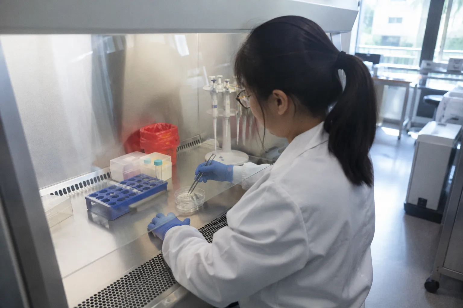
Bench teams watching the incubation timer understand something surgeons rarely see: the difference between a viable graft and a wasted donor often plays out in a five-to-eight-hour window of buffered saline and controlled temperature. Get the geometry wrong, miss the seeding window, or lose the monolayer during transfer, and the patch that looked perfect under the microscope becomes the kind of biology nobody wants to explain to a patient the morning after surgery. The bench is where subretinal vision science either earns its keep or quietly fails.
This is the ground-level reality of donor-derived RPE sheet grafting — a field where cellular manufacturing, scaffold engineering, and immunological math collide in a space measured in square millimeters. What follows is a walk through the practical steps that determine whether a patch will hold its orientation once it meets the subretinal space, drawing on documented bench workflows and the early clinical signals that have emerged from the first generation of ESC- and iPSC-derived implants.
Bench-Side Preparation: Scaling Cell Density and Scaffold Geometry
Every RPE construct that reaches the operating theater has already been shaped by two physical decisions made at the bench: how many cells fit on the delivery surface, and what kind of substrate holds them there. These choices are not interchangeable — they define what the surgeon can physically pass through a 25-gauge cannula and what the cells will do once they land on Bruch's membrane.
The two dominant formats in the published literature sit at opposite ends of a practical spectrum. Suspended cell sheets, stripped of their rigid support and delivered as a fragile monolayer, carry roughly 1 × 10⁵ cells in a footprint of 1.3–2 mm by 2–3 mm. Scaffold-based implants — patches built on polyester membranes, vitronectin coatings, or synthetic hydrogels — pack a denser payload of 1–10 × 10⁵ cells across a larger 3–3.5 mm by 6–6.25 mm area. The trade is straightforward: more cells and a sturdier delivery vehicle versus a smaller, more flexible sheet that demands near-perfect handling.
| Parameter | Cell sheet (no rigid scaffold) | Scaffold-based patch |
|---|---|---|
| Approximate cell count | ~1 × 10⁵ | 1–10 × 10⁵ |
| Typical dimensions | 1.3–2 mm × 2–3 mm | 3–3.5 mm × 6–6.25 mm |
| Handling demand | High — fragile monolayer, no mechanical support | Moderate — substrate provides structure during delivery |
| Cell–cell polarity preservation | Depends on enzymatic release integrity | Depends on coating chemistry and scaffold porosity |
| Surgical delivery | Tighter tolerance, smaller insertion footprint | Requires wider retinotomy or custom inserter |
The bench doesn't fail surgeons with dramatic errors. It fails them with monolayers that look intact but lose polarity the moment the substrate is lifted.
Bench teams running these preparations quickly learn that the assays matter more than the catalog numbers. Cell attachment confirmation, tight junction staining, and polarized phagocytosis markers are the daily language of RPE quality control — not because regulators ask for them, but because a patch that loses its apical-basal polarity in transit will not behave like retinal pigment epithelium once it reaches the subretinal space.
Preservation Dynamics: Managing the 5-to-8 Hour Viability Window
Once the patch is prepared, time becomes the most expensive reagent on the bench. Documented protocols for stem cell-derived RPE grafts describe viability windows of up to five hours in dedicated graft storage medium and up to eight hours in saline solution prior to subretinal delivery. These numbers are not theoretical — they represent the realistic transport envelope between a manufacturing clean room, a courier vehicle, and a parked surgical microscope.
What happens inside that window matters. Cells continue to metabolize. Transepithelial resistance shifts. Microvilli on the apical surface begin to flatten. None of these changes are catastrophic in the short term, but they compound, and by hour seven or eight the patch the surgeon receives is not quite the patch the manufacturing team released. Bench protocols that account for this — pre-equilibrated media, controlled temperature at around 16–20°C for most reported workflows, minimal light exposure — extend the usable window without claiming to extend it indefinitely.
The Kamao et al. work published in 2017 brought this conversation into the open by systematically tracking RPE graft viability in storage media and giving the field a reference point that operating-room coordinators could plan around. Before that report, much of what passed for "viability" in the operating theater was hopeful estimation. Now it is measured decline — and measured decline is something a logistics team can schedule against.
For eye banks and biobanking coordinators, the implication is practical: a donor-derived RPE graft is not a vial of frozen cells. It is a living patch with a delivery deadline. Procurement planning, transport packaging, and surgical booking all need to bend toward that deadline, not the other way around.
Substrate Integrity: Challenges in Seeding Human Bruch's Membrane
Even a pristine patch with perfect polarity is only as good as what it lands on. In the subretinal space, that surface is Bruch's membrane — a five-layered extracellular structure that, in the diseased eye, is exactly where the disease has done its worst work.
Human cadaveric Bruch's membrane explants harvested within 48 hours of death have given researchers a sobering view of what pluripotent stem cell-derived RPE cells will and will not do when presented with diseased substrate. Where the inner collagenous layer and deeper layers of Bruch's membrane remain intact, RPE cells can establish a monolayer with reasonable fidelity. Where deeper layers are exposed — the very geography that defines atrophic AMD — the seeded cells fail to form an intact monolayer. They cluster, they drift, they lose the tight junctions that define functional RPE.
This is not a bench failure. It is a structural reality that the bench has to acknowledge. Scaffolds and matrices in development are partially a response to this finding: rather than relying on diseased Bruch's membrane to host transplanted RPE, engineered substrates provide a clean surface that carries the cells into position and offers some degree of biological support where the native tissue cannot. The clinical question is whether such substrates integrate with, or merely sit on top of, the residual host membrane — and that question is still open in the published literature.
For the biobanking side of this work, the 48-hour post-mortem procurement window for usable Bruch's membrane explants is a meaningful constraint. Eye banks coordinating with cell-therapy programs need to align tissue recovery, preservation methodology, and downstream experimental use within that envelope. Anything past it, and the substrate itself becomes a confounding variable in the seeding experiment.
Immunological Barriers and Long-Term Graft Integration
The subretinal space enjoys a degree of immune privilege — that much is established and clinically useful. But immune privilege is not immune invisibility, and the allogeneic transplant literature makes the distinction clear.
In nonhuman primate studies, subretinal delivery of allogeneic iPSC-derived RPE cells without systemic immunosuppression led to cell graft loss by three weeks post-surgery. The mechanism was immune-mediated rejection — the kind of brisk, antibody-and-T-cell-driven response that does not care about anatomical privilege once the antigen presentation has begun. This is not an abstract finding; it shapes how clinical programs design their patient selection and consent protocols.
The autologous route, by contrast, sidesteps the rejection problem but introduces manufacturing complexity and cost that the autologous iPSC-RPE workflow first demonstrated in Kobe in September 2014. That first-in-human autologous cell sheet transplant showed what the field could do when rejection was removed from the equation — and what it cost in production time, patient-specific manufacturing, and regulatory burden. The allogeneic path, with its off-the-shelf economics, only works if the immunological math can be solved, either through donor matching, localized immunosuppression, or engineered hypoimmunity.
Three weeks is enough time for a subretinal graft to integrate — or to be cleared. The bench controls only half of that timeline.
For eye banks and procurement coordinators, this carries a downstream implication. If allogeneic programs move toward larger-scale manufacturing, donor RPE will need to be characterized for HLA profile, antigen presentation behavior, and ideally hypoimmune engineering status — quality-control steps more familiar to solid organ banking than to traditional corneal tissue distribution.
Clinical Benchmarks: Translating Patch Survival to Visual Acuity
The translation question is the only one that ultimately matters to a patient in the chair: does a surviving patch restore function? The Da Cruz et al. study released in 2018 provided one of the earliest clinical windows into that question, with two exudative AMD patients receiving ESC-derived RPE patches on polyester membranes coated with human vitronectin.
Both patches were delivered successfully. Both survived the perioperative window. At 12 months, the two patients gained 21 and 29 letters of visual acuity, respectively. These are not population-level outcomes — they are two patients — but they demonstrate the operative principle: when the bench work holds and the surgical delivery succeeds, the visual system responds. The numbers are encouraging enough to justify continued investment and cautious enough to resist any narrative of immediate clinical availability.
The honest framing is that the field has passed its first translational waypoint. Donor-derived RPE patches can be prepared, transported, delivered, and integrated with measurable visual benefit in selected patients. The unsolved chapters are real and documented: long-term survival beyond five years across diverse populations, the standardization of bench protocols that guarantee monolayer integrity regardless of donor age, and the immunological strategies that will make allogeneic products as durable as autologous ones.
Where the Bench Meets the Bedside
What strikes me most, looking across the published workflows and the early clinical signals, is how much of the field's near-term progress depends on bench discipline rather than surgical innovation. The surgeons are ready. The delivery instruments exist. The imaging to confirm subretinal placement is mature. The bottleneck is upstream — in the hours after a patch leaves its manufacturing substrate, in the geometry of the scaffold, in the integrity of the monolayer as it meets a diseased Bruch's membrane, and in the immunological accounting that determines whether a graft lives three weeks or three years.
For the eye banking and procurement community, the practical takeaway is that RPE tissue is moving from a research curiosity into a logistics problem. Procurement windows are tightening. Transport conditions are getting specified. Quality-control markers are being defined. The people who coordinate donor tissue today are quietly building the supply chain that the next generation of cell therapies will depend on, whether or not they recognize it yet.
This is not a field that needs miracles. It is a field that needs measured, repeatable bench work — and the patience to let the biology settle into a durability that five-year follow-up studies have not yet confirmed. The work is happening. The clock is honest. The bench, as always, decides.