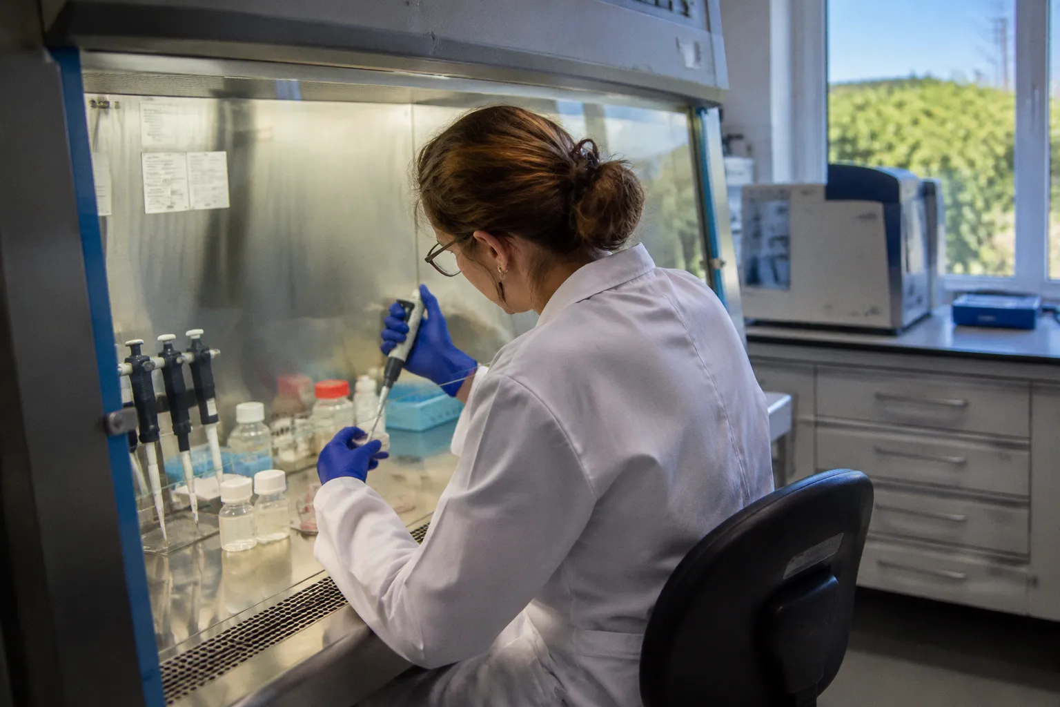
The human donor retina offers a less accommodating answer: the vector first encounters a physical barrier, then an extracellular matrix environment, then biological variability—and only after that does capsid tropism become meaningfully testable.
The inner limiting membrane (ILM) is not a minor anatomical inconvenience. It is a diffusion barrier that restricts intravitreally administered AAV from reaching the outer retina, including the outer nuclear layer and retinal pigment epithelium. In human retinal explants, even successful uptake does not translate into immediate transgene expression. With AAV2-GFP, notable GFP expression appears between days 6 and 12, while the first four days show no significant signal. The vector may be present; the therapeutic readout is not.
That distinction matters. A donor-eye model can expose the weaknesses of ocular gene delivery with uncomfortable efficiency—but only if researchers stop treating it as a miniature clinical trial and start treating it as a structured test of barriers, timing, tissue integrity, and vector biology.
The Inner Limiting Membrane Is a Gate, Not Background Anatomy
The inner limiting membrane is formed at the interface between the neural retina and the vitreous. For AAV administered by intravitreal injection, this interface is the first major obstacle. The vector does not enter an empty optical chamber and diffuse evenly toward every retinal layer. It moves through a biological environment whose matrix composition, geometry, and cell interfaces constrain access.
The basic mismatch is straightforward:
- Intravitreal administration places AAV on the vitreous-facing side of the retina.
- The ILM limits diffusion into deeper retinal tissue.
- Outer retinal targets remain comparatively difficult to reach.
- The vector’s nominal concentration does not equal the concentration delivered to the target cell population.
This sounds obvious, yet preclinical discussions often flatten the problem into a question of dose. More vector, higher titer, better outcome. Despite the consensus, that is not a complete model of ocular gene delivery. AAV capsids are approximately 25 nm in diameter, but small size does not make them biologically frictionless. They still interact with extracellular matrix components and tissue interfaces. Diffusion is not the same as access, and access is not the same as productive transduction.
The ILM barrier is particularly consequential when the therapeutic strategy depends on reaching photoreceptors or the retinal pigment epithelium. These are not simply farther away in a geometric sense. They are behind a structure that limits the movement of the vector from the vitreous-facing surface toward the outer retinal layers.
A useful way to frame the problem is to separate four stages that are too often collapsed into one:
1. Distribution — where the vector physically travels after administration.
2. Entry — whether it crosses the relevant tissue interface and reaches a target cell.
3. Cellular processing — whether the vector is internalized and processed sufficiently for expression.
4. Transgene manifestation — when the reporter or therapeutic product becomes detectable.
A vector can perform adequately at one stage and fail at the next. That is why an apparently weak reporter signal cannot automatically be interpreted as poor capsid tropism. The vector may have encountered a transport barrier, an intracellular bottleneck, or simply an observation window that was too short.
The first failure in ocular AAV delivery may occur before the vector ever gets the chance to demonstrate its biology.
Why the human donor retina changes the argument
Human donor tissue introduces the anatomy that rodent experiments can obscure. The issue is not that animal models are useless; it is that their structural barriers cannot be assumed to represent the human retina. In particular, rodent ILM thickness or permeability should not be treated as a reliable proxy for human resistance to intravitreal AAV.
That caveat is not academic. Capsid performance is often discussed as though tropism were an intrinsic property independent of tissue context. It is not. A capsid that appears efficient in one species may encounter a different ILM, a different extracellular matrix environment, and a different distribution of target cells in human tissue. The phrase donor eye viral vector transduction therefore describes more than a technical assay. It describes a test of whether the delivery system survives the anatomical conditions that matter clinically.
The Vector Cannot Carry Whatever the Therapy Requires
The second barrier is not retinal anatomy but molecular cargo. Recombinant AAV vectors have a physical packaging capacity of approximately 4.7 to 4.8 kilobases of single-stranded DNA. That limit is not a manufacturing preference. It is a constraint imposed by the vector itself.
For small therapeutic genes, the payload may fit with careful construct design. For larger genes, the arithmetic becomes punitive. The coding sequence must compete for space with regulatory elements, promoters, polyadenylation signals, and other components required for useful expression. A gene that technically approaches the limit may leave little room for the regulatory architecture needed to control where, when, and how strongly it is expressed.
The packaging problem is therefore not merely binary—inside or outside the vector. It is a design trade-off:
| Design pressure | What it demands | What the AAV limit forces |
|---|---|---|
| Larger therapeutic gene | More coding capacity | Truncation, fragmentation, or an alternative delivery strategy |
| Cell-specific expression | Promoter and regulatory sequences | Less room for the therapeutic cassette |
| Durable expression | Additional control elements | Greater pressure on the approximately 4.7–4.8 kb payload ceiling |
| Broad retinal targeting | Flexible expression architecture | A compromise between specificity and physical payload size |
| Clinical manufacturing | Consistent, intact vector genomes | Less tolerance for oversized or heterogeneous constructs |
The paradox is familiar: the retina may be difficult to reach, yet the gene may be too large to package in the vector chosen to reach it. Researchers can improve capsid diffusion and still remain trapped by the payload ceiling. Conversely, a compact transgene does not solve the anatomical problem. The two constraints are independent, and combining them does not make either one disappear.
This is where the conventional capsid-centered narrative starts to fail. If the therapeutic sequence exceeds the capacity of a single AAV, switching capsids will not rescue the construct. One can explore dual-vector or split-gene approaches, but those introduce their own dependence on coordinated delivery and expression. They also complicate interpretation in donor tissue: a low therapeutic signal may reflect inefficient co-delivery rather than inadequate tropism.
Titer is not a substitute for architecture
Experimental AAV administrations may use concentrations in the range of 10¹² to 10¹³ vector genomes per milliliter. Those figures sound forceful, and they are useful descriptors of experimental dosing. But a high titer does not erase the ILM, enlarge the capsid’s packaging capacity, or guarantee productive entry into the desired retinal cell.
This is the recurring paradigm deficit in ocular gene therapy: concentration is treated as if it were a universal answer. It is not. A high-titer preparation may increase the number of particles presented to the tissue, while the fraction reaching the relevant cell layer remains constrained by diffusion and matrix interactions. More particles can also make biological interpretation less clean if toxicity, inflammatory signaling, or nonspecific uptake becomes part of the response.
The serious question is not whether a vector was administered at a high concentration. It is whether the experimental design can distinguish:
- increased exposure from improved tissue penetration;
- particle presence from functional transduction;
- reporter expression from clinically relevant gene activity;
- cell entry from durable, correctly localized expression.
Without those distinctions, “efficient delivery” becomes a label applied to several different phenomena.
Six Days Is Not a Delay to Be Edited Out
Human retinal explant experiments expose another inconvenient fact: transgene expression is not immediate. In human retinal explants transduced ex vivo with AAV2-GFP, no significant GFP expression is detected during the first four days. Noticeable expression emerges between days 6 and 12.
That timeline should change how donor-retina studies are designed and interpreted. A short observation period may measure the beginning of intracellular processing rather than the final performance of the vector. A negative result at an early time point is not automatically evidence that the capsid failed to enter cells. It may simply be a premature verdict.
The distinction is especially important when comparing capsids or delivery conditions. Suppose one vector produces a visible signal earlier than another. That could indicate faster transduction, but it could also reflect differences in promoter activity, intracellular trafficking, genome processing, or reporter kinetics. Likewise, a vector with a weak early signal may later produce a meaningful response. Time is not a decorative variable in the assay; it is part of the biology being measured.
A credible human retinal explant design therefore needs to treat time as an experimental axis rather than a scheduling detail. At minimum, the analysis should ask:
1. Was the explant observed long enough for transgene manifestation to become detectable?
2. Was the same time window used across capsids and administration conditions?
3. Was uptake distinguished from reporter expression?
4. Were changes in tissue viability separated from changes in vector performance?
5. Did the assay examine the cell types relevant to the intended therapy rather than relying on bulk signal?
These questions are not bureaucratic additions. They prevent an early negative readout from being mistaken for a definitive failure of delivery.
The explant is informative precisely because it is incomplete
Human retinal explants provide direct access to donor tissue and allow researchers to assess capsid tropism and transgene expression in a human retinal architecture. That is their advantage. They can reveal whether a vector reaches particular retinal layers and how expression develops over time without immediately passing through the full complexity of clinical administration.
But the model does not reproduce every in vivo condition. It does not retain intact vascular flow or a complete systemic immune defense system like a living human eye. The absence of those features is not a flaw to hide; it is a boundary to report.
An explant can answer questions about tissue-level barriers, cell tropism, and expression kinetics. It cannot, by itself, establish how a vector will behave under full physiological, inflammatory, and immune conditions in a patient. Nor should it be asked to answer a question its design does not contain.
A human explant is not a clinical eye in a dish. It is a human anatomical filter—and that is exactly why its limitations must be made explicit.
The Sub-ILM Route Demonstrates the Barrier by Bypassing It
The most revealing evidence that the ILM is a primary barrier comes from an approach that avoids it. Surgically placing the vector into a hydrodissected sub-ILM space, between the ILM and neural retina, bypasses the barrier and enables transduction of retinal ganglion cells, Müller glia, ON bipolar cells, and photoreceptors in primate retinas.
The point is not that sub-ILM delivery automatically becomes the preferred clinical route. The point is diagnostic. If moving the vector past the ILM changes the spectrum of transduced cells, then the ILM was not a theoretical obstacle. It was controlling the distribution of the vector.
The comparison is stark:
| Delivery condition | Primary advantage | Main interpretive concern |
|---|---|---|
| Intravitreal AAV | Less invasive access to the vitreous-facing surface | ILM and extracellular matrix restrict deeper retinal penetration |
| Sub-ILM hydrodissection | Bypasses the ILM and exposes the neural retina more directly | Requires a surgical maneuver and changes the safety and translation profile |
| Ex vivo retinal explant exposure | Direct assessment in human donor tissue | Lacks intact vascular flow and complete systemic immune context |
| Capsid optimization alone | May alter tropism or intracellular behavior | Cannot guarantee passage through the human ILM or solve payload limits |
This is where rapid-fire comparisons are more useful than broad optimism. Intravitreal delivery is clinically familiar but anatomically constrained. Sub-ILM administration improves access but adds procedural complexity. Human explants improve species relevance but reduce physiological completeness. Capsid engineering can improve one part of the problem while leaving the others untouched.
The emerging evidence does not support a single universal route. It supports a more uncomfortable conclusion: delivery strategy must be matched to the target cell layer, and the route itself can determine which biological claims are even testable.
Bypassing the barrier is not the same as solving translation
A surgical approach that improves transduction in primate retinas demonstrates feasibility under a particular anatomical condition. It does not automatically establish safety, durability, manufacturability, or clinical practicality. The maneuver changes the exposure geometry, and therefore it changes the experiment.
That matters when comparing studies. An investigator may report stronger transduction after sub-ILM placement, while another reports limited outer retinal access after intravitreal injection. Both findings can be correct. They are not testing the same barrier configuration.
The temptation is to treat the stronger signal as proof that the vector is superior. More accurately, it may be proof that the vector was finally placed where the barrier could no longer exclude it. The distinction is not semantic. It determines whether the next engineering step should involve a different capsid, a different surgical route, a different formulation, or an altogether different delivery platform.
Neutralizing Antibodies and Matrix Composition Complicate the Picture
Physical access is only the beginning. Serum neutralizing antibodies can reduce effective vector availability, while extracellular matrix composition can influence movement through retinal tissue and interactions at the ILM. These factors make donor-tissue studies more valuable, but also harder to interpret.
Neutralizing antibodies are particularly awkward for a field that prefers clean comparisons. A capsid may appear less effective because it is intrinsically poor at transduction—or because antibodies reduce the functional particle pool. In an ex vivo system, that immune context may not reproduce the full systemic situation found in a living subject. The result is neither irrelevant nor complete. It is a defined slice of the delivery problem.
Matrix composition creates a similar issue. Two tissues may receive the same nominal vector dose but present different diffusion environments. Tissue age, donor condition, postmortem interval, preparation, and culture duration can all affect the biological state of the explant. Those variables do not need to be turned into excuses for every inconsistent result. They do need to be recorded and incorporated into the interpretation.
A strong donor-eye viral vector transduction study should therefore avoid the fiction of a single efficiency number. Instead, it should report a profile:
- which retinal layers were reached;
- which cell classes showed evidence of transduction;
- when expression became detectable;
- whether the signal was localized or diffuse;
- how the result changed across vector designs or exposure conditions;
- how tissue quality and culture duration constrained the conclusion.
This is slower than declaring one capsid a winner. It is also more useful.
Why Rodent Results Cannot Carry the Whole Translational Argument
Rodent models remain attractive because they are accessible, reproducible, and compatible with controlled experimental designs. But their convenience can become a translational liability when structural differences are treated as negligible. The human ILM should not be assumed to behave like a rodent ILM simply because both are labeled with the same anatomical term.
The issue is not a crude species hierarchy in which human tissue is always better. It is a question of relevance. A mouse study can help identify candidate capsids, promoters, dosing concepts, and toxicity signals. It cannot establish that a vector will cross the human inner limiting membrane with comparable efficiency. That specific claim requires human-relevant evidence.
The same caution applies to retinal cell distribution and target accessibility. A capsid that demonstrates a particular pattern in a rodent retina may not reproduce that pattern in a human donor retina. If the therapeutic objective depends on outer retinal transduction, the model must expose the outer retinal access problem rather than allowing it to remain hidden behind a more permissive anatomy.
A practical preclinical sequence is therefore less about replacing animal models than about assigning each model a narrower job:
1. Use rodent studies for early construct and toxicity screening, without treating them as a final estimate of human retinal penetration.
2. Use non-human primate studies to examine delivery geometry and retinal access under a more relevant anatomical context.
3. Use human retinal explants to test donor-tissue barriers, capsid tropism, and expression timing directly.
4. Compare results by mechanism, asking whether differences arise from the capsid, route, payload, tissue barrier, or observation window.
5. Carry uncertainty forward, especially where long-term explant viability and direct human in vivo efficiency remain unresolved.
The field does not suffer from a shortage of models. It suffers from model overreach—asking one system to validate a chain of assumptions it was never designed to test.
A Better Reading of Human Retinal Explant AAV Delivery
The value of the human retinal explant is not that it predicts the clinic perfectly. No ex vivo model can do that. Its value is that it removes one of the most convenient forms of denial: the denial that a human retinal barrier may behave differently from the one used in a small-animal experiment.
The model can interrogate several questions with unusual directness:
- Does the candidate vector reach the intended retinal layer?
- How much of the apparent failure is attributable to the ILM?
- Does expression emerge only after several days?
- Does bypassing the ILM change the transduced cell population?
- Is the therapeutic cassette compatible with AAV’s approximately 4.7–4.8 kb capacity?
- Are comparisons being made at equivalent time points and exposure conditions?
But the experiment must be built around those questions. A single endpoint, a bulk fluorescence image, or a nominal vector concentration will not resolve them. Nor will a rodent result erase the human anatomical barrier.
The most useful conclusion may be negative: a candidate approach does not yet solve the delivery problem. That is not a failed publication. It is a successful narrowing of the translational search space. In regenerative therapeutics, the expensive mistake is not rejecting a weak vector. It is carrying a structurally mischaracterized vector into increasingly elaborate studies because early models made the barrier look smaller than it is.
The Real Barrier Is the Combination
AAV transduction barriers in human donor retina research are often described as a list: ILM, antibodies, extracellular matrix, payload size, timing. The list is accurate but insufficient. These barriers interact.
A vector with a compact payload may still fail to reach the target layer. A capsid with promising tropism may be blocked by the ILM. A vector that crosses the barrier may express too slowly to be judged in a short experiment. A sub-ILM approach may improve cellular access while creating a less straightforward clinical procedure. A human explant may reveal the relevant anatomy while omitting systemic immune and vascular conditions.
That is why the central question should not be, “Which AAV is best?” It should be more precise:
Which delivery configuration can place an adequately sized genetic payload into the intended human retinal cells, across the relevant anatomical barrier, within a time window that permits durable and interpretable expression?
That question is less marketable than a capsid leaderboard. It is also the one that can guide actual therapeutic development.
The next phase of ocular gene therapy will not be settled by better slogans about precision delivery. It will be settled by experiments that separate diffusion from transduction, transduction from expression, and expression from therapeutic adequacy. The human donor retina is valuable because it makes those separations difficult to avoid.
Despite the consensus, the ILM is not an incidental layer, high titer is not a universal solution, and early absence of GFP is not proof of biological failure. Until preclinical programs account for all three—and respect the 4.7–4.8 kb payload ceiling—the field is not confronting a single delivery barrier. It is negotiating a cluster of them while pretending they are interchangeable.
The provocative challenge is therefore simple: stop asking whether an AAV vector works in the retina. Ask which retina, through which barrier, carrying what payload, observed when—and whether the answer survives contact with human tissue.