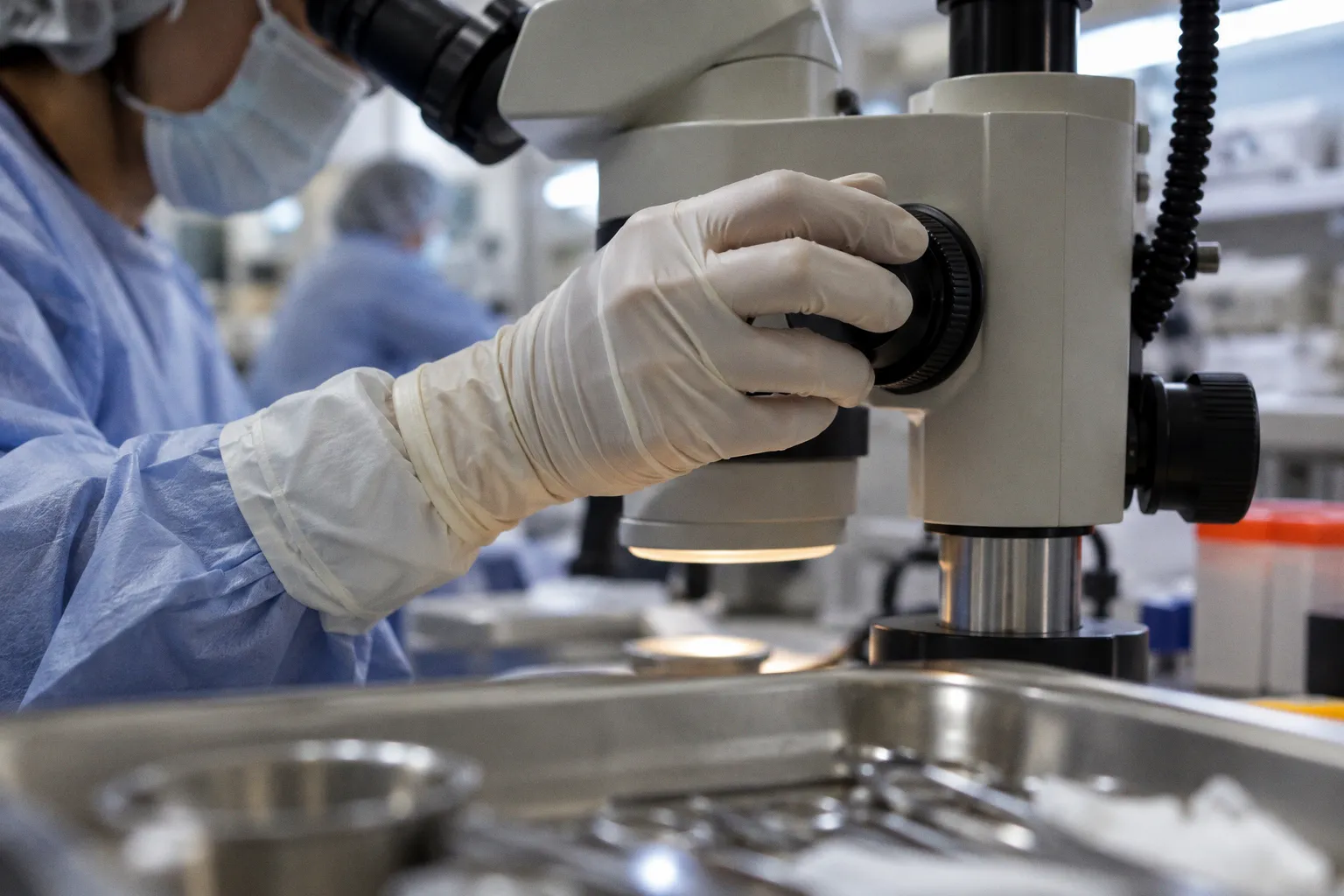
Donor Eye Procurement: Clinical Standards vs Research Needs
Research recovery prioritizes something else: short post-mortem intervals, preserved retinal architecture, intact RNA and protein profiles, and traceable pre-analytical metadata.
The same donor eye can therefore be acceptable for one workflow and unusable for another. A cornea may remain suitable for clinical evaluation while the associated retinal tissue has already exceeded a researcher’s permitted post-mortem interval. Conversely, a donor with medical contraindications for transplantation may still represent a high-value research specimen. The operational system has to distinguish these pathways before recovery begins.
This is the central difference in donor eye procurement versus research-grade tissue recovery. Clinical banking is optimized around surgical suitability. Research biobanking is optimized around information preservation.
Divergent priorities: clinical transplantation versus molecular research
Clinical and research workflows use different definitions of a successful recovery.
For transplantation, the principal question is whether the tissue can be released as a safe and functional graft. Corneal clarity, tissue structure, endothelial cell density, donor eligibility, microbiological controls, and preservation conditions form the core evaluation set. The procurement process is designed to protect the anterior segment and deliver tissue into a validated storage and assessment pathway.
Research recovery has a broader and more fragile target. The specimen may include the whole globe, retina, optic nerve, choroid, sclera, vitreous, or a coordinated set of ocular tissues. The objective is not merely to preserve anatomy. It is to preserve the molecular state of the tissue at or near the time of death.
That changes the acceptance criteria.
A clinical corneal workflow can tolerate a more extended operational sequence if the tissue remains within transplantation specifications. A retinal transcriptomics project may not. RNA degradation kinetics, ischemic stress, autolysis, and cell-type-specific molecular changes begin before the specimen enters the biorepository. Storage at the endpoint cannot fully reverse delays accumulated upstream.
The comparison can be represented as follows:
| Parameter | Clinical transplantation | Research-grade ocular tissue |
|---|---|---|
| Primary objective | Release a safe, functional surgical graft | Preserve cellular, molecular, and anatomical information |
| Dominant tissue target | Usually the cornea and its endothelium | Retina, optic nerve, choroid, sclera, vitreous, or whole globe |
| Key quality metric | Endothelial cell density, clarity, structure, eligibility | PMI, RNA integrity, cellular morphology, transcriptomic yield, metadata completeness |
| Tolerance for delay | Bounded by clinical acceptance criteria | Often constrained by a narrow researcher-defined PMI |
| Donor eligibility | Focused on transplant safety and contraindications | Can include donors unsuitable for transplantation |
| Recovery model | Standardized clinical procurement pathway | Protocol-specific recovery, dissection, freezing, fixation, or media allocation |
| Data requirement | Donor screening and graft release records | Detailed death, authorization, recovery, transport, processing, and storage timestamps |
| Failure mode | Tissue cannot be released for surgery | Tissue may be anatomically present but scientifically unusable |
The key operational error is treating research tissue as a lower-grade version of transplant tissue. It is not. It is a different product with different latency constraints and different data requirements.
A research specimen can fail because its transcriptomic yield is degraded even when its gross morphology appears intact. A transplant cornea can fail because of a low endothelial cell density grade even when the retina would still be valuable for a molecular study. These are separate quality systems.
Clinical suitability asks whether tissue can support surgery. Research suitability asks whether the specimen still contains the biological signal the study was designed to measure.
The PMI bottleneck in retinal science
Post-mortem interval is the highest-friction variable in research-grade ocular procurement.
PMI is not a single logistical number in practice. It is a chain of intervals:
- time from donor death to identification;
- time from death to authorization by the legal next of kin;
- time from authorization to recovery;
- time from recovery to eye-bank check-in;
- time from check-in to dissection or stabilization;
- time from stabilization to final storage or shipment.
Each segment adds latency. The cumulative value determines whether the specimen meets the study protocol.
In one eye-bank review of research donor recoveries, only 32% of tissues recovered specifically for research met a PMI requirement below 12 hours. That result does not indicate that the remaining tissue had no research value. It indicates that the procurement system was not configured to satisfy a narrow threshold consistently.
This distinction matters. A binary threshold such as PMI below 12 hours converts a continuous biological process into a pass-or-fail decision. Tissue recovered at 12 hours and 10 minutes may be excluded from one project, while tissue recovered at 15 hours and 45 minutes may remain useful for another project with a more permissive design. The database needs to preserve the actual timestamps, not only the eligibility label.
Retinal research is particularly exposed to this bottleneck. Neural tissue is vulnerable to ischemic and post-mortem changes that can alter cellular morphology and molecular abundance. A study measuring gene expression, RNA quality, spatial transcriptomics, proteomics, or retinal cell states may require a narrower PMI than a project focused on gross anatomy or selected structural proteins.
The result is a procurement mismatch:
1. Researchers specify a short maximum PMI.
2. Authorization frequently occurs after several hours.
3. Recovery teams operate within fixed staffing windows.
4. Transport adds another variable.
5. The tissue arrives outside the protocol even when the eye bank has acted within its normal clinical workflow.
A study of ocular donation workflow reported an average of 7.0 hours from donor death to authorization by the legal next of kin. Average death-to-recovery time was 12.6 hours, and average death-to-eye-bank check-in was 17.0 hours. These values show where the system accumulates delay. The critical point is not simply that recovery takes time. Authorization itself can consume more than half of a 12-hour research window.
Clinical workflows can absorb some of this delay because their release criteria are not identical to molecular research criteria. The research pipeline cannot absorb it without either changing the protocol, expanding staffing coverage, or accepting a smaller and less predictable supply.
Authorization is a throughput constraint, not an administrative footnote
The legal next-of-kin process is often treated as a preliminary step outside the technical workflow. Operationally, it is part of the cold-chain and quality-control system.
Until authorization is complete, the recovery team cannot proceed under the research protocol. The clock continues to run. No increase in laboratory capacity can recover the lost interval once the tissue has exceeded the study’s PMI limit.
This creates a dependency structure with three linked inputs:
- donor identification and referral;
- timely consent or authorization;
- recovery capacity at the required hour.
Failure in any one input reduces final throughput. A biobank can have sufficient storage capacity, trained processors, and active research demand while still producing few qualifying specimens because the upstream authorization window is too slow.
Targeted consent models can reduce this mismatch. In a hospital study, targeted corneal donation for research from donors with medical contraindications for transplantation did not reduce the number of corneas procured for clinical transplantation. The operational implication is specific: research recruitment can be directed toward donor groups that are unlikely to enter the clinical graft pathway, rather than competing for the same pool of transplantable tissue.
That is a procurement design choice, not a claim about donor value. The purpose is to route different inputs into different output streams.
A donor with a contraindication for clinical transplantation may still supply retina, optic nerve, sclera, or other tissue suitable for research. The recovery program can therefore increase research availability without diverting clinically usable grafts. This requires the consent form, referral criteria, recovery protocol, and database schema to reflect the intended destination from the beginning.
The data model should capture at least:
- exact time of death or the best available documented estimate;
- time of referral;
- time of authorization;
- authorization type and research scope;
- recovery start and completion times;
- tissue-specific processing times;
- transport departure and arrival;
- fixation, freezing, or stabilization conditions;
- storage location and temperature history;
- researcher-defined acceptance status.
Without these fields, retrospective quality assessment becomes approximate. A record showing only the recovery date cannot support a credible analysis of PMI-sensitive tissue. It may confirm that a specimen exists, but not whether it remains valid for a particular molecular assay.
Clinical metrics do not transfer cleanly into research biobanking
Endothelial cell density is a strong clinical metric because the corneal endothelium is central to graft function. It is not a universal proxy for research-grade ocular tissue quality.
In an eight-year report from Lions Eye Bank Jakarta, procurement time was the factor that predicted corneal endothelial cell density grade. Longer procurement times after death reduced the odds of obtaining Grade A clinical tissue. The operational message is direct: recovery latency can affect clinical release quality.
But the same metric does not answer the research questions posed by retinal scientists. A researcher studying photoreceptor degeneration, retinal vascular pathology, optic nerve injury, or cell-specific gene expression may need a complete PMI history and tissue-specific handling record. Endothelial cell density may be irrelevant to the endpoint.
This is where biobanking donor eye procurement differences become visible in database design. Clinical records are often organized around donor eligibility, graft assessment, release decisions, and surgical traceability. Research records must additionally support specimen comparability.
A research coordinator may need to distinguish:
- globe recovered at 11 hours and dissected at 12 hours;
- globe recovered at 15 hours and immediately frozen;
- retina fixed after a documented delay;
- optic nerve separated from the globe under a protocol that differs from the retinal protocol;
- tissue stored in a way that preserves morphology but compromises RNA extraction.
These are not interchangeable specimens. A repository that groups them under a single label such as “research eye” loses the variables that determine downstream utility.
The same principle applies to preservation media. Corneal storage media and research stabilization methods serve different objectives. A medium validated for maintaining corneal tissue for transplantation should not automatically be treated as suitable for preserving retinal transcriptomic profiles. The endpoint determines the preservation method.
A two-axis quality model
Research-grade ocular tissue should be evaluated across at least two separate axes:
1. Biological preservation
PMI, tissue-specific degradation, RNA integrity, protein stability, cellular morphology, and assay-specific yield.
2. Provenance integrity
Timestamp completeness, donor metadata, consent scope, processing records, storage history, and chain of custody.
A specimen with good biological preservation but incomplete provenance may be unusable for a regulated or comparative study. A specimen with perfect metadata but excessive PMI may be equally unsuitable. Quality is the intersection of the two.
The economics of a low-volume, high-specificity supply chain
Research procurement is usually a smaller-volume operation than clinical transplantation. That difference affects staffing, pricing, scheduling, and recovery economics.
At one reviewed eye bank, research tissue procurement fees represented approximately 10–20% of the fees associated with clinical transplantation tissues. This does not establish a universal price ratio. It does show the economic asymmetry facing research programs: the recovery process can require specialized coordination while generating substantially less revenue than the clinical graft pathway.
The cost structure includes more than the recovery itself. It can include:
- donor referral and case review;
- authorization handling;
- after-hours staffing;
- recovery kits and sterile supplies;
- tissue-specific dissection;
- fixation or cryopreservation;
- temporary storage;
- packaging and shipment;
- database entry and quality review;
- researcher communication;
- rework caused by incomplete metadata or protocol deviation.
A clinical workflow can justify a standing operational infrastructure because it serves a predictable and valuable throughput. Research demand is often fragmented across projects, each with different PMI limits, anatomical targets, and handling requirements. The result is lower batch efficiency.
This creates a classic capacity problem. If the eye bank staffs only for the routine clinical schedule, it may miss the narrow research recovery window. If it maintains full after-hours research capacity without reliable demand, the idle cost may be difficult to support.
The reviewed operational model found that extending research staffing by three hours and increasing the allowable researcher PMI from below 12 hours to below 16 hours could potentially increase research donor eye availability by 223% over the existing baseline. The figure is scenario-specific, not a global forecast. Its value lies in identifying the leverage points.
The intervention has two components:
- increase recovery coverage;
- align the scientific acceptance threshold with the actual supply chain.
Changing only one variable produces a weaker result. Additional staff cannot compensate for protocols that reject every specimen beyond 12 hours. A relaxed PMI threshold cannot compensate for a recovery team that is unavailable when authorization arrives.
The correct decision therefore depends on the research endpoint. A study requiring ultra-rapid molecular stabilization may have no scientific basis for extending PMI. Another study may obtain valid results at a longer interval if it models PMI as a covariate, stratifies specimens, or selects endpoints less sensitive to early degradation.
That decision belongs to the study design and the tissue-quality committee. It should not be made informally by procurement staff after recovery.
The supply gap is partly biological and partly contractual: researchers request a threshold, while eye banks operate a schedule.
Building a procurement system that can serve both pathways
A shared donor network does not require a shared acceptance standard. It requires an explicit routing architecture.
The first step is to classify the intended tissue destination before recovery. A donor may be routed toward clinical transplantation, research recovery, or a split pathway in which different tissues serve different purposes. The routing logic should be based on documented eligibility and consent, not on an assumption that all tissue from a donor has the same value.
The second step is to define protocol-specific minimum datasets. A clinical corneal release record and a retinal research record should not be forced into a single simplified template. They can share a core donor identifier while retaining separate modules for:
- clinical eligibility;
- corneal evaluation;
- whole-globe recovery;
- retinal processing;
- molecular quality;
- shipment and receipt;
- final use or rejection reason.
The third step is to make time visible at every handoff. A single death-to-recovery number is useful but incomplete. The system should preserve the duration of each interval so that bottlenecks can be assigned to the correct process owner.
A practical recovery protocol for research should therefore include the following sequence:
1. Referral screening
Determine whether the donor meets the study’s anatomical, medical, and consent criteria. Identify whether the donor is likely to enter the clinical transplant pathway.
2. Authorization escalation
Route the case through a defined contact process. Record the time of each authorization attempt and the final decision time.
3. Protocol selection
Assign the recovery procedure based on the research endpoint. Retina, optic nerve, cornea, and whole-globe protocols may have different handling requirements.
4. Recovery activation
Confirm staff availability, recovery kit readiness, transport route, and receiving laboratory capacity before the procedure begins.
5. Timestamped tissue handling
Record recovery start, recovery completion, dissection, fixation, freezing, and storage times at the tissue level.
6. Pre-analytical quality review
Compare actual PMI and handling conditions against the researcher’s acceptance criteria. Do not infer suitability from donor status alone.
7. Disposition and feedback
Record whether tissue was accepted, partially accepted, rejected, or redirected to another study. Capture the reason in a structured field.
This sequence does not eliminate the PMI constraint. It makes the constraint measurable.
The value of controlled flexibility
Rigid protocols are useful when the assay is highly sensitive to pre-analytical variation. They are inefficient when the threshold is inherited from convention rather than validated against the study endpoint.
A repository can create controlled flexibility by defining several research-use classes:
- ultra-rapid molecular tissue, with the narrowest PMI;
- standard research tissue, with a wider validated interval;
- morphology-focused tissue, where fixation and structural preservation dominate;
- exploratory tissue, released with full metadata but without a claim of suitability for high-sensitivity molecular assays.
This approach prevents the entire supply from being judged by the most restrictive protocol. It also prevents researchers from receiving tissue without a clear statement of its limitations.
The classification must remain evidence-based. A longer PMI should not be treated as equivalent to a shorter one merely because the repository needs inventory. Instead, the database should expose the difference and allow the investigator to select specimens according to the planned analysis.
Standards should follow the endpoint, not the inventory label
The phrase “research-grade ocular tissue” is too broad to function as a quality specification. It describes a destination, not a measurable standard.
A usable standard must identify:
- the ocular structure being supplied;
- the intended assay or experimental endpoint;
- the maximum PMI;
- acceptable preservation and storage conditions;
- required donor metadata;
- permitted deviations;
- rejection criteria;
- expected quality metrics.
For transcriptomic studies, this may include RNA integrity and tissue-specific yield. For histology, morphology and fixation timing may dominate. For immunohistochemistry, antigen preservation and processing conditions may matter more than total RNA. For genomic analysis, the relevant quality variables may differ again.
The repository should publish these parameters at the specimen or batch level. A researcher should not need to infer quality from a generic label or request a manual reconstruction of the chain of custody.
The same logic applies to procurement fees. A research specimen requiring after-hours recovery, whole-globe handling, rapid freezing, and specialized shipping should not be priced as though it were a routine clinical corneal recovery. Conversely, a lower-complexity specimen should not carry the full cost of a high-intensity protocol.
The exact cost threshold at which research procurement becomes financially equivalent to clinical graft procurement remains undefined across eye banks. That gap is operationally significant. Without a clear costing model, research supply may depend on cross-subsidy, temporary grants, or staff willingness to absorb complexity that is not represented in the fee schedule.
A sustainable system needs case-level costing, even if the external price remains standardized. The eye bank must know which steps consume capacity and which modifications improve availability. Otherwise, staffing decisions will be based on aggregate revenue rather than actual workflow constraints.
Closing assessment
Clinical transplant procurement and research-grade ocular tissue recovery are not competing versions of the same process. They are parallel supply chains sharing donors, facilities, and sometimes the same recovery event.
Clinical transplantation is governed by surgical suitability, donor eligibility, endothelial performance, and release controls. Research recovery is governed by PMI, molecular preservation, tissue-specific protocols, and metadata integrity. Procurement time affects both pathways, but the consequences are measured differently.
The current research supply gap is not explained by a lack of donor tissue alone. It is produced by the interaction of delayed authorization, limited after-hours staffing, narrow researcher PMI thresholds, fragmented protocols, and incomplete timestamp capture. The fact that only 32% of reviewed research recoveries met a PMI below 12 hours illustrates the scale of the mismatch. The potential 223% availability increase under a combined staffing and PMI adjustment illustrates where operational leverage may exist.
Neither figure should be generalized beyond its underlying workflow. The correct response is not to relax every standard or to treat all recovered eyes as interchangeable. It is to separate clinical and research specifications, route donors deliberately, measure latency at each handoff, and match acceptance criteria to the assay endpoint.
In a functioning ocular biobank, tissue quality is not a static property. It is the accumulated result of authorization speed, recovery coverage, preservation method, data completeness, and protocol discipline. Procurement succeeds when those variables are managed as one system rather than as disconnected administrative steps.