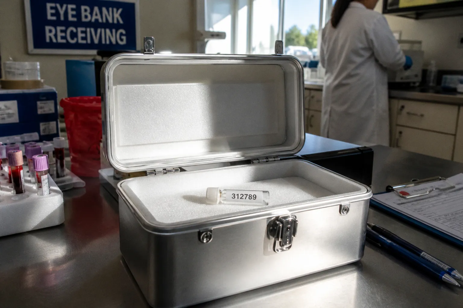
The 24-hour post-mortem clock is not a bureaucratic window but a cellular one. Within hours of circulatory arrest, corneal epithelium begins to lose tight junction integrity; microvilli retract, glycogen reserves deplete, and the endothelial pump drifts toward a viability threshold from which it will not recover. By the time a recovery technician arrives at the morgue, the donor eligibility file — assembled across hospital charts, serology laboratories, and recovery logs — has already determined whether procurement is even attempted. A single unspecified cause of death, an undocumented transfusion volume, or a missing serology timestamp, and the entire cascade halts before a scalpel touches the limbus.
This is the architecture most recipients never see: ocular tissue procurement as a sequenced forensic review, calibrated against regulatory frameworks that govern not just who may donate, but who may be allowed to receive. Eye bank donor screening criteria sit at the intersection of federal statute, accreditation standards, and the practical limits of tissue viability — a layered defense where each filter narrows the eligible pool while the cellular clock keeps ticking.
The Regulatory Floor: 21 CFR Part 1271 and FDA Oversight
Ocular tissue in the United States is regulated under the same framework as other human cells, tissues, and cellular- and tissue-based products (HCT/Ps). Subpart C of 21 CFR Part 1271 lays out the eligibility requirements that any establishment recovering, processing, or distributing such tissue must satisfy. The center of gravity is the donor eligibility determination — a documented conclusion, supported by donor screening and donor testing, that a donor is either eligible or ineligible.
For eye banks, this translates into a defined file structure. The medical and behavioral history review captures risk factors for relevant communicable disease agents — HIV, hepatitis B, hepatitis C, treponemal infection, and a defined set of additional agents with plausible ocular transmission. Required infectious disease testing, performed on a specimen collected at the appropriate interval, supplies the molecular evidence. Both halves are mandatory: a clean behavioral history cannot substitute for missing serology, and negative serology cannot rescue an undocumented exposure history. The 2007 FDA Guidance for Industry codifies the operational interpretation of these requirements.
Without a completed donor eligibility determination on file, tissue does not move. The regulation treats the document itself as a controlled product, not an afterthought.
Subpart C also defines what must be retained. Records of donor screening, testing, and the eligibility determination itself must be retained for at least ten years after the date of administration (or, for tissue not administered, after the date of disposition) — and they must be available for inspection by FDA at any point in that window. Eye banks are not laboratories in the speculative sense; they are tissue establishments under federal jurisdiction, with the corresponding audit exposure.
Medical Record Review and the Architecture of Contraindications
The first filter is the chart. Eye bank medical directors or designated personnel review the donor's hospital records, focusing not on general health status but on the specific exclusion criteria that preclude tissue release. These absolute contraindications — conditions in which the donor is categorically ineligible, without exception — form a short, deliberately rigorous list:
- Creutzfeldt-Jakob disease (CJD) and variant CJD
- Active leukemia, active disseminated lymphoma, and other actively disseminated malignancies of the hematopoietic system
- Active viral encephalitis (including active herpes simplex encephalitis, rabies, and related central nervous system infections)
- Active septicemia
- Death of unknown cause
Each exclusion rests on a transmission pathway with documented or plausible ocular involvement. CJD and other transmissible spongiform encephalopathies raise the specter of corneal inoculation of misfolded prion protein; although the magnitude of risk is debated, the precautionary exclusion is absolute. Active leukemias and disseminated lymphomas carry a risk of hematogenous cellular seeding; the corneal stroma is largely avascular but the limbal vasculature, conjunctival lymphatics, and intraocular compartments are not. Active septicemia implies bacteremia or fungemia at the moment of death, with risk of micro-abscess formation in donor tissue. Active viral encephalitis raises the possibility of viral genome in ocular fluid. Death of unknown cause is excluded as a matter of evidentiary integrity — without a cause, none of the above can be reliably ruled out.
| Contraindication | Primary transmission concern | Tissue-level risk profile |
|---|---|---|
| CJD / variant CJD | Prion transmission | Long-latency, undetectable by serology |
| Active leukemia / disseminated lymphoma | Hematogenous cellular seeding | Limbal and conjunctival vasculature |
| Active viral encephalitis | Viral presence in ocular fluid | Aqueous, vitreous, and neural tissue |
| Active septicemia | Bacterial / fungal seeding | Stromal micro-abscess potential |
| Death of unknown cause | Evidentiary incompleteness | Cannot exclude any of the above |
Reviewers are not abstracting general health; they are looking for the presence or absence of named conditions in specific clinical contexts. "Active" matters: a malignancy in long-term remission is not the same entity as active dissemination, and the chart documentation is parsed for treatment status, last recurrence date, and disease phase before the eligibility algorithm closes.
Serological Testing and the Hemodilution Problem
If the chart clears, the second filter is the blood. Required infectious disease testing — typically performed on specimen matrices collected either pre-mortem or within the post-mortem recovery window, processed in CLIA-certified or equivalent laboratories, and documented by accession number — generates the laboratory half of the eligibility determination.
The complication is hemodilution, a problem specific to post-mortem and rapid-transfusion scenarios. Plasma dilution from crystalloid infusion or blood product administration can lower circulating antibody concentrations below the detection threshold of an otherwise valid assay, producing a false-negative serology result on a specimen that does not reflect the donor's true immunological status. The threshold at which hemodilution must be assessed is 2,000 mL of crystalloids or blood products administered within the hour preceding either death or specimen collection, whichever interval applies. Beyond that volume, a pre-transfusion specimen must be sought if available, or the testing must be qualified by the documented fluid administration record.
The procedural sequence is tightly choreographed. A pre-mortem specimen, drawn during the hospital admission that preceded death, is the preferred substrate because it predates any fluid resuscitation. When that is unavailable, a post-mortem cardiac or great-vessel draw supplies the primary specimen, and the accompanying infusion record is no longer optional — it becomes part of the testing chain of custody. Each step has an audit hook: time of draw, time of death, time of fluid administration, lot numbers of administered products. Miss any of them, and the test result cannot be interpreted, regardless of how cleanly the specimen processed in the laboratory.
Hemodilution is the silent disqualifier. The serology report comes back clean, and the file still cannot be released, because the donor received three liters of crystalloid in the hour before death.
The Post-Mortem Clock and Cellular Viability
Even when serology and chart review converge on an eligible donor, the tissue itself imposes a hard schedule. Standard post-mortem ocular tissue recovery must generally occur within 24 hours of death, and the upper end of that window is not equivalent to the lower end in terms of the resulting tissue quality. Corneal endothelial cell density and epithelial integrity decline continuously after circulatory arrest; eyes recovered within the first few hours retain endothelial mosaics and tight junction architecture that eyes recovered near the 24-hour mark do not.
Cold-chain biospecimen logistics govern the transition from procurement to processing. Whole globe donor recovery — for downstream corneoscleral rim dissection — is followed by cold storage in moist chambers or in commercially available hypothermic storage media. The storage medium itself functions as a short-term metabolic substrate, supporting endothelial viability at refrigeration temperatures for an interval defined by the product specification and the tissue's intended use. Each preservation method carries a different tissue-quality profile and a different downstream eligibility filter: refrigerated moist chamber, hypothermic storage medium, and organ-culture incubation at elevated temperature in some non-US jurisdictions all yield tissue with distinct experimental and clinical characteristics.
For ocular biorepositories, the procurement math extends beyond surgical suitability. Investigators downstream may need globes suitable for retinal pigment epithelium work, neurosensory retina dissection, or optic nerve procurement — each with its own window, its own structural vulnerabilities, and its own contraindications. The RPE senescence that follows prolonged post-mortem intervals degrades the monolayer's barrier function; the outer segments of photoreceptors fragment as the autolytic cascade proceeds. An ocular tissue donor eligibility determination approved for keratoplasty does not automatically translate into RPE-grade tissue; the recovery protocol, preservation route, and downstream notification path must align with the requested research use.
Accreditation as Audit: The EBAA Layer
Beneath the federal floor sits an accreditation ceiling. Eye banks operating in the United States typically hold Eye Bank Association of America (EBAA) accreditation — a voluntary designation that, in practice, defines the operating standard for procurement, processing, and distribution. EBAA-accredited eye banks undergo comprehensive on-site inspections at least every three years, conducted by an evaluation team that includes a practicing corneal surgeon and an experienced eye banking professional.
The inspection is not a paperwork review. Surveyors trace donor files from referral through eligibility determination and into final disposition, examining chart review adequacy, serological chain of custody, tissue recovery documentation, and storage temperature logs. Deviations trigger corrective action plans with defined timelines; unresolved deviations threaten accreditation, which in turn affects the eye bank's ability to supply tissue into a research and clinical network that treats accreditation as a baseline qualification.
The documentation retention standard reinforces the audit chain. Eye banks are required to retain quality assurance records for at least ten years — including, in practice, donor charts, consent forms, recovery records, serology reports, and any deviations or adverse events. This retention period creates a longitudinal archive that supports post-distribution adverse event investigation months or years after transplantation, and supplies the evidentiary record for regulatory or accreditation review at any point within the window.
The accreditation cycle does not reset at three years; it accumulates. Every recovered tissue is a potential audit subject for the next decade, and every error is a delayed liability waiting for the next inspection.
What the Architecture Cannot See
The screening cascade filters a great deal, but it cannot resolve every edge case. Donor records occasionally present contraindicated exposures in time windows that fall outside the standard exclusion criteria — historical malignancies with undocumented remission status, serology-negative donors with relevant behavioral exposures in the distant past, or causes of death that almost but do not quite meet the threshold for the unknown-cause exclusion. In these instances, medical directors apply judgment against a documented eligibility algorithm, and the decisions — defensible in principle — still leave the residue of cases where the available evidence is incomplete.
Across jurisdictions outside the United States, age-cutoff limits for ocular donation vary considerably, with some systems accepting tissue from donors well past 80 years while others draw the line earlier — a heterogeneity that affects donor biorepository composition but, for the moment, defies harmonization. And even with rigorous tissue donor suitability standards in place, no eye bank can eliminate the residual possibility of communicable disease transmission; testing and chart review lower risk to an acceptable medical level, but the absolute-zero threshold is not a regulatory promise.
What the screening cascade does deliver, with verifiable regularity, is a documented record: a chain of evidence from donor to recipient that has been reviewed against published criteria, tested with regulated assays, and audited against accreditation standards by credentialed professionals who treat each file as a forensic object. That record is the working product of the eye bank — invisible to the recipient, but inseparable from every viable cornea that arrives under an operating microscope, and from every retinal specimen that lands in a downstream laboratory prepared for the molecular work that follows.