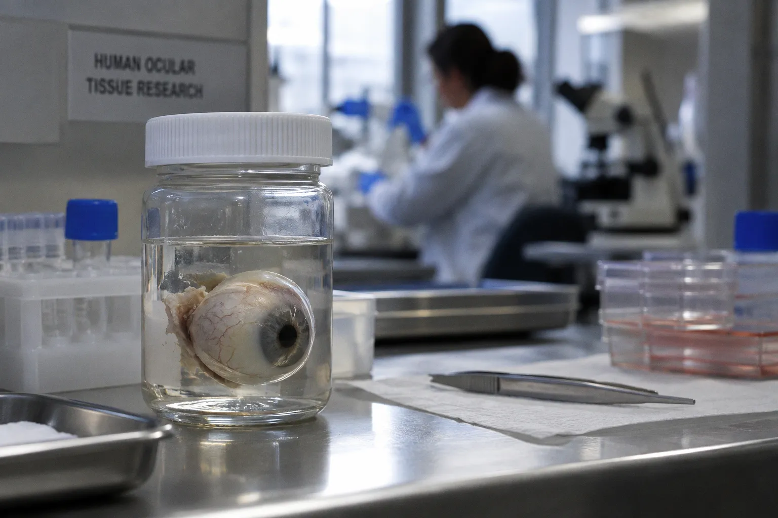
That number is a logistical ceiling, not a yield claim. It sets the denominator against which glaucomatous protein shifts, subregion-specific pathway enrichments, and post-translational modifications are normalized.
Glaucoma proteomics lives in the gap between this healthy atlas and the diseased catalog. The reference set is dense; the glaucomatous signature remains fragmented across studies, sub-microdissection protocols, biomechanical strain models, and donor tissue procurement pipelines. Reconciling the two requires a tight audit of three data streams: in vitro lamina cribrosa cell proteomics under cyclic strain, healthy donor tissue baseline profiling, and transcriptomic analyses of glaucomatous optic nerve head specimens.
The 2,711-protein baseline is not a finished atlas. It is a resolution limit that gates every cross-study comparison.
Mapping the healthy human optic nerve head proteome
The 2016 Orbitrap LC-MS/MS catalog of healthy human retrobulbar optic nerve remains the most extensive protein-level reference for this tissue. The methodology prioritized depth over subregion specificity: whole optic nerve segments rather than microdissected lamina cribrosa or prelaminar zones. This produces a high-coverage baseline but flattens the spatial resolution that the glaucomatous process actually exploits.
For downstream studies, that flattening is consequential. Glaucomatous damage is regionally heterogeneous — concentrated at the optic nerve head, propagating through the lamina cribrosa, and producing axonal loss in the retinal nerve fiber layer. A flat reference atlas cannot resolve whether a protein shift observed in a glaucomatous donor sample reflects a true disease signal or a sampling artifact from pooling across unaffected nerve segments. The logistical answer is subregion-specific microdissection with matched controls. The published record has not yet converged on this protocol.
Post-mortem interval adds a second layer of noise. Protein degradation kinetics begin at procurement. The interval between circulatory arrest and tissue preservation, typically documented in hours, defines the upper bound on transcriptomic yield and the reproducibility of post-translational modification calls. Phosphorylation and citrullination degrade faster than bulk protein abundance. Any cross-study comparison of optic nerve head proteomics must normalize for this variable before pathway-level claims can be sustained.
Mechanical strain and proteomic signaling hubs in lamina cribrosa cells
The biomechanical hypothesis of glaucoma — that elevated intraocular pressure transmits pathological strain through the lamina cribrosa — drove the 2012 iTRAQ LC-MS/MS study of cultured human ONH lamina cribrosa cells under cyclic mechanical stretch. The output: 526 proteins identified at 95% confidence from cells subjected to controlled in vitro loading. The protein count matters less than the signaling architecture it exposed.
Strain activates four convergent hubs: TGFβ1, TNF, CASP3, and p53.
- TGFβ1 drives extracellular matrix deposition and myofibroblast transdifferentiation — the fibrotic remodeling long observed histologically in glaucoma.
- TNF signals neuroinflammatory recruitment.
- CASP3 marks the apoptotic execution pathway.
- p53 integrates cellular stress and DNA damage responses.
The convergence is the analytical point. These are not independent perturbations but a coordinated stress response downstream of mechanical loading.
Downstream targets in the strain proteome include BAG5 (a BAG-family co-chaperone implicated in apoptosis regulation), NO66 (a Jumonji-domain histone demethylase linked to ribosomal biogenesis), and eIF-5A (a translation elongation factor associated with cellular stress). Their presence in the strain-responsive protein set implies that biomechanical loading alters not only ECM composition but also protein synthesis capacity and chromatin state.
A companion study of human ONH astrocytes under 3% and 12% cyclic equiaxial stretch identified 573 proteins via six-plex iTRAQ LC-MS/MS. The differential regulation pattern across strain magnitudes is informative: lower and higher strain produced distinct protein signatures, with consistent hits including ANXA4 (annexin A4, implicated in membrane trafficking and calcium signaling), GOLGA2 (a Golgi matrix protein), and alpha B-crystallin (a small heat shock protein with chaperone function). Alpha B-crystallin specifically links the astrocyte strain response to the heat shock protein axis — a known compensatory pathway in glaucomatous stress.
Extracellular matrix remodeling and inflammatory pathways in glaucoma
Transcriptomic and molecular profiling of human glaucomatous ONH donor tissue has converged on a consistent finding: ECM organization is upregulated. Specific transcripts driving this signal include LOXL1 (lysyl oxidase-like 1, a collagen and elastin crosslinking enzyme), SERPINH1 (serpin family H member 1, a collagen-specific chaperone), CHI3L1 (chitinase 3-like 1, a glycoprotein associated with tissue remodeling and inflammation), IL6 (interleukin-6, a pro-inflammatory cytokine), and COL1A1 (collagen type I alpha 1 chain).
These are not random hits. LOXL1 and SERPINH1 together imply active stiffening of the lamina cribrosa — collagen deposition coupled with enzymatic crosslinking. CHI3L1 and IL6 indicate ongoing neuroinflammatory signaling. COL1A1 marks fibrotic replacement. The signature is mechanically and immunologically coherent: a tissue under chronic stress, remodeling its structural support while sustaining a low-grade inflammatory state.
Proteomic and transcriptomic data converge on this remodeling phenotype but do not converge on its temporal ordering. ECM remodeling could be a driver of axonal damage, a consequence of it, or a parallel process with separate upstream causes. The proteomic studies of strained LC cells show that mechanical load alone is sufficient to trigger ECM-related protein expression changes. This supports a model in which biomechanical strain initiates the fibrotic cascade. It does not exclude parallel inflammatory triggers.
A 2026 IOVS presentation of transcriptomic profiling of human ONH donor tissue extends the molecular cataloging from in vitro surrogates into primary donor specimens. The methodological shift — from cell culture proteomics to whole-tissue transcriptomics — changes the question being asked. Cultured cells isolate specific signaling responses; donor tissue captures the integrated network state at the time of procurement.
The role of PAD2 and protein citrullination in axonal degeneration
The Bhattacharya et al. proteomics work in human glaucoma tissue introduced a post-translational modification into the conversation: peptidyl arginine deiminase 2 (PAD2) upregulation and enhanced optic nerve protein citrullination. Citrullination converts arginine residues to citrulline, altering protein charge, structure, and interaction surfaces. It is enzymatic, irreversible at the protein level, and implicated in autoimmune targeting when the modified proteins reach immunologically active compartments.
In the glaucomatous optic nerve, PAD2-mediated citrullination of structural and signaling proteins produces two downstream effects. First, it modifies the antigenic profile of optic nerve proteins, potentially exposing epitopes that trigger adaptive immune responses. Second, it disrupts protein-protein interaction surfaces, destabilizing normal scaffolding and signaling assemblies in the lamina cribrosa and prelaminar regions.
The presence of PAD2 upregulation in glaucoma donor tissue reframes the disease from a purely mechanical or purely inflammatory process to one with an autoimmune-accessible post-translational dimension. This matters for biomarker strategy: if citrullinated peptides enter the aqueous humor or vitreous and can be detected by targeted mass spectrometry, they become candidate biomarkers for early-stage disease, before irreversible axonal loss consolidates.
PAD2-mediated citrullination reframes glaucoma as a disease with an immunologically accessible post-translational footprint — and a quantifiable biomarker target.
The biomarker pipeline, however, has not validated this hypothesis at clinical scale. The unknown — and it is explicitly unconfirmed in the published record — is whether a single ONH protein biomarker can reliably diagnose early-stage primary open-angle glaucoma before visual field defects appear. The proteomic data support the candidate list; the validation infrastructure does not yet exist.
Bridging the gap: from in vitro strain models to human donor tissue
The central logistical challenge in ONH proteomics is the translation gap. Cultured lamina cribrosa cells under cyclic strain produce clean, reproducible signaling data but cannot replicate the multicellular architecture of the intact optic nerve head. Astrocytes, axons, capillaries, and ECM components interact in three dimensions. Cell culture collapses this into a single cell type on a deformable membrane.
The published data must be read accordingly. Proteomic shifts observed in animal models of ocular hypertension should not be projected onto human glaucoma donor ONH proteome profiles as direct equivalents. Species differences in ECM composition, astrocyte biology, and lamina cribrosa architecture introduce quantitative and qualitative offsets. Similarly, in vitro biomechanical strain replicates a subset of cellular stress signals but does not reproduce the chronic, pulsatile, multi-axis loading pattern of the in vivo optic nerve head. The 3% and 12% cyclic stretch models capture acute responses; glaucoma is a decades-long process. Protein-level signatures overlap at the pathway level — TGFβ1, TNF, ECM remodeling, heat shock response — but the temporal integration is absent.
Human donor tissue closes part of this gap. The Orbitrap catalog of healthy retrobulbar optic nerve, the transcriptomic profiling of glaucomatous ONH, and the PAD2 citrullination studies all use primary human specimens. They capture the integrated state at procurement. They cannot, however, capture dynamics. A donor tissue sample is a single time point in a process that began years or decades earlier.
| Data stream | Method | Protein/transcript yield | Primary signal |
|---|---|---|---|
| Healthy retrobulbar optic nerve (donor) | Orbitrap LC-MS/MS | 2,711 non-redundant proteins | Reference baseline |
| Strained lamina cribrosa cells (in vitro) | iTRAQ LC-MS/MS | 526 proteins at 95% CI | TGFβ1, TNF, CASP3, p53 hub activation |
| Strained ONH astrocytes (in vitro) | Six-plex iTRAQ LC-MS/MS | 573 proteins | ANXA4, GOLGA2, αB-crystallin regulation |
| Glaucomatous ONH donor tissue | Transcriptomic profiling | Pathway-level enrichment | ECM remodeling, IL6, CHI3L1 upregulation |
| Glaucoma donor tissue (Bhattacharya et al.) | Proteomic + PTM analysis | PTM-level | PAD2 upregulation, enhanced citrullination |
The logistical bottleneck is procurement latency itself. The interval between death and preservation determines what can be measured. RNA degrades faster than protein. Post-translational modifications degrade faster than bulk abundance. Labile modifications — citrullination, phosphorylation — are the first to be lost. Donor tissue proteomics is therefore biased toward stable proteins and away from the dynamic regulatory layer where early-disease signals are most likely to reside.
Assessment
The proteomic landscape of the glaucomatous optic nerve head is increasingly mapped but not yet actionable. The healthy reference atlas of 2,711 proteins provides the resolution ceiling. The in vitro strain proteomics studies — 526 proteins in LC cells, 573 in astrocytes — define the signaling hubs activated by biomechanical stress. The transcriptomic and proteomic analyses of glaucomatous donor tissue identify the ECM remodeling, neuroinflammatory, and citrullination signatures present at the time of procurement.
What remains is the validation infrastructure. No single ONH protein biomarker has been clinically validated for early-stage POAG diagnosis prior to irreversible axonal loss. The candidate list is long; the targeted assay pipeline is short. The path forward requires standardized sub-microdissection protocols, matched healthy and diseased donor cohorts, controlled post-mortem intervals, and targeted mass spectrometry assays for the specific post-translational modifications — citrullination first, phosphorylation second — that the proteomic discovery studies have flagged.
The data exist. The logistics of converting them into clinical biomarkers have not yet been solved.