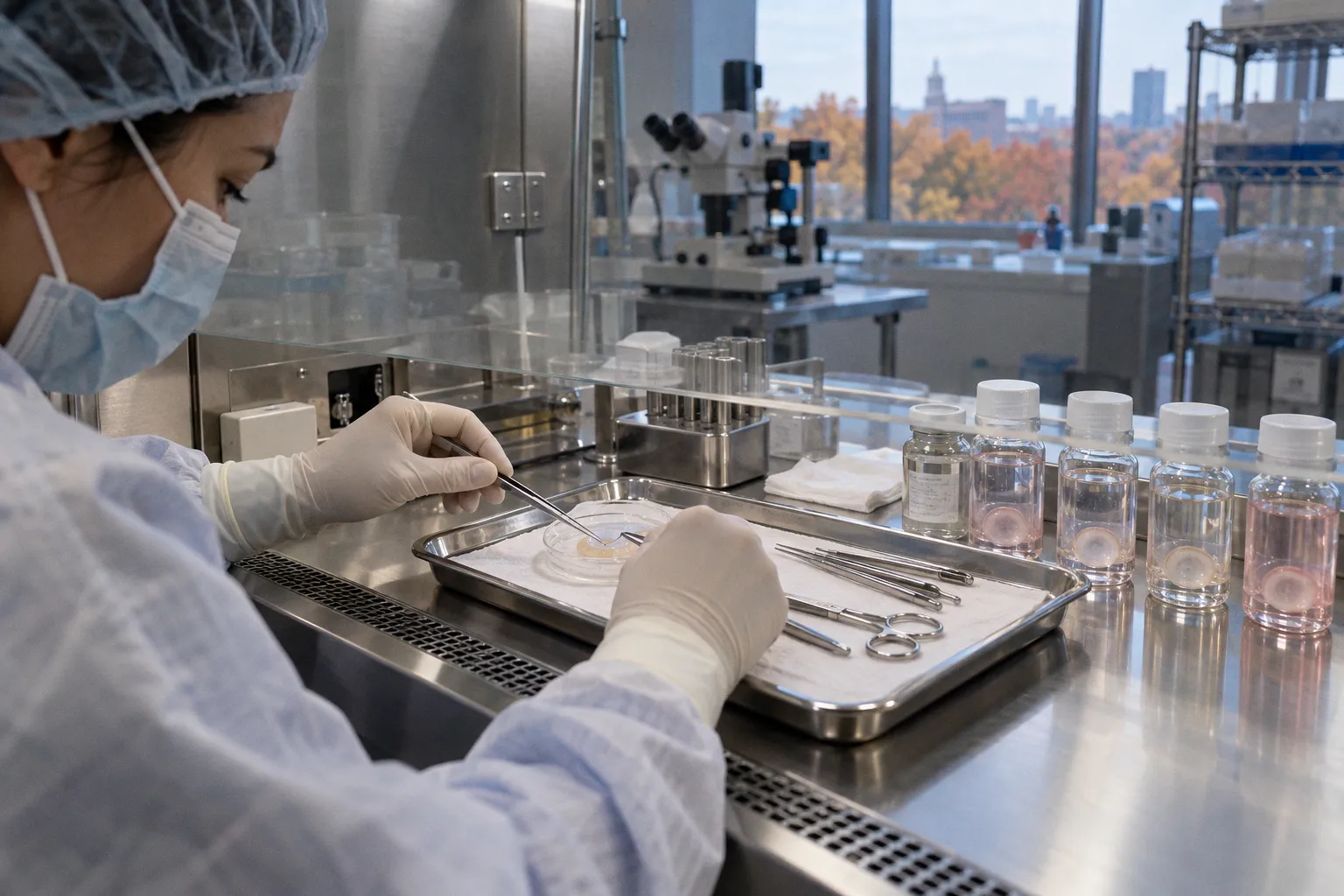
It is a tidy picture, the kind that survives in review articles and procurement protocols. The reality, as anyone who has watched a graft haze over without obvious surgical cause will tell you, is that quiescence in the corneal stroma is not a permanent condition. It is a metabolic posture, and donor tissue handling perturbs it routinely. The central question — what happens to corneal stromal keratocytes between death of the donor and implantation in the recipient — is rarely interrogated with the seriousness it deserves. We will.
The Quiescent State: What G0 Arrest Actually Buys the Cornea
Adult human corneal stroma is a remarkable tissue. It sits between Bowman's layer and Descemet's membrane, occupies roughly 90% of corneal thickness, and depends for its optical function on a population of cells that, under normal conditions, do almost nothing. Corneal stromal keratocytes (CSKs) are mesenchymal-derived cells of neural crest origin, and in the uninjured adult cornea they are arrested in the G0 phase of the cell cycle. They do not divide. They do not migrate. They do not, in any meaningful sense, behave like fibroblasts, despite sharing lineage.
What they do is synthesize: type I and type V collagens, along with the keratan sulfate proteoglycans — particularly lumican, keratocan, and mimecan — that impose the precise, quasi-periodic spacing on collagen fibrils responsible for stromal transparency. This is not idle activity. The lattice geometry is what scatters light coherently rather than diffusely, and any departure from it produces haze. The cell that maintains this lattice is, by design, metabolically expensive to maintain and developmentally expensive to replace. The cornea treats the quiescent keratocyte as an asset worth protecting.
The protection is real. Quiescent keratocytes express a characteristic surface profile: CD34-positive, keratocan-positive, Thy-1-negative, alpha-SMA-negative. They sit in a low-serum, low-cytokine environment between precisely organized collagen lamellae. They are, in effect, an anti-inflammatory reservoir that happens to also be a transparency machine. Strip away any of those conditions — serum exposure, mechanical strain, cytokine insult — and the reservoir activates. This is where donor tissue handling begins to matter.
Quiescence in the corneal stroma is not a default state. It is an actively maintained posture, and every step of donor tissue procurement tests it.
Mechanisms of Phenotypic Transdifferentiation: From Keratocyte to Myofibroblast
The transformation is well documented in animal models and increasingly confirmed in human donor tissue, yet the eye banking community continues to discuss it as if it were a peripheral concern. It is not. The cascade runs roughly as follows. A keratocyte receives an activation signal — serum exposure during organ culture, mechanical strain during trephination, post-mortem enzymatic stress, or simply the trauma of excision — and exits G0. It begins to express markers it should not have. Keratocan and CD34 fall away. The cell flattens, extends processes, and assumes the morphology of a stromal fibroblast (SF). It proliferates — the first proliferative activity these cells have undergone since embryogenesis — and begins secreting a different extracellular matrix: fibronectin, tenascin, and disordered type III collagen.
If the insult is sustained or sufficiently severe, a subset of these fibroblasts undergoes a second transition into myofibroblasts (MyoFBs), expressing alpha-smooth muscle actin (α-SMA), acquiring contractile capacity, and laying down the disorganized, highly reflective matrix that produces persistent stromal scarring. This is the cell type responsible for the dense, white opacities that follow some keratoplasties — the ones that do not respond to steroids, that persist beyond the expected healing window, and that ultimately compromise graft optical function regardless of how cleanly the surgery was performed.
The transdifferentiation is, by all current evidence, considered irreversible in vivo once established. This is the part of the literature that deserves more emphasis than it usually receives. We are not talking about cells that have temporarily shifted behavior and can be coaxed back. We are talking about cells that have committed to a new phenotype, reorganized their cytoskeleton, and begun producing extracellular matrix that is functionally incompatible with transparency. Treating this as a reversible process — as some in the field have done when discussing steroid rescue — is a paradigm deficit of considerable clinical consequence.
The keratocyte that has become a myofibroblast is not a confused keratocyte. It is a different cell with a different job, and its job is incompatible with transparency.
Storage Modalities and the Cellular Bill That Comes Due
The choice of preservation method is the single largest variable an eye bank controls, and it is also the variable most likely to trigger activation. Two paradigms dominate contemporary donor corneal storage: hypothermic storage in the 2–8 °C range, typically allowing 7–14 days of preservation, and organ culture at 28–37 °C, allowing up to 30 days. The numbers are well established; the cellular consequences are routinely underdiscussed.
Hypothermic storage slows metabolism. Cells in G0 arrest tolerate this reasonably well, because G0 arrest means they were already not doing much. The technique buys time without imposing the serum-rich, cytokine-active environment that organ culture requires. Keratocyte activation under proper hypothermic conditions is minimized, though — and this is important — not eliminated. The threshold at which post-mortem time or mechanical insult during retrieval triggers activation despite cold storage is not precisely defined in the literature, which is precisely the problem.
Organ culture is something else entirely. It preserves corneas in conditions that support ongoing metabolic activity, including a serum-containing medium. This is the trade. You get longer storage — up to 30 days, roughly twice the hypothermic window — and you get a tissue that arrives at the operating room with more verifiable cellular viability. But you also get an environment that contains exactly the signals that activate quiescent keratocytes: serum, growth factors, and the absence of the low-cytokine milieu that defines its native state. Organ cultured corneas arrive with measurable Thy-1 expression and measurable α-SMA positivity in a non-trivial fraction of cases. Whether this is acceptable depends entirely on what the recipient cornea is being asked to do.
| Parameter | Hypothermic storage (2–8 °C) | Organ culture (28–37 °C) |
|---|---|---|
| Typical storage duration | 7–14 days | Up to 30 days |
| Metabolic activity of resident cells | Substantially suppressed | Actively maintained |
| Serum exposure of keratocytes | Minimal (storage medium typically serum-free or low-serum) | Continuous (medium supplemented to support viability) |
| Keratocyte activation risk | Lower baseline, but not zero | Measurable; Thy-1 and α-SMA positivity documented |
| Stromal haze risk at graft | Generally lower, dependent on retrieval and shipping variables | Variable; some studies suggest higher baseline activation markers |
| Pre-implantation viability assessment | Limited; relies heavily on slit-lamp and endothelial criteria | More dynamic; allows extended microbiological and viability testing |
The table is not a verdict. It is a description of the trade-off, and the eye banking community has, by historical accident, organized itself regionally around one paradigm or the other without rigorously comparing downstream outcomes at the keratocyte level. North American eye banks historically favor hypothermic storage; many European programs have built decades of infrastructure around organ culture. Both work. Neither is innocent of cellular consequence.
Molecular Markers of Activation: The Fingerprints of an Already-Changed Cell
The diagnostic question — has this graft's stroma already activated before it ever touched a recipient — has an emerging answer, and the answer involves markers that should not be there. Two in particular deserve attention.
The first is Thy-1, a cell surface glycoprotein absent from uninjured human corneal stroma. When quiescent keratocytes transition to repair phenotypes — fibroblasts and, eventually, myofibroblasts — Thy-1 expression appears. It is not subtle. It is a binary shift in surface profile, and it has become one of the more reliable indicators that an activation cascade has begun. Eye banks do not routinely assay for Thy-1 on outgoing tissue, despite the fact that a simple immunohistochemistry or flow cytometry panel could meaningfully stratify grafts by activation status before they reach the surgeon. The omission is, in the critical reading, a logistical one dressed as a scientific one.
The second marker is the loss of keratocan. Quiescent keratocytes secrete keratan sulfate proteoglycans in a carefully balanced ratio; keratocan is the species most closely associated with the small-diameter, evenly spaced collagen fibrils of a transparent stroma. As keratocytes activate, keratocan expression drops, and the collagen fibrils that result from the new synthetic program are larger, more heterogeneous, and more reflective. CD34, another quiescence marker, follows the same trajectory. The combination — Thy-1 positive, keratocan negative, CD34 negative, with rising α-SMA — describes a cell that has fully committed to the repair phenotype, regardless of whether the original insult was serum, strain, or surgeon.
What is striking is that these markers can be detected in donor tissue before transplantation. They are not retrospective findings. They are present at the point of implantation, in tissue that has been certified by the eye bank as suitable for surgery. The implication is that the certification process — slit-lamp evaluation, endothelial cell counts, optical clarity assessment — is blind to stromal activation status at the cellular level. This is not an indictment of eye banks; it is a description of what current inspection paradigms can and cannot see.
Clinical Implications: What the Recipient Cornea Inherits
The clinical endpoint matters more than the molecular pathway, and here the data are uncomfortable. A graft that goes in with measurable stromal activation does not necessarily haze. Many do not, at least not visibly in the early post-operative period. But the cellular program has begun, and the conditions of recipient corneal healing — surgical trauma, sutures, topical medications, the recipient's own cytokine milieu — are precisely the conditions that push an already-primed fibroblast further down the activation cascade. The recipient provides the second hit.
This is the model: donor tissue handling provides the first activation signal, sometimes small and sometimes large. Recipient healing provides the second. Persistent stromal haze, interface scarring in lamellar procedures, and the chronic opacification that compromises graft clarity at six to twelve months are downstream consequences of this two-hit process. They are not always surgical failures. They are not always rejection episodes. They are, with uncomfortable frequency, the predictable result of a cellular program that began before the surgeon ever opened the operative field.
The therapeutic response — topical steroids, sometimes prolonged — is imperfect. Steroids reduce inflammation but do not reverse α-SMA expression or restore keratocan production in cells that have already committed. This is why the assertion that myofibroblast activation is reversible in vivo warrants skepticism. The cellular machinery that has been remodeled is not, on current evidence, simply switched off by pharmacologic intervention.
Where the Evidence Actually Stands
Here is the uncomfortable summary. Donor corneal stromal keratocytes do not remain quiescent through procurement and storage as a rule. They activate in measurable numbers under both hypothermic and organ culture conditions, with the latter imposing a higher baseline activation load due to its inherent design. The molecular markers of that activation — Thy-1 positivity, keratocan loss, α-SMA expression — can be detected before transplantation in tissue that passes current certification. The downstream consequence is a population of grafts entering the operating room with a partial wound healing response already underway, primed for further activation by the recipient environment.
The dogmatic position — that eye bank certification guarantees a quiescent stromal cellular compartment — does not survive critical interrogation of the literature. Paradoxically, the more sophisticated our preservation methods become, the more clearly we see that storage itself is an activation event, and that the window of quiescence preservation is narrower than the procurement literature implies.
The eye banking community has a choice. It can continue to certify tissue on endothelial and slit-lamp criteria while treating stromal activation as a recipient-side problem, or it can begin to assay what it is actually shipping. Thy-1, keratocan, α-SMA, and CD34 are measurable markers with clear biological meaning. Adding them to the pre-release panel would cost time and money. It would also, for the first time, allow surgeons to know what they are implanting at the cellular level rather than the optical level.
The research community has a parallel obligation: to stop describing myofibroblast activation as a reversible process in vivo without producing the data that would support that claim. The current evidence base does not support reversibility once α-SMA is expressed. Pretending otherwise in service of therapeutic optimism is not a kindness to patients.
Donor tissue is a finite, irreplaceable resource. Every graft that goes into a recipient cornea carries the molecular history of its retrieval, its storage, and its handling. It is time the field treated that history as something to be measured rather than assumed.