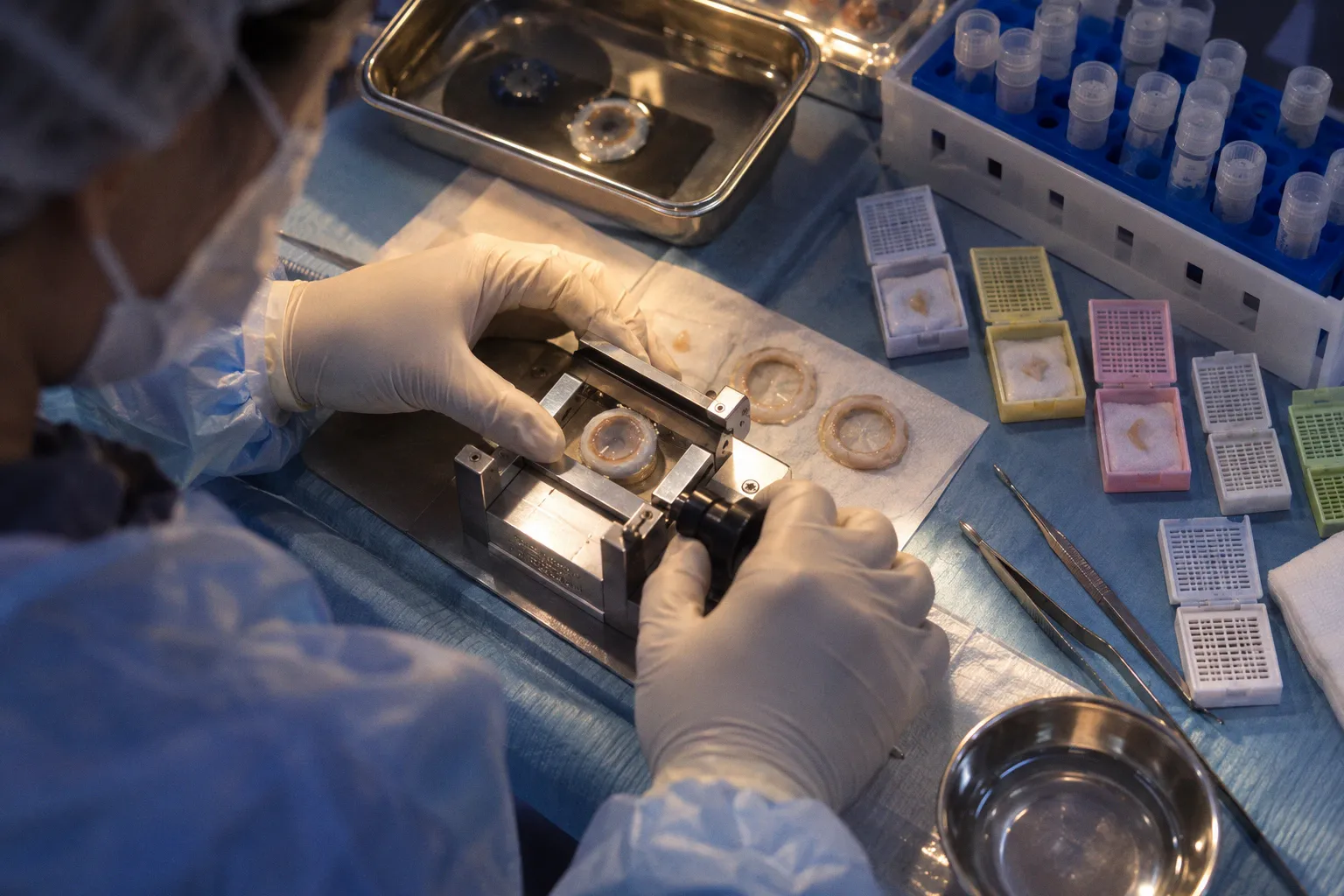
Human donor corneoscleral limbus is not merely a border zone between transparent cornea and vascularized conjunctiva; it is a layered anatomical niche whose folds, crypts, stromal projections, and extracellular matrix appear to influence which epithelial cells persist, divide, and repopulate the ocular surface.
That distinction matters because donor tissue can be handled as if its value were determined only by cell yield. It is not. A limbal graft may contain epithelial cells and still fail to reproduce the microenvironment that maintains them. Conversely, a small biopsy can be biologically useful when it preserves the right architecture. The practical question is therefore not simply how many cells can be removed from a donor cornea. It is which features of the limbal niche survive procurement, processing, culture, and transplantation.
The limbus is an anatomical niche, not a painted boundary
The human corneoscleral limbus performs at least two jobs that are often discussed separately but cannot be separated in tissue biology.
First, it acts as an anatomical barrier. The organized transition between corneal epithelium and adjacent ocular surface tissues helps prevent uncontrolled conjunctival epithelial encroachment onto the cornea. Second, it provides the microenvironment in which limbal epithelial stem and progenitor cells are maintained.
This is where the familiar description of the limbus as a narrow ring becomes inadequate. Histological studies identify specialized structures including the Palisades of Vogt, limbal crypts, and focal stromal projections. These are not decorative landmarks for an atlas. They create local variations in epithelial support, stromal contact, extracellular matrix composition, and access to surrounding tissue compartments.
The resulting niche is spatially uneven. A cell harvested from one limbal position is not automatically equivalent to a cell harvested from another. That point sounds obvious once stated, yet many experimental workflows still treat the limbus as a uniform strip of donor material. The paradigm deficit is built into the sampling strategy.
Why structure changes the interpretation of donor tissue
A conventional cell-isolation workflow tends to reward dissociation. Tissue is enzymatically processed, cells are released, and the resulting suspension is assessed by viability, proliferation, colony formation, or marker expression. Those measurements are useful, but they answer only part of the question.
They can tell us whether cells survived isolation and whether they retain selected properties in culture. They do not necessarily tell us whether the native niche was preserved, whether the original stromal signals remain available, or whether the most clinically relevant subpopulation was preferentially removed during processing.
Human donor limbus therefore needs to be considered at two levels:
- Cellular composition: which epithelial, stromal, and progenitor populations are present.
- Spatial organization: how those populations are arranged within crypts, palisades, epithelial layers, and underlying stroma.
The first is easier to quantify. The second is more difficult, and consequently easier to neglect.
A limbal sample is not defined only by the cells it contains; it is also defined by the architecture those cells lost—or retained—during processing.
This matters particularly in post-mortem donor tissue. Corneal limbus histology after death can reveal preserved structures, but histological presence should not be confused with full biological function. The exact microarchitectural changes that occur during prolonged organ culture storage remain insufficiently defined. A scaffold may preserve the outline and matrix composition of a niche while no longer reproducing every signaling relationship present in living tissue.
That is not a reason to dismiss architecture. It is a reason to measure it more honestly.
Palisades, crypts, and stromal projections: the working parts of the niche
The Palisades of Vogt are commonly used as a visual proxy for limbal stem-cell organization. They are important, but the niche cannot be reduced to the palisades alone. Limbal crypts and focal stromal projections add depth to the model by creating epithelial-stromal interfaces that may support the retention and renewal of limbal epithelial populations.
The critical issue is not whether one named structure is present. It is whether the donor tissue retains the coordinated microenvironment in which several structures operate together.
That includes:
1. A defined epithelial compartment. The limbal epithelium contains basal cells with stem and progenitor characteristics, but these cells are not interchangeable with central corneal epithelial cells in long-term stemness or niche signaling.
2. A specialized stromal interface. The underlying stroma contributes extracellular matrix and physical organization. It is not a passive floor beneath the epithelium.
3. Crypt-like and palisade-associated geometry. Local folds and invaginations increase the complexity of epithelial-stromal contact and may help maintain cellular states that are not reproduced on a flat culture surface.
4. A regional distribution of cell behavior. Donor site influences colony-forming efficiency and the proportion of slowly cycling, N-cadherin-expressing cells. The tissue is not biologically uniform from superior to inferior or from vertical to horizontal limbus.
This is why “limbal stem cell niche maintenance” should not be treated as a synonym for keeping epithelial cells alive. Viability is necessary. It is not sufficient. A population can remain metabolically active while losing the positional cues that regulate self-renewal and differentiation.
The distinction is familiar in other stem-cell systems, but corneal research has often applied a more permissive standard: isolate cells, demonstrate growth, detect a marker, and infer preservation of the niche. That inference is too generous. Growth in culture proves adaptability to culture. It does not prove faithful retention of the donor microenvironment.
Decellularized human limbal scaffolds: preservation without biological nostalgia
Decellularized human limbal scaffolds offer a more disciplined way to address the architecture problem. Derived from cadaveric peripheral donor corneas, decellularized human limbal, or DHL, scaffolds are designed to remove cellular and nuclear material while preserving native limbal tissue architecture and extracellular matrix composition.
The attraction is obvious. If the niche contains instructive information in its geometry and matrix, stripping the cells while retaining the structural framework may provide a surface more faithful to the human limbus than a generic culture substrate.
The difficult question is whether the scaffold preserves the right information.
Efficient removal of cells and nuclear material is an important processing result, but it is not the same as functional equivalence to native tissue. Decellularization can preserve architecture while altering matrix-associated molecules, mechanical properties, or residual signaling capacity. The fact that a scaffold looks anatomically convincing does not establish that every relevant biological cue remains intact.
This is where the field should resist two opposite mistakes.
The first is to treat decellularized tissue as inert. That ignores the possibility that extracellular matrix organization and preserved limbal geometry influence epithelial attachment, proliferation, and stem-cell maintenance.
The second is to present DHL scaffolds as a clinically completed replacement for standard donor corneoscleral grafts. They are not. Their significance is experimental and translational: they provide a model for reconstructing the niche and testing which elements are necessary for long-term epithelial identity.
A useful comparison is therefore less dramatic and more precise:
| Feature | Native donor limbus | Decellularized human limbal scaffold | Flat culture substrate |
|---|---|---|---|
| Cellular content | Contains epithelial, stromal, and other resident cells | Cellular and nuclear material removed | Usually lacks native donor tissue cells |
| Tissue architecture | Preserves palisades, crypts, and stromal organization when adequately maintained | Designed to preserve native limbal architecture | Typically simplified and planar |
| Extracellular matrix | Native composition and organization, subject to post-mortem and storage changes | Matrix retained to a degree through processing | Composition depends on added coating or synthetic material |
| Stem-cell interpretation | Reflects cells within their original niche | Tests whether preserved structure can support introduced or remaining cells | Primarily tests cell behavior outside the native niche |
| Translational status | Established donor tissue source, with quality constraints | Experimental niche-reconstruction platform | Useful research control, not a limbal equivalent |
The table exposes the central issue: scaffold preservation is a biological hypothesis, not a ceremonial label. The question is not whether the scaffold is “natural.” The question is which properties of the natural niche it preserves, which it modifies, and which it cannot reproduce.
Vertical donor sites outperform the convenient average
A 2020 study of human limbal organoids added an especially inconvenient detail to the standard donor-tissue model: the location of the harvested tissue matters.
Organoids generated from superior and inferior, or vertical, donor limbus showed higher colony-forming efficiency than organoids derived from horizontal donor sites. They also contained a greater proportion of slow-cycling cells expressing N-cadherin. That finding complicates any pooled analysis in which tissue from different clock-hour positions is combined and treated as a single biological sample.
The difference is not merely academic. Colony-forming efficiency is often used as a practical readout of progenitor potential. If vertical donor sites produce more colonies and a larger fraction of cells associated with slow-cycling behavior, then donor position becomes a variable in the experiment, not a detail for the methods section.
A pooled limbal sample may still be useful, but it creates an interpretive problem. If vertical and horizontal sites are mixed, the resulting culture can conceal regional differences. The average may be reproducible while remaining biologically vague.
What donor-site comparisons actually tell us
They do not establish that every slow-cycling, N-cadherin-expressing cell is a definitive long-term stem cell. Nor do they prove that vertical sites are universally superior for every transplantation strategy. Those claims would outrun the evidence.
They do establish a narrower and more valuable point: the human limbus contains regional variation, and that variation can be detected through organoid behavior and marker distribution.
This should change how donor corneal limbus viability is discussed. A viability score alone cannot describe the biological value of the sample if it ignores the anatomical origin of the tissue. Two samples can contain similarly viable cells while differing in the abundance of the subpopulation most capable of colony formation or prolonged maintenance.
For procurement and research coordination, the implications are practical:
- Record the clock-hour or regional origin of the limbal tissue rather than labeling it only as “limbus.”
- Keep vertical and horizontal sites separate when the research question concerns stem-cell enrichment or organoid efficiency.
- Treat superior and inferior tissue as biologically informative categories, not interchangeable leftovers.
- Report whether tissue was processed as intact limbal architecture, fragmented tissue, or fully dissociated cells.
- Distinguish post-mortem interval and storage conditions from intrinsic donor-site biology; they may affect the same assay through different mechanisms.
The last point is particularly important. A weak organoid result can reflect poor donor tissue preservation, suboptimal processing, regional sampling, or genuine biological variation. Without location data, these explanations collapse into one unhelpful number.
From extensive limbal harvests to SLET: less tissue, fewer avoidable injuries
The clinical history of limbal transplantation is often told as a story of technical refinement. The more revealing story is about recognizing that the donor limbus is itself a finite stem-cell compartment.
Traditional limbal stem-cell harvesting techniques could require removal of up to 50% of the donor limbus. That approach may provide a substantial amount of tissue, but it also raises the risk of iatrogenic limbal stem-cell deficiency in an otherwise healthy donor eye. The donor is not an abstract source of biological material. It has its own epithelial barrier, its own niche, and its own limited capacity for regeneration.
Simple limbal epithelial transplantation, or SLET, moved in the opposite direction by using a much smaller donor sample—typically 1 to 2 clock hours of limbus. The logic is not complicated: if a small amount of appropriately selected tissue can expand on the recipient surface, then removing a larger segment from the donor may be unnecessary exposure to risk.
The contrast is sharp:
| Harvest strategy | Approximate tissue burden | Biological assumption | Principal concern |
|---|---|---|---|
| Extensive traditional autograft | Up to 50% of the donor limbus | More tissue provides more regenerative material | Donor-site depletion and iatrogenic limbal stem-cell deficiency |
| SLET | 1–2 clock hours of limbus | A limited, viable sample can expand after transplantation | Whether the small sample contains sufficient high-quality niche-associated cells |
| Organoid-based research workflow | Site-dependent and culture-oriented | Donor-site biology can be amplified and studied ex vivo | Culture may select for cells that do not fully represent the native niche |
SLET should not be misread as proof that tissue quantity is irrelevant. Quantity still affects the starting population. The more important correction is that quantity is not the only variable—and may not be the dominant one once regional biology and niche preservation are considered.
A small graft from a biologically favorable site may be more informative or clinically useful than a larger, poorly characterized sample. That is an uncomfortable conclusion for workflows built around yield. It also aligns with the basic architecture of the tissue: the limbus is not a reservoir that can be emptied without consequence.
The most sophisticated harvest may be the one that removes less, records more, and stops pretending that donor tissue is biologically interchangeable.
Markers are evidence of identity, not a substitute for the niche
Limbal epithelial stem cell markers help identify whether cultured cells retain features associated with limbal progenitor populations. Engineered epithelial sheets derived from single limbal organoids have been shown to express Keratin 15, or K15, and p63, both widely used in the characterization of stem and progenitor cell states.
These markers are valuable. They are also easy to overinterpret.
K15 and p63 expression can support the conclusion that an engineered epithelial sheet contains cells with limbal stem or progenitor-associated characteristics. It does not, by itself, establish durable stemness after transplantation. Nor does it prove that the cells have preserved every function supplied by the native limbal niche.
This is the recurring problem in marker-driven biology: a detectable protein becomes a proxy for an entire biological identity. The proxy is convenient, measurable, and insufficient.
A stronger assessment combines several kinds of evidence:
1. Phenotype: expression of markers such as K15 and p63.
2. Behavior: colony-forming efficiency, proliferative capacity, and the presence of slow-cycling populations.
3. Regional context: the anatomical site from which the cells were obtained.
4. Structural context: whether the cells were maintained within native or reconstructed limbal architecture.
5. Functional persistence: whether epithelial integrity and regenerative behavior are sustained beyond short-term culture.
No single category settles the question. A culture can express appropriate markers yet behave poorly. It can form colonies while losing the spatial relationships that regulate long-term maintenance. It can survive on a scaffold that resembles native tissue without proving that the scaffold reproduces all relevant signals.
The strongest experiments therefore resist the temptation to produce a single triumphant readout. Stem-cell identity is a pattern assembled from structure, phenotype, behavior, and durability.
What tissue procurement must preserve—and what it must document
For human ocular tissue procurement, limbal architecture is not a decorative addition to the sample description. It determines what downstream conclusions are defensible.
A donor cornea intended for limbal biology should be documented in a way that allows researchers to distinguish tissue quality from tissue geography. At minimum, the sample record should clarify:
- whether the limbus was collected with adjacent sclera and peripheral cornea;
- which clock-hour regions were included;
- whether vertical and horizontal donor sites were pooled;
- how much tissue was removed;
- whether the specimen remained structurally intact before processing;
- what preservation or organ-culture interval preceded isolation;
- whether the final assay measured viability, colony formation, marker expression, or scaffold architecture.
These details matter because the same phrase—“viable limbal tissue”—can describe materially different specimens. One may preserve a recognizable niche with attached stroma. Another may be a dissociated epithelial suspension. Both can be viable. They are not interchangeable research inputs.
The field also needs to separate three claims that are often bundled together:
- The tissue structure is preserved.
- The cells remain viable.
- The niche remains functionally competent.
The first can be assessed histologically. The second can be measured through viability and culture assays. The third requires more demanding evidence and cannot be inferred automatically from either of the first two.
That distinction becomes essential when working with decellularized scaffolds or stored donor tissue. Preserved architecture may support cell attachment and organization, yet the long-term effects of storage and processing on niche microarchitecture remain incompletely defined. The unknown is not a minor technical footnote. It sits at the center of translational reliability.
The remaining question is not whether the niche exists
The evidence supports a clear conclusion: human limbal stem-cell biology depends on more than a peripheral epithelial strip. Palisades of Vogt, limbal crypts, stromal projections, extracellular matrix, and regional donor-site differences all complicate the idea of a uniform limbal reservoir.
Work on DHL scaffolds shows that native architecture can be retained after cellular and nuclear material is removed. Organoid studies show that vertical donor sites can display greater colony-forming efficiency and a higher proportion of slow-cycling, N-cadherin-expressing cells than horizontal sites. SLET demonstrates the clinical value of minimizing donor tissue removal. K15 and p63 expression helps characterize engineered epithelial sheets, but does not eliminate the need for functional and structural validation.
The old model asks whether enough cells were harvested. The better model asks what biological system was harvested, from where, in what condition, and with how much of its organizing context intact.
That is a more demanding standard. It is also the only one that fits the tissue. If corneal research continues to reduce the limbal niche to cell count, marker positivity, or short-term culture growth, it will keep producing tidy answers to the wrong question. The next advance will not come from declaring the niche preserved. It will come from proving which parts of its architecture actually remain biologically operative after donation, storage, processing, and transplantation.