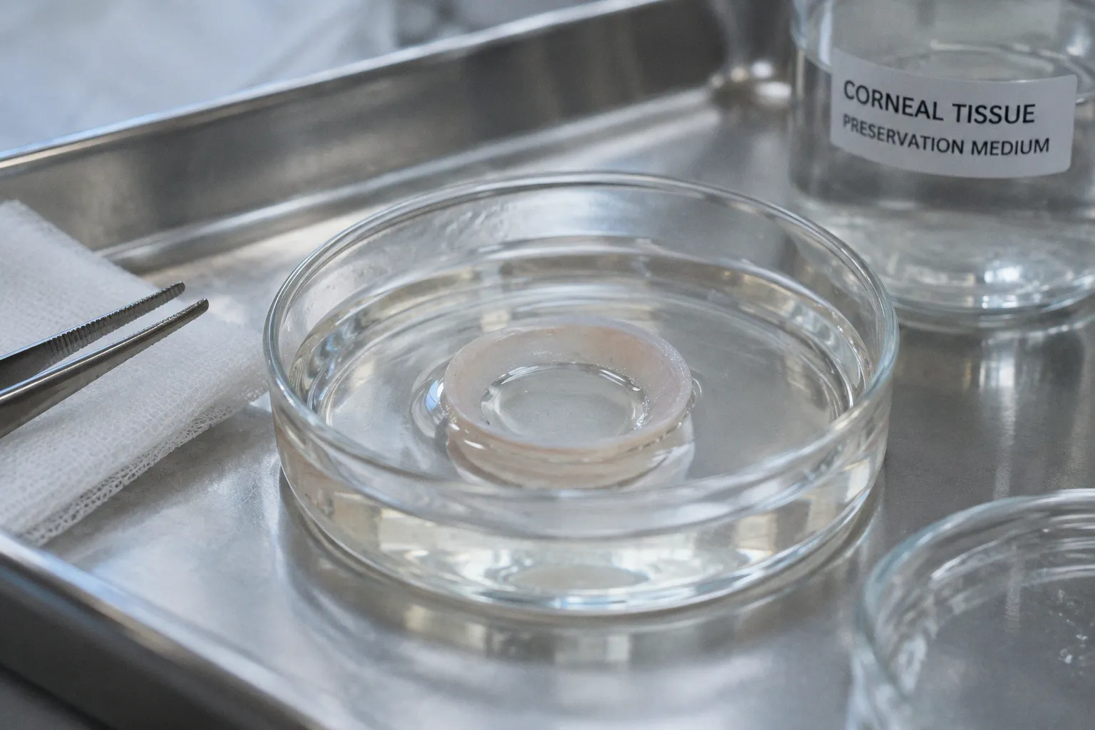
Donor age, death-to-dissection interval, storage medium, anatomical clarity, and the precision of the angle dissection can each shift the outcome before a single cell reaches culture.
That is the first uncomfortable fact of human trabecular meshwork research: the decisive variables are often treated as administrative details. A rim arrives late, the donor history is incomplete, or the dissection crosses into adjacent tissue, and the resulting culture is then interpreted as if it were a clean model of native human TM. It is not. At best, it is a selected and technically filtered population whose identity still has to be demonstrated.
For researchers working on aqueous outflow, glaucoma biology, anterior chamber drainage histology, or corneal transplantation workflows, trabecular meshwork cell isolation from donor eyes is therefore less a matter of finding one perfect protocol than of controlling avoidable sources of variability.
Donor selection begins before the tissue reaches the bench
The most productive source of primary human trabecular meshwork cells is often not an intact research globe collected specifically for the purpose. It is the discarded corneal rim remaining after transplant surgery. That material is valuable precisely because it has already passed through a clinical workflow, but that same origin creates constraints: the rim may have been exposed to preservation conditions, handling delays, or mechanical distortion during trephination.
The practical question is not whether a donor rim is usable in principle. It is whether the available metadata support a defensible interpretation of the resulting culture.
At minimum, the tissue record should allow the laboratory to reconstruct:
- donor age and relevant ocular history;
- time of death and time of tissue recovery;
- time of dissection or receipt by the laboratory;
- preservation medium and storage duration;
- whether the rim was generated during penetrating or lamellar corneal surgery;
- whether the tissue shows tearing, desiccation, heavy manipulation, or visible contamination.
These variables do not carry equal weight. Age is particularly important because the anterior segment does not remain anatomically static across the lifespan. Older donors may offer clearer tissue planes for dissection, but their cells may also be more vulnerable to post-mortem delay and culture-associated selection. Very young donors create a different problem: donors younger than five years may have softer tissue and less distinct anatomical margins, which makes the angle harder to isolate cleanly and increases the likelihood of collecting neighboring cell populations.
This is not a minor technical inconvenience. A contaminated primary culture can still attach, proliferate, and acquire a convincing cobblestone appearance. Morphology alone will not rescue a poorly defined dissection.
The post-mortem window is a biological variable, not a courier metric
Consensus guidance reported by Keller and colleagues in 2018 recommends obtaining donor tissue within 24 hours post-mortem for older donors, particularly when the aim is to preserve a high likelihood of useful TM cell recovery. That window applies most directly to unpreserved tissue. Corneal rims stored in Optisol may remain suitable for viable recovery for up to seven days, but “up to seven days” is not equivalent to “seven days without consequence.”
The longer the tissue remains outside the physiological environment, the more the laboratory inherits a problem of attrition. Some cells will be lost. Others will survive but behave differently after attachment. The remaining population may be biased toward cells with greater resistance to ischemia, handling, or enzymatic stress. Yet these changes are often invisible in the sample log, where the tissue is simply marked as received and processed.
A defensible workflow should treat the post-mortem interval as a continuous variable rather than a pass-or-fail label. Record the actual interval, not merely whether it falls inside a nominal limit. If several donor rims are being compared, do not quietly pool samples with radically different recovery histories and then describe the resulting culture as a single biological condition.
The donor rim is not a neutral container of cells. It is already an experimental history.
The storage medium matters as well. Optisol can extend the period during which viable recovery remains possible, but preservation does not freeze the cellular state. It changes the starting point of the experiment. A seven-day-stored rim and a freshly processed rim may both yield attached cells, while producing different outgrowth kinetics, stress responses, and proportions of surviving subpopulations.
That distinction becomes especially important in studies of glaucoma-associated mechanisms, extracellular matrix remodeling, mechanotransduction, or glucocorticoid responses. If the study endpoint is sensitive to cellular stress, the storage interval belongs in the biological model, not in a footnote.
Anatomical dissection is where most contamination is decided
Trabecular meshwork tissue occupies a narrow and technically unforgiving region of the anterior segment. The goal is to remove the TM while minimizing carryover from the corneal endothelium, scleral tissue, ciliary body, and other neighboring structures. The phrase “micro-dissection” can make this sound tidy. It is not tidy. It is a judgment exercise performed on fragile tissue under a microscope, often after preservation has softened or distorted the landmarks.
The central risk is not failure to remove enough tissue. It is removing too much of the wrong tissue.
A dissection that remains too superficial may yield little material. A dissection that becomes too deep or too broad may generate a culture that looks healthy but contains cells from adjacent anterior segment compartments. The resulting problem is not always obvious in routine culture. Endothelial and scleral contaminants may attach under similar conditions, while later assays detect signals that are mistakenly attributed to hTM biology.
A practical dissection logic
The laboratory should establish a consistent anatomical sequence and document deviations from it. The exact instruments and handling arrangement will differ between facilities, but the logic should remain stable:
1. Inspect the rim before manipulation.
Identify tears, irregular edges, drying, compression, and regions where the angle anatomy may have been disrupted during surgery. Do not assume that the entire circumference is equally useful.
2. Orient the tissue anatomically.
The limbal region and anterior chamber angle must be recognized before tissue removal begins. Orientation errors at this stage can turn a technically clean dissection into an anatomically meaningless one.
3. Separate the intended angle region from adjacent structures.
Work conservatively. The objective is not to maximize the amount of excised tissue but to enrich for the TM while reducing the chance of corneal endothelial or scleral carryover.
4. Divide the sample only when division improves control.
Smaller fragments can make explant placement and observation easier, but excessive fragmentation also increases handling, surface injury, and the number of opportunities for cross-contamination.
5. Track the origin of each fragment.
If one region is visibly damaged or difficult to orient, record it. Pooling everything immediately removes the possibility of explaining later differences in attachment or morphology.
This approach may sound excessively cautious for a small tissue fragment. That is precisely why it is useful. Small samples offer little room for analytical forgiveness.
Why age changes the dissection problem
In donors younger than five years, the tissue can be softer and the anatomical borders less distinct. The standard adult mental map of the anterior chamber angle becomes less reliable. A researcher who relies on visual similarity rather than anatomical orientation may collect a broader mixture of neighboring tissues.
In older donors, the concern shifts. The tissue may be easier to define mechanically, but the cells have experienced longer biological aging and may be more vulnerable to post-mortem loss or selective survival. These are not interchangeable disadvantages. The young donor presents a problem of anatomical specificity; the older donor may present a problem of recovery and cellular state.
That contrast should influence experimental design. If a project compares donors across age groups, it cannot treat “donor age” as the only relevant variable. Dissection confidence, tissue softness, storage interval, and culture behavior may all move with it.
Explant culture and enzymatic dissociation answer different questions
There is a persistent temptation to ask which method is best: enzymatic digestion or explant outgrowth. That question is too blunt. The methods impose different selection pressures, and the better choice depends on whether the experiment prioritizes speed, anatomical traceability, cell yield, or preservation of a closer relationship to the original tissue fragment.
Explant processing
Explant culture keeps the tissue fragment physically present while cells migrate out and expand. The method is comparatively conservative in the sense that it avoids immediate dissociation of every cell-cell and cell-matrix association. It can also make the origin of the culture easier to document, particularly when the fragment is placed and monitored individually.
Its disadvantages are equally clear. Outgrowth may be slow or uneven. A fragment that contains non-TM tissue can seed the culture with exactly the contaminants the dissection was meant to exclude. The apparent simplicity of placing a fragment in culture therefore does not remove the need for anatomical control; it merely moves the selection event from the enzyme step to the migration step.
Enzymatic processing
Enzymatic dissociation can release cells more rapidly and may produce a more immediately dispersed starting population. It is attractive when the downstream experiment requires a relatively uniform seeding distribution or when explant outgrowth is inconsistent.
But enzymatic processing is not a neutral extraction. Digestion imposes stress, and the outcome depends on tissue condition, dissection precision, and how aggressively the tissue is handled. If the starting specimen includes adjacent corneal endothelial or scleral material, the enzyme will not politely isolate only the desired population. It may release contaminants more efficiently.
The appropriate comparison is therefore not “fast versus slow.” It is “which source of selection is more visible and controllable in this experiment?”
| Feature | Explant outgrowth | Enzymatic dissociation |
|---|---|---|
| Relationship to original tissue | Fragment remains traceable during early culture | Tissue architecture is disrupted immediately |
| Initial cell distribution | Uneven and dependent on migration from the explant | More dispersed at seeding |
| Main contamination risk | Outgrowth from incorrectly dissected neighboring tissue | Co-release of cells from adjacent structures |
| Main technical advantage | Conservative handling and clearer fragment provenance | Faster access to dispersed cells |
| Main interpretive concern | Slow or selective outgrowth | Enzyme- and handling-associated stress |
| Best use case | Studies where tissue origin and early outgrowth matter | Studies requiring distributed primary cells for downstream assays |
Neither approach creates a native trabecular meshwork model by itself. Both create a culture that must be validated.
Culture passage is a biological decision, not merely a scheduling detail
Primary hTM cultures are commonly used within passages 2 to 6 for in vitro studies. That range is practical, but it should not be misunderstood as a guarantee of preserved identity. Passage number is a crude proxy for the amount of selection, adaptation, and phenotypic drift that has occurred. Two cultures at the same passage can differ substantially if their donor ages, post-mortem intervals, or starting tissue compositions differ.
Early passage is often preferred because it limits the time available for culture adaptation. Yet early does not automatically mean authentic. A contaminating population can remain present at passage 2. A stressed TM population can already have altered expression before the first passage. The relevant question is not simply how many times the cells have been subcultured, but what the culture has become by that point.
Researchers should therefore record more than passage number:
- time from tissue receipt to initial attachment;
- whether the culture originated from explant or dissociated tissue;
- the proportion and distribution of attached cells;
- morphology during early outgrowth;
- evidence of delayed growth, overgrowth, or mixed colony appearance;
- donor and preservation metadata linked to the culture line.
This is where many protocols develop a paradigm deficit. They standardize the medium and passage schedule while leaving the donor tissue history underdescribed. The resulting cultures appear technically comparable because the culture conditions are comparable. Biologically, they may not be.
Validating hTM identity requires more than a familiar shape
The classic cobblestone morphology is useful. It is also insufficient.
Morphology can support an hTM identity assessment, particularly when it is observed consistently across the culture and accompanied by appropriate growth behavior. But neighboring anterior segment cells may produce overlapping visual patterns, especially after adaptation to plastic. A culture should therefore be treated as provisionally identified until functional or molecular features support that interpretation.
One established validation approach is to examine dexamethasone-induced upregulation of myocilin expression. The response provides a functional marker associated with TM cell identity and glucocorticoid responsiveness. It is more informative than appearance alone because it tests whether the cells behave in a manner expected of hTM under a defined stimulus.
Even here, the conclusion should remain proportionate. A positive myocilin response supports the presence of TM-like cells; it does not certify that the entire culture faithfully reproduces native human trabecular meshwork. Nor does a single marker settle questions about donor-related variation, contaminating subpopulations, or culture-induced changes.
A serious validation package should combine:
1. Morphological assessment across early culture, not a single representative image.
2. A marker or response associated with TM identity, including dexamethasone-induced myocilin upregulation where appropriate.
3. Negative evidence against obvious neighboring-cell contamination, selected according to the tissues most likely to have entered the preparation.
4. Documentation of culture history, including passage, donor interval, preservation, and processing route.
5. Replication across donors, rather than treating multiple wells from one donor as independent biological replicates.
The last point is routinely weakened by convenience. Six wells from one rim are not six donors. They may be six technical replicates of one donor-specific and one dissection-specific event. That distinction matters when interpreting endothelial stress, extracellular matrix responses, or pharmacological effects.
A cobblestone monolayer is an observation. It is not an identity certificate.
The culture can become less representative as it becomes easier to work with
Long-term primary hTM culture creates a particularly important interpretive problem. Single-cell transcriptomic analysis has shown divergence in cell composition and reduced heterogeneity in long-term cultured primary hTM cells compared with native in vivo tissue.
That finding should alter how experiments are framed. The culture may become more uniform, more expandable, and easier to assay while simultaneously losing features that made native tissue biologically informative. This is the paradox at the center of many in vitro models: technical convenience improves as physiological complexity declines.
The loss is not necessarily catastrophic. A reduced-complexity culture can be useful for mechanistic experiments, controlled perturbations, and initial screening. The error lies in treating it as an uncomplicated substitute for the tissue from which it originated.
The longer the cells remain in vitro, the more carefully researchers should distinguish between:
- donor-derived biology;
- selection during isolation;
- selection during early attachment and outgrowth;
- adaptation to a two-dimensional substrate;
- changes associated with repeated passaging;
- loss of rare or fragile subpopulations.
This is particularly relevant when studying heterogeneity within the TM. If a long-term culture shows a strong, reproducible response, that response may reflect the biology of the surviving and expanding population rather than the full range of native TM states. A clear result can still be a narrowed result.
Designing around transcriptomic divergence
There is no single corrective maneuver that restores native heterogeneity after prolonged culture. The sensible response is to design experiments that acknowledge the limitation.
Use lower passage cultures when the scientific question depends on cellular diversity. Compare findings across more than one donor. Avoid presenting a single expanded line as representative of the human TM as a whole. If the study concerns cell-state transitions, perform the comparison explicitly rather than assuming that the culture remains stable.
For projects involving Fuchs dystrophy donor corneas, corneal biomechanics, endothelial biology, or anterior chamber drainage pathology, the risk of conceptual spillover is even greater. A culture derived from a corneal rim may be highly useful for one question and poorly suited to another. Tissue availability does not determine tissue relevance.
A workflow that preserves interpretive value
A reliable trabecular meshwork tissue processing workflow should be designed backward from the final claim. If the experiment aims to describe hTM-specific glucocorticoid response, the culture must be identity-validated. If it aims to compare donors, post-mortem and storage histories must be balanced or modeled. If it aims to investigate cell heterogeneity, long-term expansion should not be treated as an invisible convenience.
A practical sequence is:
1. Screen the donor metadata before accepting the tissue.
Establish whether the post-mortem and storage history is known well enough for the planned experiment.
2. Separate usable from compromised rim regions.
Record visible damage and avoid treating the rim as biologically uniform.
3. Perform anatomically oriented micro-dissection.
Prioritize specificity over gross tissue quantity, particularly near the corneal endothelium and scleral boundary.
4. Choose explant or enzymatic processing according to the endpoint.
Do not select the method solely because it is familiar or faster.
5. Maintain fragment-level or donor-level traceability.
Pooling may simplify handling but can erase the information needed to interpret a mixed result.
6. Monitor early attachment and morphology.
Mixed morphology, unexpected overgrowth, or unusually delayed outgrowth should trigger review rather than automatic expansion.
7. Validate hTM identity before committing to downstream assays.
Cobblestone morphology should be paired with a biological response such as dexamethasone-induced myocilin upregulation.
8. Keep passage and donor effects visible in the analysis.
Passage 2 through 6 may be a practical working range, not a declaration that phenotype is preserved throughout.
9. Report what the culture cannot represent.
Long-term cultures with reduced heterogeneity should not be presented as transcriptomically equivalent to native tissue.
The details of medium, substrate, enzyme selection, and incubation conditions still matter, but they cannot compensate for an ambiguous donor source or a contaminated dissection. Better culture conditions do not repair a bad anatomical decision. They may simply produce a healthier version of the wrong cells.
The real bottleneck is not yield; it is confidence
The exact baseline yield from a donor corneal rim is difficult to standardize because donor-to-donor variability remains high. That uncertainty is not a failure of laboratory discipline. It is a property of the material. Tissue age, storage, surgical handling, anatomical integrity, and cell survival all contribute to an outcome that cannot be reduced to a fixed number per rim.
This is why “maximizing yield” should not be interpreted as collecting the largest possible cell population. A larger population with uncertain identity is not an improved preparation. It is a more persuasive confounder.
The better target is usable yield: enough viable cells to support the planned experiment while preserving confidence that the population came from the intended structure and retained the relevant biology. Sometimes that means accepting fewer cells, discarding a questionable fragment, or repeating a donor rather than expanding an ambiguous culture indefinitely.
The established paradigm prefers clean-looking cultures, standardized passage numbers, and convenient tissue access. The biological evidence is less accommodating. Donor history changes the starting population. Dissection changes its composition. Culture changes its heterogeneity. Validation changes the strength of the claim.
Trabecular meshwork cell isolation from donor eyes is therefore not solved by a more aggressive enzyme step or a more elaborate growth protocol. It is solved by refusing to treat the donor rim as interchangeable material. The field does not need another culture that merely expands. It needs cultures whose provenance, identity, and limitations remain visible long after the cells have attached.