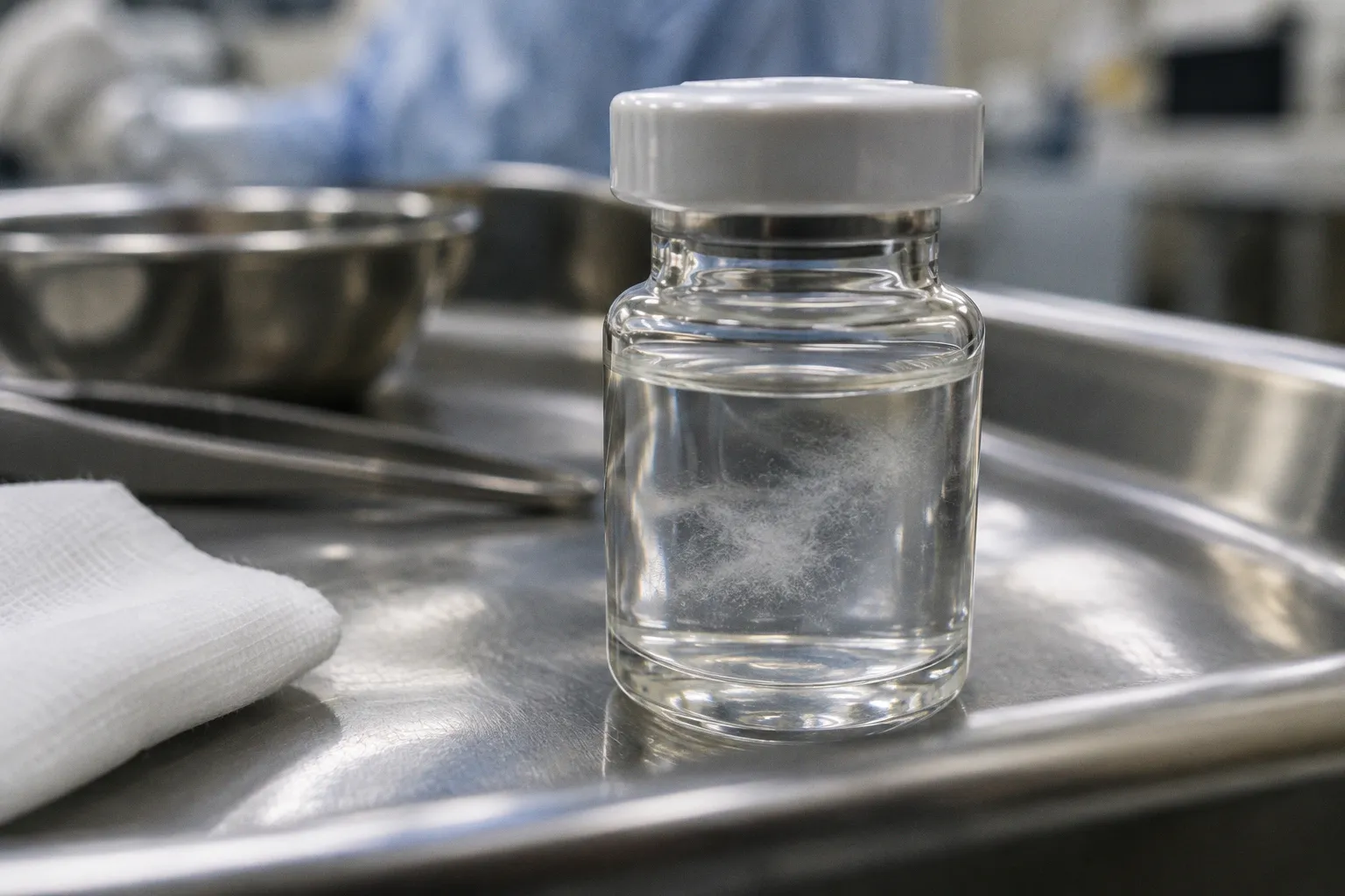
For ocular regenerative programs built on donor-derived retinal pigment epithelium, that changes the procurement conversation. A culture can look clinically usable at the bench while carrying biological contamination capable of altering cell behavior, undermining assay results, or disqualifying the material during quality control.
A pristine monolayer under the scope is necessary but nowhere near sufficient. For donor RPE, contamination can live below the resolution of ordinary visual inspection.
This is not a theoretical concern. Donor RPE cultures sit at the hinge between eye-bank logistics and the operating room. They may become the starting material for subretinal cell suspensions, scaffold-based RPE patches, or research assays used to decide whether a candidate therapy should advance. When those cultures contain hidden organisms or unwanted cell types carried over from the donor eyecup, every downstream step has to be interpreted with caution.
The important distinction is between a risk that has been detected and a risk that has been missed. Contamination does not automatically translate into a clinical event, and a contaminated culture is not evidence of a particular postoperative outcome. It does mean that the material may no longer behave as expected and must be identified, contained, and excluded through validated pre-use testing before it can be considered for further development or administration.
The Invisible Threat: Mycoplasma and Sub-Microscopic Contamination
Mycoplasma is the contaminant that creates the most persistent problems for translational cell biology because it often gives the laboratory no obvious visual warning. These organisms are smaller than the structures routinely resolved by an inverted microscope. They can remain unnoticed while the culture continues to grow, and they do not necessarily produce the dramatic turbidity or rapid collapse associated with more familiar bacterial contamination.
Their lack of a conventional cell wall is central to the problem. Many routine antibiotics used in cell culture are designed to act on bacterial cell-wall synthesis. Mycoplasma does not offer that target. A medium containing Penicillin-Streptomycin may therefore suppress some incidental bacterial growth while leaving Mycoplasma unaffected.
The prevalence figures commonly cited for cell-culture contamination are a warning rather than a guarantee about any individual laboratory. They indicate that the baseline risk is substantial enough to require a dedicated detection strategy. Donor-derived RPE is particularly sensitive to that requirement because each primary preparation is linked to a specific donor and a finite procurement event. If a preparation fails screening, the loss is not limited to a single flask. It can affect the associated tissue record, the planned experiment, the production schedule, and the interpretation of data already generated from the culture.
Contamination changes the biology, not just the appearance
The central problem is not simply that Mycoplasma is present. Infection can alter the biology of the cells carrying it. In RPE cultures, contamination has been associated with changes in metabolism, cytokine secretion, growth behavior, and antigen-presentation-related processes. Those changes matter because RPE is not being evaluated only as a visible layer of cells. Its value depends on a set of functions: maintaining epithelial organization, supporting the outer retina, handling phagocytic and metabolic tasks, and interacting with the surrounding immune and extracellular environment.
A culture affected by Mycoplasma may therefore produce data that are difficult to interpret. A potency assay can be weakened by altered cell physiology. A differentiation or maturation readout can shift. A transcriptomic profile can reflect the presence of an unrecognized stressor rather than the intended experimental variable. Even when the cells continue to form a visually convincing monolayer, their behavior may no longer represent a clean donor RPE preparation.
This is why human RPE models such as ARPE-19 are commonly screened with molecular methods rather than cleared by microscopy alone. The model may be familiar and well characterized, but familiarity does not make it immune to contamination. The same principle applies to primary donor-derived cultures, where biological variability already makes interpretation more demanding.
PCR is not a substitute for a complete quality system, but it provides a level of sensitivity and specificity that visual inspection cannot. A positive result should trigger investigation, segregation of the affected material, review of related cultures, and a documented decision about whether the preparation can be used for research. For material intended to move toward a therapeutic application, release criteria must be defined in advance rather than improvised after a suspicious result appears.
Cellular Purity Challenges: Fibroblasts and Choroidal Melanocytes
Mycoplasma is only one part of the contamination problem in donor RPE cultures. The other part is cellular: unwanted human cells carried through the isolation process and allowed to expand alongside the RPE.
The donor eyecup is not a clean, isolated sheet of pigment epithelium. The choroid beneath it contains melanocytes and fibroblasts. The tissue above it may contribute neural retinal material and glial cells. During dissection, small amounts of neighboring tissue can remain attached even when the work is performed under magnification by an experienced technician. The challenge is not to achieve a visually perfect separation at every moment. It is to prevent residual cells from gaining a proliferative advantage once the culture is established.
Fibroblasts are especially troublesome because they can expand efficiently under conditions that are intended to support RPE. Their morphology may become obvious only after the population has begun to change. By then, the culture can contain a mixture rather than a stable RPE population. Fibroblast overgrowth also complicates interpretation of marker expression, barrier-function assays, extracellular-matrix studies, and any experiment that assumes the cells are predominantly RPE.
Choroidal melanocytes create a different kind of problem. Their pigment can make cultures look biologically active while masking the fact that more than one cell population is present. They also introduce cellular markers and antigenic characteristics that are not part of the intended RPE preparation. In a research setting, that can confound the result. In a translational setting, it creates an additional identity and purity question that cannot be resolved by appearance alone.
Isolation method influences the starting population
The choice between explant outgrowth and enzymatic dissociation is therefore not a minor technical preference. It changes the way the starting population is assembled.
With the explant approach, small pieces of RPE-containing tissue are placed directly into culture and allowed to grow out. The method can be practical and familiar, but it gives neighboring cells an opportunity to migrate from the tissue fragment and establish themselves. The resulting culture may be useful for some exploratory work, yet its cellular composition requires careful characterization.
Enzymatic dissociation separates cells before plating and can reduce the amount of attached tissue carried into the culture. A protocol using 0.25% trypsin, with controlled exposure and immediate neutralization, is one example of how laboratories attempt to improve the starting purity profile. The exact protocol still needs to be validated for the tissue source, donor material, operator, and intended use. Enzymatic treatment is not automatically benign: excessive exposure can reduce RPE viability or damage surface proteins, while insufficient dissociation can leave aggregates and unwanted cells together.
The relevant question is not whether one technique is universally correct. It is whether the chosen method produces a population whose identity and purity can be demonstrated at the next stage.
| Isolation or control step | What it can help establish | What it cannot establish on its own |
|---|---|---|
| Stereo dissection under magnification | Gross tissue identity and removal of visible neural-retinal material | Absence of microscopic organisms or all neighboring cells |
| Explant outgrowth | Recovery of cells from tissue fragments with limited manipulation | A defined cellular composition or reliable exclusion of fibroblasts |
| Enzymatic dissociation | More controlled release of cells and reduced tissue carryover | Absence of microbial contamination or complete cellular purity |
| Morphology during culture | Presence of an epithelial-like population and major changes in appearance | RPE identity, pathogen status, or therapeutic suitability |
| Marker and functional testing | Evidence supporting cell identity and activity | A substitute for microbial testing and batch-release criteria |
Enzymatic dissociation is not a refinement by itself. It is useful only when the resulting population is then characterized, screened, and accepted against defined criteria.
Purity should consequently be treated as a layered question. It includes the proportion of cells with the intended RPE identity, the absence or control of unwanted cell types, the consistency of the culture over time, and the absence of microbial agents that could alter the preparation. A culture can pass one of these tests and fail another.
Pathogenic Risks: Chlamydia trachomatis and Signaling Alterations
A third category of risk receives less attention in routine procurement discussions: intracellular bacterial contamination. Chlamydia trachomatis has been documented in retinal pigment epithelial cultures derived from human donor eyes, and its significance is not limited to the organism's physical presence.
Because C. trachomatis occupies an intracellular niche, its detection requires a method appropriate to that biology. A culture may not show the visual signs associated with an acute bacterial overgrowth. Testing the culture supernatant alone may also be insufficient if the organism is present within a subset of cells. The screening strategy therefore has to match the suspected contaminant rather than rely on a single generic sterility result.
The other concern is altered cell signaling. Infected RPE cells can show changes in pathways connected with proliferation, inflammatory mediators, and extracellular-matrix production. These changes are important even when the culture remains attached and apparently viable. A signaling profile shaped by infection is not equivalent to the profile of an uncontaminated RPE preparation.
For a laboratory studying barrier function or inflammatory response, the consequences are immediate: the contaminant becomes an unplanned experimental variable. For a translational program, the issue is broader. Any change in cell behavior can affect identity testing, potency interpretation, comparability between lots, and the decision about whether a preparation is suitable for further development.
It is tempting to describe such contamination as a problem that will simply travel forward with the cells. That is too categorical. The evidence supports infection and signaling changes in donor-derived RPE cultures; it does not, by itself, establish that the organism will remain present through every expansion step or be carried into a final therapeutic product. The correct operational conclusion is more demanding and more cautious: the possibility must be addressed before use through validated, appropriately timed molecular testing and documented material controls.
Donor screening is one barrier, not the whole barrier
Donor eyes may be screened for selected pathogens at intake, but the exact panel, specimen type, timing, and analytical sensitivity determine what that screening can actually exclude. A negative donor result does not automatically prove that every derived culture is free of contamination. Conversely, a culture-level finding should not be dismissed merely because the donor passed an upstream screen.
The workflow needs checkpoints at both levels:
- At eye-bank intake: document donor eligibility, available infectious-disease information, tissue traceability, and the tests that were actually performed.
- During isolation and culture: maintain segregation, aseptic technique, and records that connect every vessel to the donor and processing event.
- Before experimental interpretation: confirm that microbial status is compatible with the planned assay.
- Before any translational use: apply pre-use or release testing that has been validated for the organisms and matrices relevant to the product.
This layered approach also makes failures easier to investigate. If a result appears during culture, the team can ask whether the signal is linked to the donor material, the processing environment, a reagent, cross-contamination from another culture, or a later handling event. Without that traceability, a positive result becomes a general warning rather than actionable information.
Beyond Microscopy: Molecular Screening and Enzymatic Protocols
The operational picture for a donor RPE program is therefore built from complementary controls rather than one decisive technique. Careful dissection limits gross tissue carryover. Enzymatic dissociation can improve control over the starting population. Morphological review provides an early warning for obvious changes. Identity and purity assays characterize the cells. Molecular screening addresses organisms that microscopy and conventional sterility methods may miss.
The order matters, but so does the timing. Testing only at the beginning can miss contamination introduced during processing or culture. Testing only at the end can allow a compromised preparation to consume time, reagents, and analytical capacity. A risk-based program defines points at which material is held, sampled, or rejected before it becomes difficult to isolate the source of the problem.
| Step in the workflow | What it controls | What it does not control |
|---|---|---|
| Stereo dissection under magnification | Gross tissue identity and removal of visible neural-retinal material | Sub-microscopic organisms or complete exclusion of neighboring cells |
| Enzymatic dissociation, such as controlled 0.25% trypsin treatment | Tissue carryover and part of the cellular admixture risk | Mycoplasma, intracellular bacteria, viruses, or every unwanted cell type |
| Routine inverted microscopy | Morphology, confluence, debris, and obvious microbial changes | Low-level contamination, organisms below visual resolution, or reliable cell identity |
| RPE identity and purity assays | Evidence that the population is predominantly the intended cell type | Microbial status unless the assay is specifically designed to detect it |
| PCR-based Mycoplasma screening | Detection of Mycoplasma genetic material within the validated test scope | Every other bacterial, fungal, or viral contaminant |
| Broader molecular pathogen testing | Selected organisms that routine sterility methods may miss | Organisms outside the panel, sampling errors, and poorly validated matrices |
| Standard sterility testing | Many cultivable bacteria and fungi under the test conditions | Mycoplasma, intracellular pathogens, slow-growing organisms, and contaminants not recovered by the method |
The table is deliberately less comforting than a single “sterile” label. Each test answers a different question. A negative standard sterility result does not erase a positive Mycoplasma result. A clean PCR result for Mycoplasma does not establish the absence of C. trachomatis. A convincing RPE morphology does not prove that fibroblasts or melanocytes are absent.
What validated testing needs to address
For donor-derived material, a meaningful testing strategy should define at least:
1. The target of the assay. The laboratory should know whether it is screening for Mycoplasma, selected bacterial pathogens, fungi, viruses, or a broader panel.
2. The sample being tested. Cells, supernatant, tissue fragments, and process samples do not necessarily provide the same detection opportunity.
3. The timing of collection. A result at intake, during expansion, and before use answers different questions.
4. The assay's analytical limits. A method should be evaluated for the relevant matrix, controls, inhibitors, and risk of false-negative results.
5. The action linked to a result. A positive or inconclusive finding requires predefined rules for quarantine, repeat testing, investigation, and disposition.
6. The relationship to cell identity and function. Microbial clearance cannot be considered separately from evidence that the remaining culture is still the intended RPE population.
This is also where documentation becomes part of the science. If a contaminated vessel is removed but its data are quietly folded into the same analysis, the problem has not been contained. Affected passages, assays, images, and derived conclusions need to be traceable. In a research database, that record may be as important as the final result because it prevents later users from treating compromised material as a clean reference.
The aim is not to make contamination impossible. No tissue procurement system can promise that. The aim is to ensure that a contaminated or questionable preparation is identified before it is used to support a decision it cannot reliably support.
Limitations of Standard Antibiotics in Ocular Tissue Procurement
One operational assumption deserves particular attention because it is often inherited from general cell biology protocols: that adding antibiotics to the medium is equivalent to controlling contamination.
Routine Penicillin-Streptomycin is not designed to eliminate Mycoplasma. Its presence may reduce the growth of susceptible bacteria and make a culture appear stable, but it does not address organisms that lack the relevant cell-wall target. Stability under antibiotic-containing conditions is therefore not evidence of microbial clearance.
Antifungal agents have a similarly limited scope. Amphotericin B and related compounds may suppress many yeasts and molds, but they do not provide a universal barrier against intracellular bacteria, viruses, or Mycoplasma. They can also complicate interpretation if a laboratory begins to rely on the absence of visible fungal growth as proof that the culture is clean.
There is a further reason to avoid treating routine antimicrobials as a safety system: they can change the ecology of a culture without revealing what remains. Suppressing one group of organisms may reduce visible signs while leaving another contaminant unaffected. In a donor RPE workflow, that creates false reassurance rather than reliable control.
The more defensible approach combines:
- qualified donor intake and tissue traceability;
- controlled dissection and isolation;
- validated enzymatic processing where appropriate;
- disciplined aseptic handling;
- cell-identity and purity characterization;
- molecular screening for Mycoplasma and other relevant pathogens;
- standard sterility testing within its actual limitations;
- and clear quarantine and release decisions before material is used.
None of these controls replaces the others. They address different failure modes. Antibiotics may have a place in a validated culture protocol, but they should not be presented as evidence that a donor-derived RPE preparation is free of hidden contamination.
Routine antibiotics protect against some of the contamination they were designed to suppress. They do not answer the questions that molecular screening is meant to answer.
Closing
The promise of donor-derived RPE transplantation is real, but it depends on more than obtaining viable pigmented cells from a donor eye. The preparation has to be identifiable, interpretable, and screened against the contaminants that ordinary microscopy and routine antibiotics cannot reliably exclude.
That standard is demanding because the material is biologically complex. A donor RPE culture may fail through microbial contamination, through the expansion of fibroblasts or choroidal melanocytes, or through a combination of altered identity and altered signaling. These are not interchangeable problems, and they cannot be solved with one universal test.
The practical lesson is equally specific. Use the procurement record to establish traceability. Use the isolation protocol to limit unwanted cellular carryover. Use morphology as an early observation, not a release decision. Use identity and functional assays to determine what the cells are doing. Use molecular screening and validated sterility methods to address organisms that visual inspection cannot see. Most importantly, place those controls before the point at which the material is used for a consequential experiment or considered for administration.
Contamination may be detected during culture, during quality-control testing, or through an investigation of an unexpected assay result. A responsible program is designed so that the first two possibilities occur before clinical use. The relevant measure of maturity is not the claim that contamination never occurs. It is whether the team can detect a compromised preparation early, segregate it, document the finding, and prevent uncertain material from moving forward.