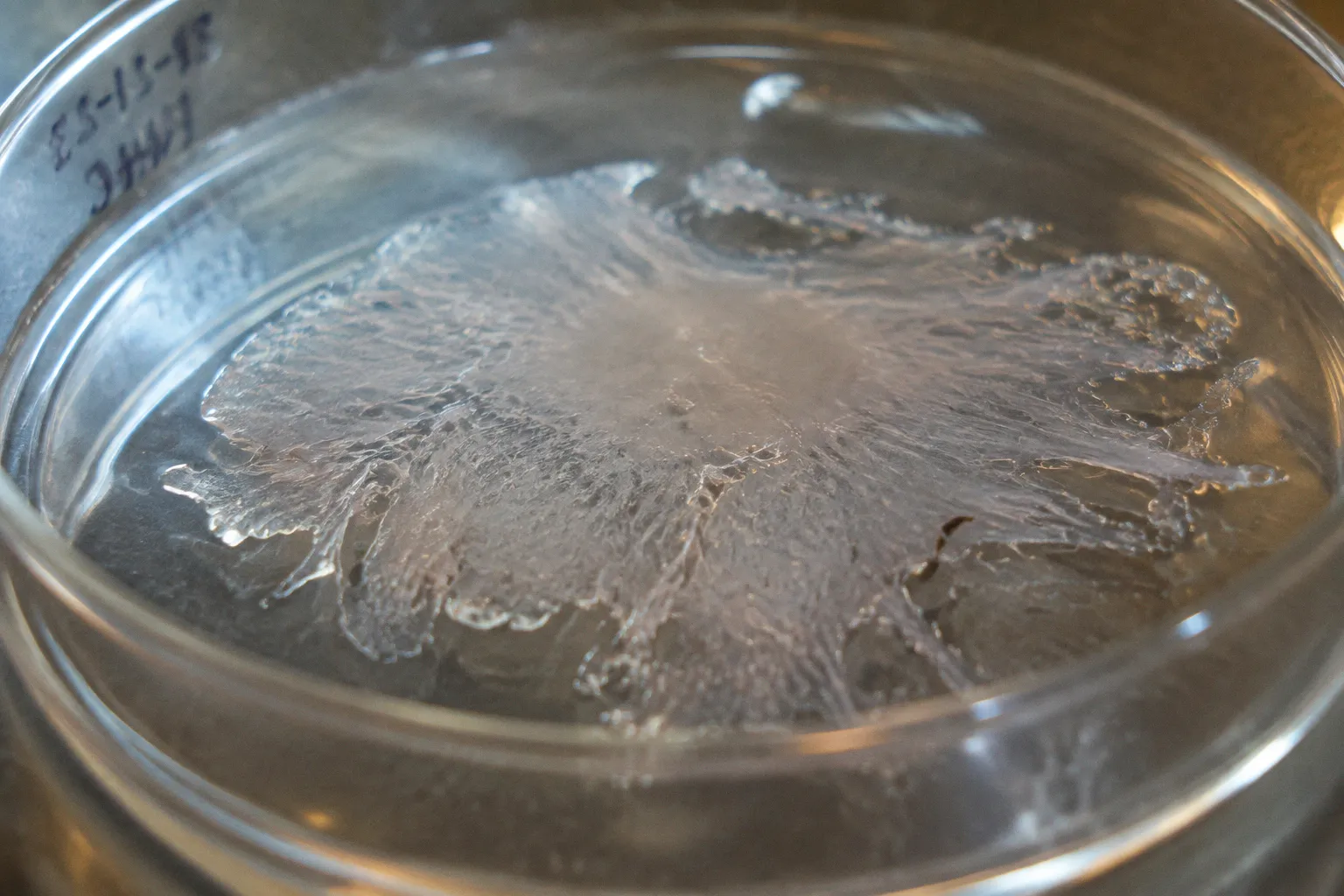
Run the incubation twenty minutes beyond the validated window and the clean, hexagonal sheet of retinal pigment epithelium (RPE) you expected to plate does not arrive. What arrives instead is a suspension of damaged cells whose tight junction belt has already been chemically pried apart, whose phenotype has begun drifting toward a mesenchymal state, and whose ZO-1 stain will come back patchy within the week. This is the silent failure mode of primary human RPE isolation, and it rarely announces itself until the first passage is plated and the morphology starts to slip.
For clinical translation programs that depend on donor tissue — RPE replacement strategies for geographic atrophy, subretinal transplantation scaffolds, in vitro disease modeling for age-related macular degeneration — the integrity of the starting material is not a downstream concern. It is the foundation. Enzymatic overdigestion is one of the most underreported variables in the procurement-to-culture pipeline, and the cost shows up months later as a failed differentiation, an unexplained epithelial-to-mesenchymal transition, or a transplantable sheet that never forms a polarized monolayer in the first place.
The Enzymatic Threshold: Balancing Protease Concentration and Tissue Integrity
The first decision a tissue procurement lab makes when processing a human eyecup for primary RPE isolation is which dissociation enzyme to reach for, and how long to leave it on. That decision — more than any subsequent culturing refinement — determines whether the cells arrive on the dish as an intact epithelial population or as a partially degraded suspension that has already begun to forget its identity.
The published literature describes a relatively narrow toolkit: papain, dispase, collagenase, hyaluronidase, and trypsin in carefully titrated combinations. Each carries a different risk profile, and the protocols that have stood up to peer review tend to converge on a tight concentration range and a strict incubation window. Twenty units per milliliter of papain, roughly two percent dispase, and a stock preparation of collagenase around nineteen and a half units per milliliter are common reference points. The digestion window for most of these enzymes sits between thirty and sixty minutes, and that window is not a guideline — it is the difference between a clean Bruch's membrane and a choroid that disintegrates into the supernatant.
Overdigestion is a chemical decision with a biological price tag: once the tight junction belt breaks, the epithelium does not reform the same way in culture.
The temptation, when working with scarce donor tissue, is to push for higher yield by extending incubation. In practice, the opposite usually happens. Cells exposed to active protease beyond the validated window begin to lose their membrane proteins first — the cadherins, the integrins, the channel proteins that hold an epithelial sheet together. Yield appears to improve because more cells detach, but the cells that detach are no longer the cells you wanted. They are partially digested fragments of an epithelium whose functional architecture has been chemically pried apart.
A practical way to think about the enzymatic threshold is in three zones. In the first zone — short exposure — the RPE sheet stays adherent and the enzyme has not done enough work. The yield will be low, but the cells that do release will retain their phenotype. In the second zone, the validated window, the sheet releases cleanly and the cells arrive intact, polarized, and ready to reform a monolayer. In the third zone, the over-digestion zone, everything that can come off does come off — including choroidal melanocytes, stromal fibroblasts, and damaged RPE fragments that contaminate the downstream culture. The third zone is where most published failures begin, even when they are reported as "low yield" or "poor attachment" rather than as overdigestion.
Molecular Consequences of Over-Exposure: From Tight Junction Degradation to EMT
What happens at the molecular level when an RPE cell spends too long in active protease is well documented in the donor tissue literature, but it is rarely described in the language clinicians and procurement coordinators use. The cleanest place to start is the tight junction belt — the ring of proteins, anchored by ZO-1, that seals adjacent RPE cells into a polarized monolayer and gives the tissue its barrier function.
Trypsin, collagenase, and hyaluronidase at unoptimized concentrations all attack this belt. Over-exposure damages the cell surface receptors that hold the cells to Bruch's membrane and to each other, disrupts ZO-1 localization, and induces one of two outcomes: cell death during the dissociation itself, or, more insidiously, loss of adhesion in the days after plating. Either way, the monolayer does not reform. What plates down is a sparse population of cells that have already lost the cues they need to recover an epithelial identity.
The second consequence, and the one that quietly undermines most primary RPE cultures past passage two, is epithelial-to-mesenchymal transition. EMT is the cellular equivalent of forgetting what you were. Pigmented, hexagonal epithelial cells transform into spindle-shaped, fibroblastic-looking cells that proliferate faster, attach more loosely, and express none of the markers that make an RPE cell an RPE cell. The high-threshold L-type calcium channels that characterize a functional RPE — the channels that underpin its role in outer segment phagocytosis and visual cycle support — are among the first features to disappear.
EMT in primary RPE is rarely caused by the cells themselves; it is almost always caused by what we did to them on the way into the dish.
The mechanism is not mysterious. Protease damage to surface receptors removes the integrin and cadherin signaling that maintains an epithelial program. Conventional subculturing on uncoated tissue culture plastic, with serum-containing medium and no maintenance of apical-basal polarity, removes the contextual cues that would otherwise hold the cells in an epithelial state. Together, the two insults converge on the same endpoint: a culture that looks RPE-derived under low magnification but has lost the molecular architecture that makes RPE RPE. For any lab modeling AMD, geographic atrophy, or photoreceptor support, this is an unusable result — and it often goes unrecognized for several passages.
Variable Donor Susceptibility: Why Post-Mortem Time Dictates Digestion Windows
One of the persistent frustrations in ocular biobanking is that the same dissociation protocol can produce excellent results on a fresh donor eye and a borderline failure on an eye that arrived twelve hours later. Donor age, post-mortem interval, cause of death, and the cold-chain history of the globe all modify how Bruch's membrane responds to enzymatic digestion, and none of those variables are visible to the technician holding the pipette.
The general pattern is that older donor tissue, and tissue with longer post-mortem intervals, becomes more susceptible to protease damage rather than less. Bruch's membrane thickens with age, accumulates lipid deposits in advanced AMD donor eyes, and changes its collagen crosslinking profile over decades. A young donor eye may tolerate a sixty-minute dispase incubation with the RPE sheet still adherent at the end; an eighty-year-old donor eye from a similar procurement may show choroid disintegration at forty minutes. There is no published universal enzyme digest duration that holds across all human donor eyes, and the literature is candid about that gap.
The practical implication for procurement coordinators is that donor metadata has to travel with the tissue. A standardized dissociation protocol applied uniformly across a donor cohort will systematically over-digest the older, longer-post-mortem specimens and systematically under-digest the younger, fresher ones. The result is a population of cultures with mixed phenotypes, mixed viabilities, and inconsistent EMT risk — and a downstream experimenter who has no way to trace the variance back to the procurement step.
This is where the biobanking infrastructure matters as much as the bench protocol. Cold-chain documentation, post-mortem interval recording, donor age annotation, and cause-of-death metadata are not administrative overhead. They are the variables that allow a lab to titrate its digestion window per specimen. A lab that receives tissue without these metadata is essentially running blind, applying a one-size-fits-all enzymatic exposure to a biologically heterogeneous input. The failures that result are real, but they are almost impossible to diagnose after the fact.
Optimizing Dissociation Protocols: Moving Beyond Standardized Incubation Times
The path forward, increasingly, is not to optimize a single dissociation protocol but to build a responsive one. Published protocols from groups with strong translational track records — including work reported in Frontiers in Cellular Neuroscience and recent tissue procurement literature — tend to converge on a small set of practical adjustments that reduce over-digestion risk without sacrificing yield.
The first is shorter total incubation, achieved by combining enzymes rather than extending any single one. A brief dispase step to loosen the RPE sheet from Bruch's membrane, followed by a short, gentle collagenase or papain step to release the cells, typically outperforms a long single-enzyme incubation. Each enzyme does less work, and the cumulative chemical insult to surface proteins is lower. Reported working windows often sit closer to thirty minutes than sixty.
The second adjustment is temperature. Most dissociation enzymes have sharply reduced activity at room temperature compared with thirty-seven degrees, and several groups now run the initial incubation at reduced temperature to slow the digestion and extend the operator's margin of error. The RPE sheet still releases, but it releases over a longer effective window, giving the technician time to monitor the digestion and stop it at the right point.
The third adjustment is mechanical assistance, used sparingly. A gentle stream of medium over the RPE surface, or a soft brush, can release a sheet that has been partially enzymatically loosened without extending the chemical exposure. The goal is to let the enzyme start the process and let the pipette finish it, rather than relying on the enzyme to do all the work.
The fourth, and often the most important, is matching the protocol to the donor specimen. A lab that has access to donor age, post-mortem interval, and cold-chain time can adjust its digestion window per eye. A lab that does not has to fall back on the most conservative possible protocol and accept the lower yield that comes with it.
| Enzyme | Typical Working Concentration | Practical Window | Primary Risk if Overdone |
|---|---|---|---|
| Papain | ~20 U/mL | ~30 min | Membrane protein degradation, loss of epithelial markers |
| Dispase | ~2% | ~30–45 min | Choroid disintegration, melanocyte contamination |
| Collagenase | ~19.5 U/mL stock | Variable, often stepped | Stromal contamination, RPE fragmentation |
| Trypsin | Low, brief | <15 min typically | Tight junction stripping, ZO-1 disruption |
| Hyaluronidase | Low, adjunct | Brief | Limited damage alone; risk comes from combination |
These numbers reflect commonly reported working ranges rather than universal standards; donor-to-donor variability remains the dominant source of outcome variation, not the choice of enzyme itself.
Phenotypic Drift: Identifying Mesenchymal Transformation in Primary Cultures
The most underappreciated moment in a primary RPE culture is the point at which the cells stop being an RPE culture and start being a mesenchymal culture. This transition is gradual, often invisible under standard phase contrast for the first passage or two, and it is the dominant reason primary RPE work has a reputation for inconsistency.
The earliest visible sign is pigmentation loss. A healthy primary RPE culture retains the brown melanin granules that mark a functional pigment epithelium for at least the first two passages. Cells that have begun drifting toward a mesenchymal state shed pigment rapidly, often within days of plating, and the culture takes on a pale, refractile appearance under phase contrast. By passage three or five, the morphology has usually followed: the hexagonal cobblestone pattern gives way to elongated, spindle-shaped cells with prominent stress fibers and loose intercellular contacts.
The molecular signs arrive in parallel. ZO-1 staining, which should localize cleanly at cell-cell borders in a polarized monolayer, becomes patchy and cytoplasmic. E-cadherin expression falls. The high-threshold L-type calcium channels that characterize functional RPE drop out. Marker panels that include RPE65, CRALBP, and bestrophin begin to fail, often in that order. By the time the morphology has clearly shifted, the molecular signature has been drifting for passages.
The practical question for a clinical translation lab is when to accept the culture and when to abort it. The honest answer is that most published guidance recommends aborting earlier than feels comfortable. A primary RPE culture that has lost its pigmentation by passage two, that does not form a clean monolayer within a week of plating, or that shows patchy ZO-1 staining on initial immunocytochemistry is almost certainly already drifting. Continuing to passage such a culture does not produce more RPE cells — it produces more mesenchymal cells with RPE-derived DNA. For downstream applications, including transplantation scaffolds, drug screening, and AMD disease modeling, this is a hard stop.
The diagnostic workflow matters. The strongest programs stain for ZO-1, E-cadherin, and RPE65 at the first confluence, photograph the morphology under phase, and compare across donor preparations. Cultures that fail any of these early checkpoints are flagged and either repeated with fresh tissue or noted as drift controls. The discipline is not glamorous, but it is the difference between a translational RPE program that produces reproducible data and one that does not.
The Procurement-to-Culture Handoff
The deeper point of all of this is that RPE isolation is not a bench protocol. It is the end of a procurement chain that starts at the donor, the recovery team, the eye bank, the cold-chain logistics, and the metadata that travels with the globe. Every one of those handoffs shapes what the enzyme sees when it arrives, and the enzyme does not discriminate between a thirty-year-old donor eye and an eighty-year-old one. It simply digests what is in front of it.
For clinical translation programs that depend on primary human RPE — geographic atrophy studies, subretinal transplantation development, in vitro modeling of AMD and diabetic retinopathy — the practical implication is that procurement quality and dissociation protocol design have to be developed together. A standardized protocol applied to uncharacterized tissue will produce uncharacterized cultures. A responsive protocol applied to well-characterized tissue can produce cultures that retain their epithelial phenotype through passage two or three and support the kind of reproducible in vitro work that translational science depends on.
The enzymatic window is narrow, the donor variability is real, and the cost of overdigestion is paid months later as a mesenchymal drift that no downstream protocol can fully reverse. Knowing that — and designing procurement and dissociation workflows around it — is the difference between a translational RPE program that holds up under peer review and one that quietly does not.