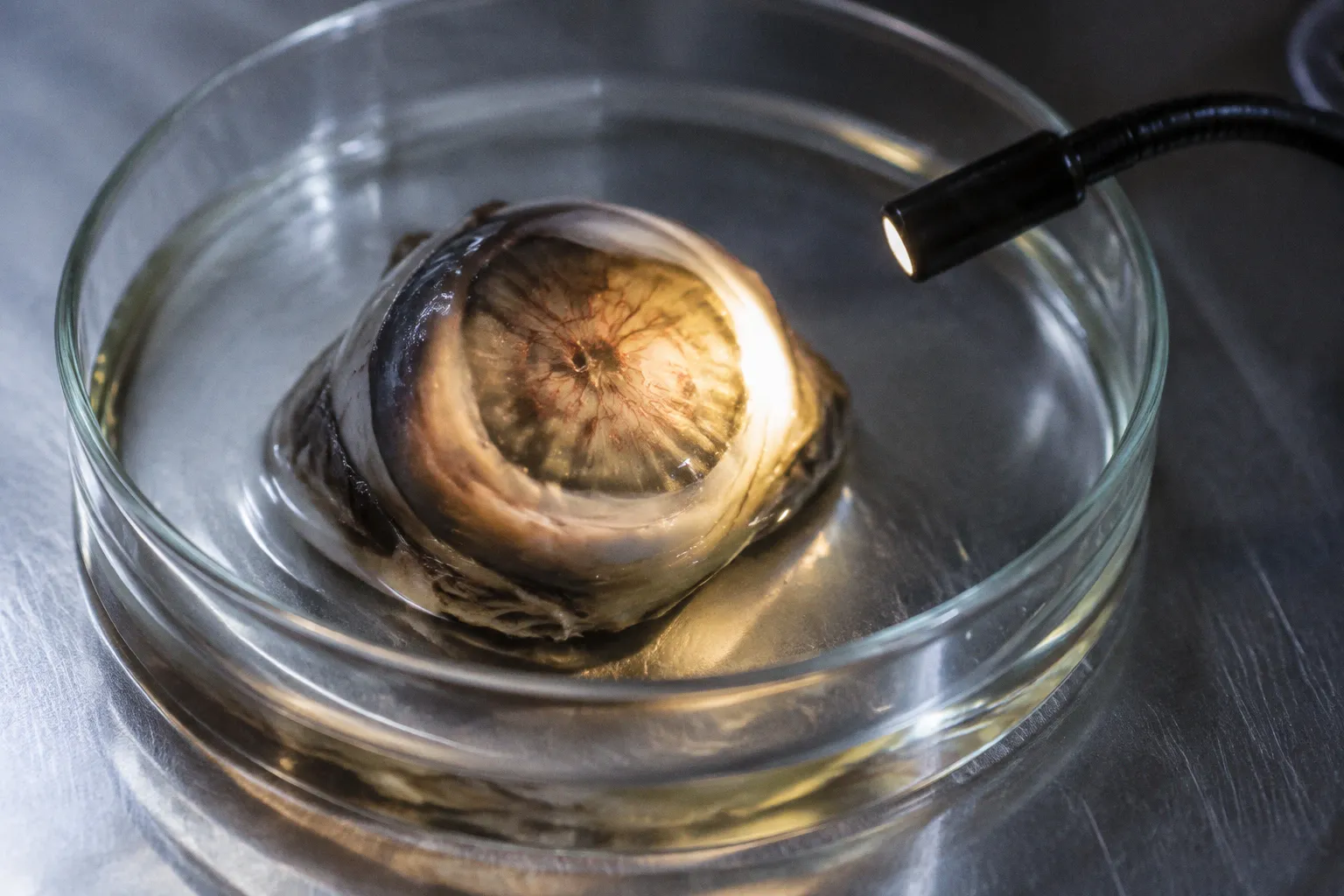
The tissue may show sub-RPE growth, surviving RPE, fibrous remodeling, patent vascular channels, and adjacent choriocapillaris loss in the same specimen. Treating these findings as interchangeable produces incorrect disease staging.
Human donor retina and choroid are therefore not passive confirmation of clinical diagnoses. They are endpoint datasets. The value lies in reconstructing lesion architecture, tissue response, and vascular context while accounting for procurement latency, fixation, sectioning plane, treatment history, and the difference between clinically visible and clinically silent disease.
In AMD donor eyes, clinically undetected choroidal neovascularization is typically associated with surviving retinal pigment epithelial cells in both macular and peripheral regions. That association is operationally important. RPE survival is not a marginal observation surrounding the lesion. It is part of the lesion’s structural environment and may determine how the neovascular complex is classified.
Donor-eye CNV is an architecture problem before it is a counting problem.
Histopathological patterns of sub-RPE neovascular growth
The phrase “wet AMD donor choroid histology” covers several distinct tissue configurations. A lesion can occupy the space beneath the RPE, disrupt or traverse Bruch’s membrane, extend through the RPE layer, or become embedded in fibrotic tissue while retaining vascular channels. These configurations should not be reduced to a single binary label such as CNV present or absent.
The most consequential distinction is between the clinical phenotype recorded before death and the structural phenotype recovered after fixation. A donor eye may carry a clinical history of AMD without a documented exudative event. Conversely, histology may identify sub-RPE vascular growth corresponding to occult CNV even when classic clinical features were not recorded. The Submacular Surgery Trials donor program, for example, confirmed sub-RPE growth patterns corresponding to angiographic occult CNV without classic CNV features.
That distinction affects both tissue annotation and downstream model selection. A specimen labeled simply as “AMD with CNV” loses information about whether the lesion was:
- clinically detected or clinically silent;
- sub-RPE, subretinal, or trans-RPE in its dominant location;
- associated with preserved, displaced, attenuated, or absent RPE;
- surrounded by intact or remodeled Bruch’s membrane;
- accompanied by choriocapillaris narrowing or broader vascular loss;
- treated before death or treatment-naive.
These variables are not interchangeable. They describe different stages of lesion organization and different levels of confidence in the reconstructed disease process.
Bruch’s membrane and the limits of a two-dimensional section
Bruch’s membrane rupture is often used as a structural marker in human donor retina CNV pathology. The limitation is geometric. A histologic section samples a narrow plane through a complex lesion. If the section misses the point of origin or crosses only the lateral margin, the observed relationship between CNV and Bruch’s membrane may be incomplete.
This is one reason why lesion grading should use serial sections, orientation records, and block-level mapping where available. A single section can establish the presence of vascular proliferation. It may not establish the full trajectory of the lesion.
The relevant questions are procedural:
1. Does the section pass through the lesion center or its edge?
2. Is the RPE continuous over the lesion, displaced, fragmented, or missing?
3. Is Bruch’s membrane intact, discontinuous, thickened, or difficult to resolve because of processing?
4. Are vascular channels patent, collapsed, or filled by post-mortem artifact?
5. Is fibrosis part of the lesion itself or a neighboring response?
6. Does the lesion align with the documented macular region and clinical imaging?
A grading system that ignores section geometry will generate false differences between specimens. One donor eye may appear to contain a compact sub-RPE complex. Another may appear to contain a dispersed lesion simply because the cutting plane intersects a different portion of the same three-dimensional structure.
RPE survival as a spatial variable
The association between clinically undetected CNV and surviving RPE cells is especially relevant to tissue sampling. If the RPE is assumed to be uniformly absent in advanced AMD, sampling strategies will preferentially select damaged areas and exclude the microenvironments where residual RPE interacts with vascular proliferation.
In practical terms, the RPE should be mapped as a field, not recorded as a yes-or-no attribute. Useful annotations include local cell continuity, pigmentation, epithelial thickness, displacement, and the relationship between surviving cells and the vascular complex. Macular and peripheral regions should remain separate in the dataset. The presence of surviving RPE in the periphery does not provide a substitute for macular characterization.
This matters for transcriptomic work. A punch containing residual RPE, Bruch’s membrane, choriocapillaris, and CNV can produce a mixed molecular signal. Without precise spatial annotation, the resulting expression profile may be assigned to RPE biology while containing substantial vascular and fibrotic contributions.
Vascular density and choriocapillaris regression in geographic atrophy
Geographic atrophy adds a second structural axis to donor-eye analysis. CNV is a proliferative vascular event. Geographic atrophy is associated with tissue loss and choriocapillaris rarefaction. These processes can coexist in the same eye, and their spatial relationship may be more informative than either diagnosis alone.
In donor eyes with geographic atrophy, choroidal blood vessel density in affected areas is reduced by approximately 30% to 50% compared with control eyes. In submacular regions, the reduction can reach 53%. The choriocapillaris is not uniformly erased. Residual narrowed vessels remain. That distinction is essential when interpreting both histology and molecular data.
The dataset should therefore distinguish among:
- reduced vessel density;
- narrowed residual vessels;
- complete loss of identifiable vascular profiles in a sampled section;
- fibrotic replacement;
- vascular remodeling adjacent to atrophic zones;
- vascular proliferation associated with CNV.
These states have different biological meanings. “Absent in section” is not equivalent to “absent in tissue.” A section may fail to capture small residual channels, or processing may compromise the visibility of endothelial structures. Conversely, a low-density region may represent a true atrophic field rather than technical under-sampling.
A compact comparison of lesion environments
| Parameter | CNV-dominant region | Geographic atrophy-dominant region |
|---|---|---|
| Primary structural event | Proliferation of choroidal vascular tissue, often beneath or around the RPE | Loss and remodeling of outer retinal and choroidal structures |
| RPE relationship | Surviving RPE may envelop or border the neovascular complex | RPE loss can be extensive; approximately 90% loss has been observed in the posterior pole of geographic atrophy donor eyes |
| Choriocapillaris | May contain proliferative or remodeled vascular channels | Vessel density reduced by 30% to 50%, with reductions up to 53% in submacular regions |
| Main grading risk | Confusing occult sub-RPE growth with nonspecific fibrosis | Treating residual narrowed vessels as complete vascular absence |
| Molecular interpretation | Mixed RPE, endothelial, inflammatory, and fibrotic signal is likely | Signal may be dominated by tissue loss, stress responses, and residual vascular compartments |
The table describes dominant environments, not mutually exclusive categories. A donor eye can contain an atrophic macular field, a neighboring CNV complex, and peripheral RPE survival. The correct unit of analysis is often the region or lesion rather than the eye as a single clinical label.
Geographic atrophy changes the denominator. A vascular count means little unless the sampled tissue area and surviving tissue compartments are defined.
The denominator problem in donor tissue
Vascular density is a ratio. The numerator is the number or area of identifiable vessels. The denominator is the tissue field selected for comparison. In advanced atrophy, the denominator may contain extensive loss of RPE, photoreceptors, and choroidal structures. A lower vessel count can reflect biological regression, tissue collapse, section selection, or all three.
For this reason, donor-eye studies should preserve the original field boundaries and document whether measurements were taken from submacular, parafoveal, peripheral, or lesion-adjacent tissue. Comparisons between GA and control eyes become unstable if one group is sampled from a narrow atrophic core and the other from intact macular tissue.
The same issue affects spatial transcriptomics. A low-transcript region may indicate genuine cellular depletion. It may also reflect reduced capture from a thin or damaged section. Transcriptomic yield must be interpreted against histologic cellularity, not in isolation.
Post-PDT structural remodeling: patent vessels and fibrous proliferation
Treatment history introduces a separate confounder. A donor eye examined after photodynamic therapy does not provide a simple untreated CNV specimen. It provides a record of lesion remodeling under treatment exposure, followed by an interval of biological change and post-mortem preservation.
Histopathological examination of donor eyes after PDT identified average CNV lesion dimensions of approximately 550 × 280 micrometres. The lesions were enveloped by RPE cells. Light-microscopic analysis also showed fibrous proliferation and patent vascular channels within the CNV. There was no evidence of permanent vascular occlusion or thrombus formation as a general interpretation of these findings.
That last point sets a boundary on causal language. PDT-associated fibrosis does not equal complete vascular shutdown. A lesion can be structurally remodeled and still contain open vascular channels. The presence of patent vessels should be recorded as a distinct endpoint rather than treated as a failure of treatment or proof of active exudation at death.
Treatment history as a metadata field
PDT exposure should be treated as a high-value metadata variable. At minimum, the tissue record should separate:
- documented treatment from unknown treatment status;
- interval between treatment and death, where available;
- lesion location;
- fibrosis distribution;
- vessel patency;
- RPE coverage;
- evidence of hemorrhagic or exudative sequelae;
- quality of fixation and section preservation.
The long-term vascular regression dynamics beyond 17 months after treatment remain unresolved in the available evidence base. A donor-eye study should not convert a single post-treatment endpoint into a longitudinal regression curve. Histology supplies a state. It does not automatically supply the trajectory that produced that state.
This is a recurring issue in biobanking datasets. A tissue sample is temporally precise at the moment of death but temporally incomplete with respect to disease evolution. Clinical records, treatment intervals, imaging history, and pathology must be joined before the lesion can be interpreted as part of a sequence.
Lesion dimensions are not disease burden
The 550 × 280 micrometre average describes the dimensions of examined post-PDT CNV lesions. It should not be used as a universal size estimate for CNV in donor eyes, nor as a direct surrogate for clinical lesion area. Post-mortem tissue selection, section plane, shrinkage, and lesion heterogeneity all influence the measured dimensions.
For lesion grading, dimensions should be paired with architecture:
- maximum observed length and width;
- estimated section depth or serial-section extent;
- vascular channel density;
- degree of fibrosis;
- relationship to the RPE;
- Bruch’s membrane status;
- presence of hemorrhage, exudate, or inflammatory infiltrate when preserved;
- distance from the foveal center or other anatomical reference point.
A measurement without anatomical context has low transfer value. It cannot reliably support comparison across donor programs using different fixation, embedding, or sectioning protocols.
Molecular signatures in the RPE–Bruch’s membrane–choriocapillaris complex
The RPE, Bruch’s membrane, and choriocapillaris form a coupled analytical compartment. Separating them conceptually is useful. Separating them physically is often difficult. The complex contains epithelial, vascular, extracellular-matrix, inflammatory, and complement-related signals that shift with AMD status and local tissue damage.
In AMD eyes, post-mortem studies have reported elevated C-reactive protein and decreased complement factor H in the RPE/Bruch’s membrane/choriocapillaris complex compared with healthy controls. These findings support the use of the complex as a molecularly informative unit. They do not establish that every CNV lesion has the same inflammatory or complement profile.
The correct interpretation depends on sampling architecture. A specimen containing fibrotic CNV, surviving RPE, and adjacent atrophic choriocapillaris may show a composite signature. That composite can be biologically meaningful, but it should not be assigned to a single cell type without spatial or cellular deconvolution.
Linking histology to transcriptomic yield
Spatial transcriptomics and single-cell approaches are especially sensitive to procurement and tissue quality. The relevant variables are not limited to RNA preservation. They include:
- post-mortem interval and cold ischemia exposure;
- time to dissection;
- retinal detachment or mechanical distortion;
- fixation duration;
- section thickness and tissue integrity;
- preservation of the RPE monolayer;
- lesion enrichment versus mixed-tissue sampling;
- anatomical registration between histology and molecular capture.
These factors influence transcriptomic yield and degradation kinetics. A low-yield sample may be biologically depleted, technically compromised, or both. Without paired morphology, those explanations remain confounded.
A useful study design separates at least three spatial classes:
1. Lesion core. The region containing the highest concentration of CNV-associated vascular and fibrotic structures.
2. Lesion margin. The interface between the complex and neighboring RPE, Bruch’s membrane, and choriocapillaris.
3. Matched non-lesional tissue. A region from the same eye selected for anatomical comparability rather than simply physical distance.
The margin is often the most informative compartment. It captures the transition between surviving RPE and pathological vascular growth. The core may be dominated by fibrosis and vascular channels. A distant control region may differ because of macular specialization, peripheral anatomy, or unrelated atrophic change.
Data integration without false precision
A biorepository should not treat histology, clinical imaging, and molecular data as independent attachments. They should share a common coordinate system. At minimum, the record should preserve:
- donor-level identifiers;
- eye-level laterality;
- macular and peripheral location;
- block and section identifiers;
- lesion boundaries;
- treatment exposure;
- clinical CNV status;
- GA status;
- RPE preservation score;
- choriocapillaris density category;
- molecular assay type and quality metrics.
This structure allows researchers to distinguish a true biological difference from a procurement or sampling difference. It also makes negative findings more useful. A specimen with no detected CNV can be interpreted only if the sampled region, section quality, and lesion detection sensitivity are known.
Translational implications for donor-tissue biorepositories
The immediate value of donor-eye CNV pathology is not the production of another disease label. It is the improvement of tissue selection. Translational studies need specimens that match a mechanistic question. A model of RPE–endothelial interaction requires preserved lesion margins. A study of atrophic vascular regression requires defined choriocapillaris fields. A fibrosis study requires treatment history and structural documentation. A transcriptomic study requires cellular preservation and spatial registration.
Procurement latency is therefore a biological variable, not a warehouse metric. As latency increases, tissue quality becomes more heterogeneous. The effect will not be uniform across retina, RPE, choroid, and optic nerve. A sample may remain suitable for morphology while becoming unsuitable for high-yield RNA work. Another may preserve broad anatomy but lose the fragile RPE layer required for cell-specific analysis.
The repository should expose these distinctions rather than collapse them into a single quality grade. A practical data model can use separate scores for:
- anatomical integrity;
- RPE continuity;
- vascular preservation;
- lesion visibility;
- nucleic-acid suitability;
- treatment metadata completeness;
- spatial registration confidence.
This approach supports query-based procurement. Researchers can request donor eyes with clinically occult CNV, preserved RPE at the lesion margin, documented PDT exposure, or GA-associated choriocapillaris reduction. They do not need to accept a broad AMD category whose internal heterogeneity is unknown.
What should be retained in a CNV pathology record
A high-value donor record should retain the following elements:
- Clinical phenotype: AMD diagnosis, documented CNV, occult or classic imaging features where available, and GA status.
- Anatomical map: macular, submacular, parafoveal, peripheral, or lesion-adjacent location.
- Histologic architecture: sub-RPE growth, trans-RPE extension, fibrosis, Bruch’s membrane disruption, and RPE coverage.
- Vascular state: density, calibre, patency, narrowing, and relationship to surrounding choriocapillaris.
- Treatment exposure: PDT status, known treatment interval, and uncertainty where records are incomplete.
- Preservation variables: procurement latency, fixation conditions, section integrity, and tissue-specific quality.
- Molecular linkage: assay platform, sampled compartment, transcriptomic yield, and matched histology.
- Uncertainty flags: clinically undetected lesions, incomplete treatment history, ambiguous section geometry, and unresolved lesion boundaries.
This is not administrative overhead. It is the minimum metadata required to interpret a donor-eye observation without overextending it.
The boundary between pathology and inference
Post-mortem examination can establish structural relationships. It can show that CNV is present beneath or around the RPE, that RPE cells survive around a lesion, that fibrous proliferation coexists with patent vascular channels after PDT, or that choroidal vessel density is reduced in GA tissue.
It cannot, by itself, establish the exact incidence of subclinical CNV across all donor repositories. It cannot reconstruct long-term vascular regression beyond the observed endpoint. It cannot prove that a molecular signal originated from one cell population when the sampled compartment contains several. Those conclusions require population-level sampling, longitudinal clinical data, or spatially resolved methods.
The distinction is operational. A repository that records inference as fact will contaminate future analyses. Once a lesion is labeled as fully occluded, completely avascular, or biologically inactive, later researchers may treat that label as ground truth even when the histology shows otherwise.
A stricter framework for choroidal neovascular lesion grading
A useful grading system should be hierarchical. It should begin with observation and add interpretation only where the evidence supports it.
Level 1: Structural detection
Record whether abnormal vascular tissue is identified and where it is located relative to the RPE, Bruch’s membrane, and choroid. This is the foundation. It should not depend on clinical evidence of exudation.
Level 2: Architectural classification
Describe the dominant pattern:
- sub-RPE;
- subretinal or trans-RPE;
- fibrotic and vascular;
- predominantly fibrotic;
- lesion with patent vascular channels;
- lesion adjacent to extensive atrophy.
The categories can overlap. The aim is to preserve structure rather than force a single label.
Level 3: Tissue response
Record RPE survival, RPE displacement, epithelial loss, inflammatory features, fibrosis, and choriocapillaris remodeling. This level connects the lesion to its local microenvironment.
Level 4: Clinical correspondence
Link the histology to available clinical records. A lesion may be clinically detected, clinically occult, or clinically indeterminate. The last category is preferable to a forced conclusion when records are incomplete.
Level 5: Treatment interpretation
Add treatment exposure only after confirming that the record is adequate. In post-PDT tissue, describe fibrous proliferation and vascular patency directly. Do not translate those observations into permanent occlusion unless the evidence specifically supports it.
This grading sequence reduces the risk of importing clinical assumptions into morphology. It also makes datasets more interoperable. Different laboratories may use different stains and imaging systems, but they can still exchange the same core variables.
Final assessment
Choroidal neovascularization donor tissue is most valuable when treated as a spatially indexed endpoint rather than a generic AMD specimen. The strongest findings are relational: CNV with surviving RPE, occult sub-RPE growth without classic clinical features, fibrous remodeling with patent vessels after PDT, and choriocapillaris loss within geographic atrophy that remains incomplete rather than absolute.
The infrastructure determines how much of that biology survives into a usable dataset. Procurement latency, block selection, section geometry, treatment metadata, and molecular quality are not secondary documentation. They define the confidence limits of the pathology.
The evidence supports a strict conclusion. Human donor eyes can resolve CNV architecture and its surrounding tissue response at a level unavailable to routine clinical imaging. They cannot justify universal assumptions about lesion size, vascular occlusion, subclinical incidence, or long-term regression. The correct workflow is therefore conservative: preserve the anatomy, separate observation from inference, and make every tissue-quality variable queryable.