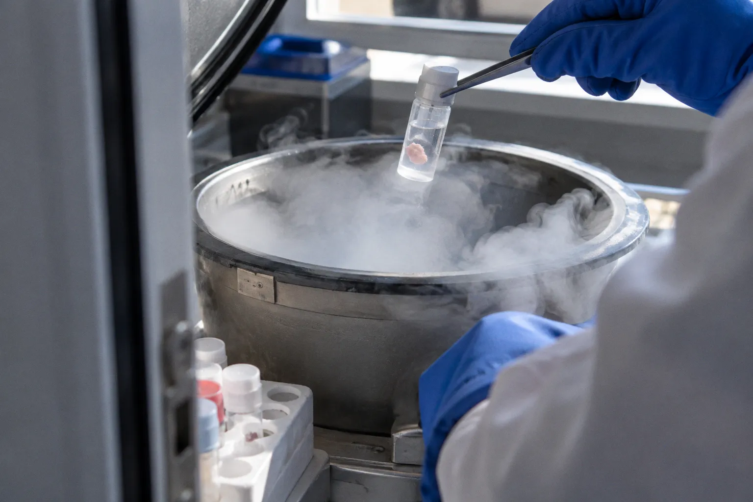
Cryopreservation of intact retinal sheets, as opposed to dissociated cell suspensions, introduces a structural problem that has nothing to do with viability assays: the multilayered neural tissue can fracture along its laminae during the freeze-thaw cycle. For programs working on retinal progenitor cell grafting, donor-derived RPE transplantation, or scaffold-based ocular bioengineering, this is the kind of failure that does not show up in a trypan blue count but absolutely shows up under the surgical microscope.
This is the case of cellular fracturing in donor retinal tissue cryopreservation — a quiet, persistent challenge that sits between biobank logistics and the operating room. It is not a problem of cell survival per se. It is a problem of structural survival, and the two have to be solved separately.
The Mechanics of Cellular Fracturing in Multilayered Retinal Tissue
A whole donor retina is not a uniform block of cells. It is a stratified structure — outer nuclear layer, outer plexiform layer, inner nuclear layer, ganglion cell layer — separated by synaptic interfaces that behave mechanically like weak cleavage planes. When ice forms during freezing, water expands by roughly 9% as it transitions to the crystalline phase. In a single-cell suspension, that expansion has somewhere to go; the cells float freely in cryoprotectant. In an intact sheet, the expansion must be absorbed within the tissue itself, and it preferentially relieves along the path of least mechanical resistance. For laminated neural tissue, that path runs between the cellular layers.
The result is what tissue bank staff and ocular researchers describe as fracturing — visible splits in the retinal architecture that do not heal on rewarming. The photoreceptor layer can cleave from the inner nuclear layer. The outer limiting membrane, the band of junctions that seals the photoreceptor somata, can rupture. The retinal pigment epithelium, if still attached to the donor tissue, can delaminate from Bruch's membrane, the thin layer that separates the RPE from the choroidal blood supply. None of these are necessarily visible on a standard post-thaw viability count using dissociated cells, because the standard assay involves enzymatic digestion of the tissue — which destroys exactly the structural information the protocol was trying to preserve.
Fracturing in an intact retinal sheet is the kind of failure that does not show up in a trypan blue count. It shows up under the surgical microscope.
This is one of the central reasons retinal cryopreservation protocols bifurcate. Programs either dissociate the tissue into cell suspensions before freezing, accepting the loss of architecture, or attempt to preserve intact sheets, accepting the risk of fracturing. A third path — vitrification, in which ice is avoided entirely by pushing the sample into a glassy state — works in principle but is technically demanding and introduces its own constraints, particularly around cryoprotectant concentration and cooling rates that must be optimized for tissue geometry rather than for cells in suspension.
Thermal Stress and the Glass Transition Threshold in Ocular Biologics
Every cryopreservation protocol operates in two regimes: the freezing regime above the glass transition temperature, and the storage regime below it. The glass transition — the temperature range during which unfrozen water solidifies into a non-crystalline glassy state and molecular diffusion effectively stops — falls roughly between -90°C and -130°C for most biological tissues. Above that range, biochemistry is still moving: solutes can redistribute, ice can grow or recrystallize, and cryoprotectant continues to act on cells. Below it, the tissue is, in practical terms, metabolically paused.
Most ocular tissue banks store donor biologics in liquid nitrogen at -196°C, comfortably below the glass transition. The challenge is getting there without traversing the dangerous zone in a way that introduces thermal stress fractures. A whole retinal sheet does not cool uniformly; the edges and the thinner pericentral regions drop below the glass transition before the thicker central macula does, creating a transient thermal gradient across the tissue. If that gradient is steep enough, the differential contraction between already-vitrified and still-unfrozen regions generates mechanical stress that can crack the tissue along its laminae. The underlying physics is the same as a glass cracking when one side is heated faster than the other — except the working temperature here is below -100°C and the material is biological.
Cooling rate therefore matters as much as final temperature. Slow enough to allow cryoprotectant to permeate the tissue and intracellular water to exit before extracellular ice nucleates. Fast enough to avoid prolonged exposure of the unfrozen fraction to toxic solute concentrations. Too slow, and cells sit in hyperosmotic conditions for minutes, accumulating cryoprotectant damage. Too fast, and ice nucleates intracellularly before the cells have a chance to dehydrate. For dissociated cell suspensions, the acceptable range is broad. For intact multilayered retinal sheets, it is narrow — and for human donor tissue specifically, the critical cooling rates that would prevent macroscopic fracturing in full-thickness sheets are not yet standardized.
Comparative Viability: Dissociated Cell Suspensions Versus Intact Organoids
The published data on post-thaw survival in retinal tissue presents an apparent paradox that, on closer inspection, is really a question of geometry. Purified CD73+ photoreceptor precursor cell suspensions — that is, cells expressing a surface marker associated with rod photoreceptor lineage, isolated from donor retinas and frozen as single cells — show high cell mortality after freeze-thaw. Whole retinal organoids, the much larger and structurally complex three-dimensional aggregates, achieve in published studies relatively better post-thaw survival rates than the dissociated suspensions they are derived from.
The contradiction resolves once the two failure modes are separated. Dissociated cell suspensions take the brunt of cryoprotectant toxicity and osmotic stress at the single-cell level: every cell is exposed directly to the freezing solution, and there is no surrounding architecture to shield any of them. Organoids, despite being larger and more complex, have a three-dimensional structure that buffers interior cells from the worst of the CPA exposure; surface cells may die, but interior populations survive at higher rates because they are partially insulated. The same geometry that makes organoids fragile to mechanical fracture under some protocols makes them robust to chemical toxicity under others.
| Parameter | Dissociated cell suspensions | Intact sheets / organoids |
|---|---|---|
| Primary failure mode | Uniform cell death (CPA toxicity, osmotic stress) | Structural fracturing along laminae |
| Visibility in viability assay | Direct, quantifiable | Often missed in dissociative counts |
| Geometry effect on CPA exposure | Uniform across population | Surface-biased damage, interior buffered |
| Geometry effect on ice expansion | Cells free to move in suspension | Stress relieved along cleavage planes |
| Clinical translation maturity | Mature, well-characterized | Largely experimental |
For translational work, the practical consequence is that the choice between a dissociated cell product and a sheet- or organoid-based product is not simply a question of which has better survival. They fail in different ways. A dissociated RPE or photoreceptor precursor product will tend to fail through uniform cell death that is detectable in a viability assay. A sheet-based or organoid-based product will tend to fail through structural fracturing that may preserve local viability while destroying the architecture required for surgical integration. Eye banks preparing material for retinal progenitor cell grafting or donor-derived RPE transplantation have to choose which failure mode they can manage downstream — and most clinical programs today lean toward the one they can measure.
Navigating Cryoprotectant Toxicity and Post-Thaw Recovery Standards
Cryoprotective agents — DMSO, glycerol, ethylene glycol, sucrose, and a growing list of proprietary blends — are themselves the second major source of damage in ocular biologics cryopreservation. CPAs work by depressing the freezing point of intracellular water, reducing ice nucleation, and stabilizing membranes during dehydration. They are also, in concentrations sufficient to do that job, directly cytotoxic. The therapeutic window between enough CPA to prevent ice damage and so much CPA that the cells die anyway is the central engineering constraint of every retinal cryopreservation protocol.
For research-grade retinal tissue, CPA choice is a tuning problem. For clinical-grade products intended for human transplantation, it is a regulatory one. The FDA's cellular therapy framework requires that therapeutic cell products intended for clinical use maintain a minimum post-thaw recovery rate of 70% — meaning that at least seven of every ten cells in the dose must be viable and functional after the freeze-thaw cycle. That threshold applies regardless of whether the product is a vial of dissociated RPE or a stack of retinal progenitor cell sheets, and it has shaped nearly every commercially oriented retinal cryopreservation workflow over the past decade.
Meeting that threshold is harder than the headline number suggests. The 70% figure is measured on the final formulated product, after thaw, dilution of CPA, and any required recovery culture. If the starting population is donor RPE — and the macula alone requires on the order of 60,000 RPE cells to cover the central visual field — then a 30% post-thaw loss does not merely shrink the dose; it can drop it below the threshold needed for functional recovery after transplantation.
The arithmetic of cell loss in retinal grafts is unforgiving in a way that dermal fibroblast banking is not, because the retina has no regenerative reserve to compensate for under-dosing.
This is why post-thaw recovery culture periods — sometimes several hours to a full day, in optimized media with growth factors and substrates that support attachment — have become standard in clinical-grade retinal banking. The 70% number is a snapshot, but the tissue that meets it is not necessarily the same tissue that went into the freezer. Recovery culture is, in effect, a triage step that lets the cryopreservation protocol be slightly more aggressive than it otherwise could be, on the assumption that damaged cells will be cleared or selectively lost during the recovery window before the dose is released.
Current Limitations in Standardizing Full-Thickness Retinal Grafting
The unsolved problem in donor retinal cryopreservation is not dissociated cells. Those protocols are mature, if imperfect, and there are workable answers for many cell types including RPE and purified photoreceptor precursors. The unsolved problem is intact, full-thickness, donor-derived retinal sheets suitable for surgical grafting — the kind of transplant material that, in principle, could restore layered retinal architecture in advanced disease states.
No standardized, clinically approved protocol exists today for cryopreserving intact whole donor human neural retinas without microstructural fracturing. That is not a criticism of any individual laboratory; it reflects the genuine difficulty of the problem. The mechanical mismatch between retinal layers, the narrow acceptable cooling rate window, the CPA toxicity ceiling, and the 70% post-thaw recovery threshold together constrain the design space so tightly that any one of them, taken alone, would be a hard engineering problem. Taken together, they explain why most translational programs in retinal sheet grafting have moved toward either fresh, time-limited hypothermic storage for short-haul logistics, or entirely different preservation strategies — including vitrification approaches that avoid ice altogether but introduce their own loading and unloading constraints.
Programs working on scaffold-based ocular bioengineering, synthetic corneal hydrogels, and extracellular vesicle ocular therapy have largely sidestepped the intact-sheet problem by working with products that do not require intact donor retinal architecture. But for any program whose surgical endpoint is layered retinal reconstruction — and there are several such programs in preclinical and early clinical development — the fracturing question remains the central logistical bottleneck. Until the geometry problem and the thermal stress problem are solved together, eye banks can ship dissociated retinal cells at clinical grade, but they cannot reliably ship a whole retina at clinical grade.
What This Means at the Bench and at the Bedside
Retinal tissue cryopreservation is not a single problem with a single solution. It is a portfolio of related problems, each tied to a specific translational product and a specific surgical endpoint. The freezing protocol for a dissociated RPE suspension going into a subretinal injection is not the same problem as the freezing protocol for a full-thickness sheet going into a subretinal transplant. The field has been steadily sorting these problems into separate workstreams over the past two decades — early photoreceptor precursor work that established baseline viability protocols, organoid cryobiology that revealed geometry-dependent survival patterns, the regulatory clarification that anchored clinical translation to a 70% post-thaw threshold, and more recent vitrification and recovery-culture refinements that have begun to push intact-tissue survival upward. Each step has narrowed the gap between what eye banks can ship and what surgeons actually need at the table, but none of them has closed it.
For eye banks, the practical question is no longer "can we freeze this?" It is "can we freeze this product in the form the surgical protocol actually requires?" For dissociated RPE, photoreceptor precursors, and retinal progenitor cell suspensions, the answer today is generally yes, with the caveats that CPA choice, cooling rate, and recovery culture all matter, and that any given lot must clear the post-thaw viability gate before release. For intact donor neural retinas, the honest answer is not routinely — not because no one is working on it, but because the structural barrier is real and is not going to be solved by a better cryoprotectant alone. It will be solved, if it is solved, by advances in geometry-aware cooling, controlled vitrification of laminated tissue, and recovery protocols that rebuild what freezing disrupts.
Until then, retinal sheet cryopreservation remains a case of cellular fracturing — a known failure mode, a known cost, and a known target for the next generation of biobank engineering.