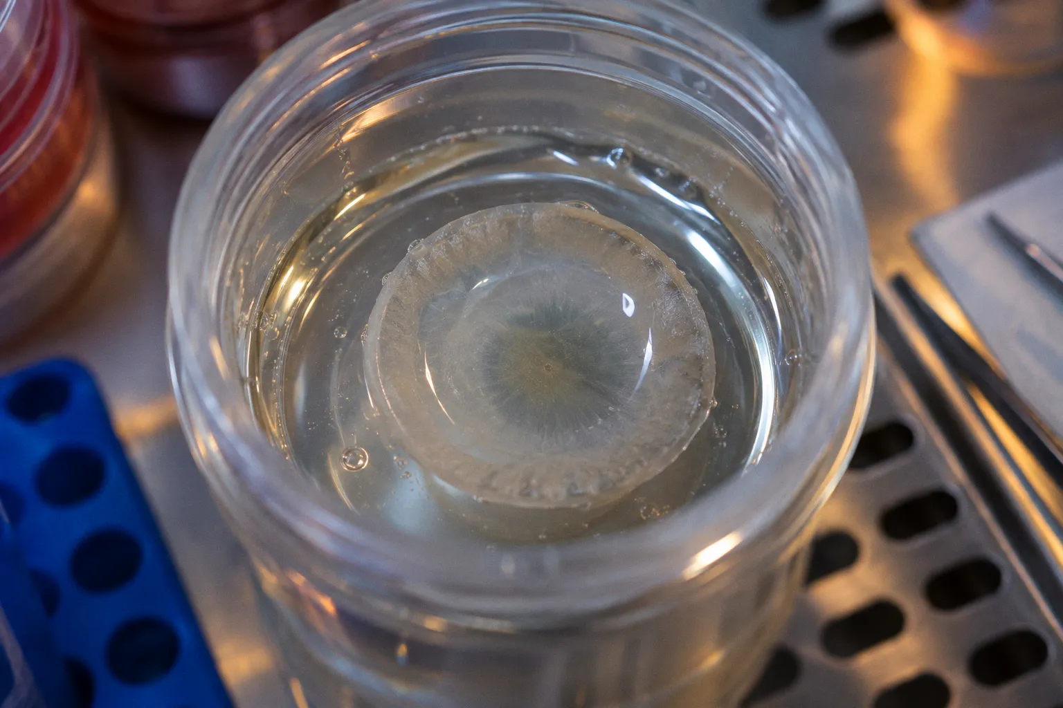
But those boxes answer different questions. A tissue may meet a clinical threshold and still be a poor fit for a particular procedure or research purpose. The useful record is not just a clearance decision; it is a description of what the cornea is likely to support.
That distinction matters in anterior-segment work, where shape, cell condition and tissue architecture can steer a cornea toward very different destinations. A count from specular microscopy is informative, but it does not describe scroll behavior, stromal structure or suitability for a specific assay. A step-by-step walkthrough should make those differences visible rather than compress them into a single pass-or-fail judgment.
Standardizing donor procurement and structural assessment
Before a cornea enters preservation, it already has a geometry that shapes the options downstream. Horizontal diameter, curvature and thickness are not decorative metadata. They affect how a specimen can be trephined, which portions remain available for research, and how confidently a surgeon can plan a graft.
Adult donor corneal diameter is reported at about 11.71 ± 0.42 mm, with a modest average difference between male and female donors in the measurements cited here. Such population averages provide context, not a substitute for measuring the individual tissue. A small difference may matter when planning a trephine or balancing a corneal button against a limbal rim intended for other work. The procurement record should capture the actual measurement where it is available, rather than invite the next person in the chain to infer it.
Shape matters for optical reasons as well. The cornea contributes roughly 40 to 44 diopters of refractive power, accounting for most of the eye’s total refractive power. That does not mean every procurement program needs to turn a donor record into a topography report. It does mean that significant curvature irregularity, scarring or previous refractive surgery should be recorded in terms of the intended use. A feature that complicates one application may be irrelevant to another.
A practical structural record can bring together three kinds of information:
- Dimensions: horizontal diameter and, where relevant, central and peripheral thickness. These help with trephination planning, tissue allocation and interpretation of imaging.
- Surface and curvature: slit-lamp findings and available topographic or tomographic measurements, especially when optical outcomes or refractive geometry matter.
- Preservation and handling history: the conditions and timing documented by the eye bank, so downstream users can interpret measurements in context.
The point is not to collect every possible measurement. It is to preserve the measurements that let a clinical or research team judge whether this particular tissue suits its intended use. A record that lists only endothelial density and serology may be adequate for a narrow clearance workflow, but it leaves important questions unanswered for a broader biologics pipeline.
Endothelial viability: the metrics that actually decide
Endothelial cell density (ECD) is often treated as a gate: above a threshold, the tissue proceeds; below it, the tissue is declined or diverted. In practice, ECD is a measurement at one point in time, not a guarantee of long-term performance. Specular microscopy gives the procurement team an important snapshot, but interpretation also depends on cell morphology, the quality of the image and the planned procedure.
Clinical thresholds vary across programs and indications. Values around 2400 to 2500 cells/mm² are commonly used as a reference range for transplantation, but a threshold should not be mistaken for a forecast. A cornea near the threshold may call for a more careful review of its morphology and intended use; a higher count does not erase concerns about uneven cell distribution or compromised tissue. Endothelial loss after transplantation is part of the clinical picture, but the amount and course should not be projected from a single screening value as if they were known for an individual graft.
A cell count is a snapshot. Its meaning depends on the cells around it, the tissue they occupy and the work the graft is being asked to do.
A more useful procurement conversation asks what the endothelial assessment actually shows:
- Cell density: record the measured value and the method used, rather than reducing the result to an unexplained pass or fail.
- Cell appearance and distribution: note whether the image supports a confident count and whether the endothelial mosaic appears sufficiently uniform for the intended application.
- Tissue condition: interpret the endothelial result alongside thickness, signs of edema and preservation history.
- Destination: distinguish a clinical graft assessment from a research allocation. Tissue that falls short of a particular clinical threshold may still be valuable for a well-defined research question.
The assessment should avoid false precision. A single cell-density band cannot sort every cornea into universal categories of optimal, acceptable or unusable, because procedure, local standards and the rest of the tissue record matter. It can, however, prompt the right next question: whether the measured endothelium is fit for the planned use, and what uncertainty remains.
Microstructural integrity: from epithelium to stroma
The cornea’s layers do different jobs, so a procurement assessment should not treat them as one undifferentiated transparent surface. The epithelium is only a small part of total thickness, but its condition matters to surface integrity and postoperative recovery. The stroma makes up most of the cornea and is central to optical quality, biomechanics and many research applications.
For surgical use, an epithelial defect may be relevant without automatically deciding the fate of the whole tissue. Its importance depends on the defect’s extent, the planned procedure and the ability to interpret the tissue’s condition. For research, the question changes with the assay. A specimen intended for biomechanics needs a different structural description from one intended for histology or preservation studies. The procurement record should say enough about the layers to let a downstream team decide whether the tissue can answer its question.
Three observations help keep that assessment grounded:
- Epithelial surface: record denudation, erosion or other visible irregularity, and distinguish localized damage from broader compromise.
- Stromal clarity and architecture: note scars, infiltrates, haze or other findings that may affect imaging, optical work or biomechanical interpretation.
- Thickness profile: compare central and peripheral measurements where available. A central cornea is generally thinner than the periphery; unexpected thickening or an unusual gradient can be a reason to investigate edema or other pathology.
These observations are not interchangeable with a histological examination. Slit-lamp assessment and imaging can identify features worth flagging, but they do not reveal every microscopic alteration. That is especially important when research users need to compare samples: a concise note about what was examined, and by which method, is more useful than a broad claim that the tissue is structurally normal.
The same principle applies to allocation. If the central stroma is scarred but the endothelium is suitable for a specific endothelial procedure, the cornea may not be a single yes-or-no proposition. It may be unsuitable for one destination and still informative or usable for another. Recording the reason for diversion preserves that distinction and helps avoid discarding material that could answer a narrower question.
Optimizing graft preparation for DMEK and DSAEK procedures
DMEK brings tissue handling into sharp focus. The graft is a thin Descemet membrane with its endothelium, and its tendency to scroll affects preparation and unfolding. Donor age has a specific role here: tissue from younger donors tends to form tighter scrolls, which can make unfolding more challenging. This is a handling consideration, not a verdict on overall tissue quality. Younger tissue may still be appropriate; it simply calls for preparation and surgical expectations that account for its behavior.
That is why age should not be flattened into either “younger is better” or “age does not matter.” For DMEK, age can help anticipate scroll characteristics. The relevant question is whether the tissue’s endothelial condition and handling profile suit the procedure, alongside the eye bank’s acceptance standards and the surgeon’s approach. A preference for looser scrolling tissue in some settings does not establish one universal optimal age for every DMEK graft.
DSAEK has a different preparation profile. Its graft includes posterior stroma as well as Descemet membrane and endothelium, so the stromal lenticule changes the handling and the tissue specifications that matter. Central thickness and the intended microkeratome cut become more prominent considerations. A thinner graft may support optical goals, but preparation depends on reliable tissue assessment and control of the cut; age-related scroll behavior is less central than it is for DMEK.
| Parameter | DMEK | DSAEK |
|---|---|---|
| Graft composition | Descemet membrane and endothelium | Descemet membrane, endothelium and a stromal lenticule |
| Handling consideration | Younger donor tissue tends to form tighter scrolls, which may be harder to unfold | The stromal component changes graft handling and reduces the emphasis on scroll behavior |
| Useful procurement focus | Endothelial condition, tissue integrity and expected scroll characteristics | Endothelial condition, central thickness and preparation requirements |
| Interpretation of donor age | Relevant to anticipating scroll handling, but not a stand-alone quality judgment | Less directly tied to scroll handling |
A well-designed walkthrough keeps those pathways distinct. If a cornea is being considered for DMEK, the record should make its handling characteristics legible without implying that age alone predicts success. If it is being prepared for DSAEK, the focus shifts toward the lenticule and the measurements needed to guide preparation. In both cases, endothelial viability remains important, but it is not the only decision point.
Navigating donor eligibility and systemic comorbidity profiles
Eligibility decisions can be too broad in either direction. A long list of exclusions may remove tissue that could serve a valid purpose; an overly casual review can overlook a genuine safety or quality concern. The more defensible approach is to distinguish systemic history from direct ocular findings and to apply the relevant eye-bank policies to the tissue’s intended destination.
Cardiovascular disease or a history of malignancy does not, by itself, establish that a cornea is unsuitable. The question is whether the donor history, cause of death, ocular involvement or applicable safety requirements affect the proposed use. Similarly, prior cataract surgery or LASIK is not a universal exclusion. Altered corneal geometry may make tissue inappropriate for some optical or full-thickness applications, while leaving other clinical or research uses open. The record should describe the history and its implications rather than collapse them into an unqualified yes or no.
Eligibility is not a contest to produce the longest exclusion list. It is a reasoned match between donor history, tissue condition and intended use.
Some findings do require clear escalation or exclusion under the relevant program’s standards. Active ocular infection, significant stromal scarring from prior keratitis, and relevant communicable-disease findings can affect whether tissue is accepted and where it can go. Preservation timing also matters, but the applicable limits depend on the eye bank’s documented protocols and the tissue destination. There is no reason to imply a single universal interval where programs set their own requirements.
A useful eligibility record makes three distinctions explicit:
- Systemic history versus ocular involvement: note the condition, then identify whether it has a direct bearing on the cornea or on required safety screening.
- Prior ocular procedures versus current tissue quality: document procedures such as cataract surgery or LASIK, and describe the resulting features that matter for the proposed use.
- Clinical allocation versus research allocation: explain whether a limitation rules out a particular graft or also affects the research question under consideration.
Research tissue is not a loophole around donor-safety standards. It is a different destination with its own requirements, documentation and scientific purpose. The same cornea can be inappropriate for an optical graft yet useful for a study of preservation or tissue structure, provided the study’s criteria and the relevant governance requirements are met. Recording why a tissue was diverted helps both the clinical and research teams understand what they have received.
The procurement record as a forecasting instrument
A procurement document should not pretend to predict a graft’s entire future. It should give the next person in the chain enough information to make a careful decision: what was measured, what was observed, how the tissue was handled and which limitations may matter for the intended procedure or study.
Age belongs in that record, but its meaning is specific rather than absolute. It can help anticipate DMEK scroll behavior, since younger donor tissue tends to form tighter scrolls and may be more challenging to unfold. It does not replace endothelial assessment, structural examination or the judgment required to match a cornea to a destination. The strongest walkthrough is therefore not one that dismisses age, nor one that lets age stand in for tissue quality. It treats age as one useful part of a broader, procedure-aware forecast.
For anyone asking how to check a step-by-step walkthrough of anterior-segment procurement, the practical test is whether the record helps distinguish one tissue’s possible uses from another’s. Endothelial morphology, stromal architecture, preparation requirements and donor history each contribute a different piece. Put together carefully, those details support better allocation without promising more than the measurements can show.