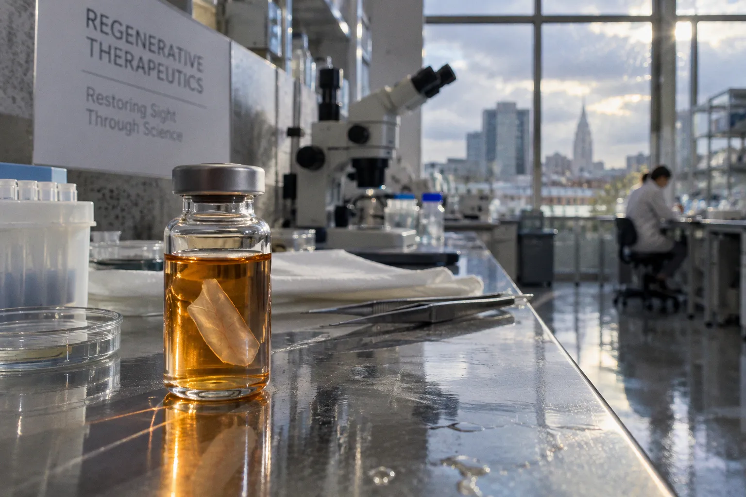
Cells that have been maintained in nutrient-rich culture are suddenly placed into the relatively nutrient-deprived subretinal space, where they must adapt to a very different metabolic environment while also responding to surgical injury, local inflammation, and limited physical support.
That early transition sets the ceiling for everything that follows. A graft cannot form meaningful synapses if too few cells remain alive. And even surviving cells face a second set of obstacles: loss of photoreceptor identity during extended culture, glial scarring, poor anatomical placement, and the difficulty of aligning immature cells with the host retina in a way that produces functional recovery rather than biological persistence alone.
The first bottleneck is metabolic, not immunological
The subretinal space is an attractive delivery site for photoreceptor precursors because it places donor cells close to the outer retina, where damaged photoreceptors would normally reside. In practical terms, however, the surgical target is also a demanding biological niche.
Cells expanded in vitro are accustomed to controlled media containing carefully balanced nutrients, growth factors, and oxygen conditions. After subretinal delivery, that support disappears. The graft must adjust rapidly to the local supply of nutrients and oxygen, to the extracellular matrix, and to the physical constraints of the space between the photoreceptor layer and retinal pigment epithelium. This abrupt change creates acute metabolic stress during the initial post-transplantation period.
The consequence is rapid cell death among subretinally grafted photoreceptor precursor cells. This can happen before the immune system becomes the dominant concern. It is a crucial distinction for therapeutic development because an intervention aimed only at immune suppression may leave the earliest and largest source of cell loss untouched.
From a clinical translation perspective, the problem begins before the cannula enters the eye. The state of the cell product matters: its developmental stage, time outside controlled culture conditions, concentration, suspension medium, and readiness for the target niche all influence how much stress the cells experience at delivery. The surgical maneuver is only one part of the transplant. The graft arrives with a metabolic history.
Early graft loss should not be read as a single immune-rejection event. The cells may be metabolically overwhelmed before they have time to engage with the host retina.
This is why post-transplantation retinal cell survival cannot be judged only by whether cells are detectable weeks or months later. A study may show long-term persistence in a surviving fraction while still losing a substantial number of cells during the first days. Those two observations describe different stages of the same treatment problem.
What the subretinal space asks of a graft
Once delivered, donor cells must cope with several simultaneous changes:
- They leave a stable, nutrient-rich culture environment and enter a comparatively constrained tissue compartment.
- They encounter local inflammatory signals associated with the procedure and tissue injury.
- They must survive without the same exogenous support used during expansion and differentiation.
- They need to remain close enough to the outer retina to receive relevant physical and molecular cues.
- They must avoid being dispersed, trapped, or deposited in a position where structural integration is unlikely.
The practical implication is straightforward: improving retinal progenitor cell engraftment requires more than increasing the number of injected cells. A larger dose may compensate for loss only superficially. If the delivery environment remains hostile, the additional cells may increase debris, local stress, or variability without producing a proportional improvement in functional grafting.
Cell identity can weaken before the transplant begins
A second hurdle develops during manufacturing. Retinal progenitor cells are not biologically static during culture. With extended passaging, they can lose photoreceptor-differentiation potential and reduce expression of markers associated with photoreceptor identity, including rhodopsin. At the same time, they may increase expression of more primitive neural stem cell markers such as Sox2 and nestin.
For a cell-therapy program, this is not a minor characterization issue. It changes what the product is capable of doing after implantation.
A population described broadly as retinal progenitor cells may contain cells at different developmental stages. Some may still retain a credible path toward photoreceptor maturation. Others may have shifted toward a less committed neural state. That mixture can affect survival, migration, secretion of neuroprotective factors, and the probability of forming the right contacts with host tissue.
In a laboratory workflow, passage number therefore becomes a functional variable rather than an administrative detail. Extended expansion may make manufacturing easier by producing more cells, but the extra yield can come at the cost of developmental quality. The relevant question is not simply whether the culture remains viable or continues to express a general progenitor marker. It is whether the cells retain the characteristics needed for photoreceptor maturation in the subretinal space.
A more useful way to evaluate the product
Before asking whether a graft survives, translational teams need to establish what kind of graft they are actually delivering. That assessment should connect identity to the intended clinical task:
- Developmental state: Are the cells committed enough to respond to retinal cues, or have they moved back toward a more primitive neural phenotype?
- Photoreceptor potential: Do they retain markers and functional characteristics associated with the desired lineage?
- Culture history: How many passages have they undergone, and did differentiation capacity change during expansion?
- Suspension readiness: Can the product tolerate the period between harvest, loading, delivery, and tissue exposure?
- Dose interpretation: Does the administered dose describe total cells, viable cells, or cells meeting the desired identity criteria?
This distinction is particularly important when comparing donor-derived retinal graft viability across studies. Two preparations may receive the same nominal dose but have very different proportions of cells capable of contributing to photoreceptor repair.
The manufacturing process also affects how results should be interpreted. If a study reports poor integration, the cause may lie in delivery or host anatomy. But it may also reflect a cell population that had already lost part of its photoreceptor potential before implantation. These variables can reinforce each other: less differentiated cells may be less prepared for the subretinal niche, while stressed cells may be less able to complete maturation after delivery.
Placement is not the same as integration
The physical route of administration determines where cells end up, but it does not guarantee what they do there. Intravitreal delivery is relatively accessible from a procedural standpoint, yet progenitor cells delivered into the vitreous often become trapped there or adhere to the vitreal surface. In many cases, they do not achieve definitive structural integration into the host neural retina.
That outcome illustrates a recurring problem in regenerative ophthalmology: presence is not incorporation.
A cell can remain detectable near the retina, release beneficial factors, or influence the local environment without replacing a lost photoreceptor or forming a functional synaptic circuit. These effects may still be therapeutically relevant, particularly when the cells provide neuroprotective support. But they should not be described as equivalent to structural replacement.
Subretinal delivery places the graft closer to the intended outer retinal compartment, but it introduces its own technical and biological demands. The injection must create a localized space without causing unacceptable retinal injury. The cells must distribute within that space rather than forming an obstructive aggregate. Their position must remain compatible with contact with the host outer retina, retinal pigment epithelium, and the surrounding extracellular matrix.
In the operating room, this is where the difference between a viable protocol and a merely plausible one becomes clear. The target is not a point on an imaging scan. It is a delicate tissue interface with limited room for error.
| Delivery context | Main practical advantage | Dominant integration concern | What survival may mean |
|---|---|---|---|
| Subretinal delivery | Places cells near the damaged outer retina | Acute metabolic stress, tissue injury, poor alignment, and glial response | Cells may remain close to the target but still fail to mature or form synapses |
| Intravitreal delivery | Familiar access route and broad vitreous distribution | Cell trapping or adherence to the vitreal surface without retinal incorporation | Persistence may reflect paracrine support rather than structural replacement |
| In vitro co-culture model | Allows controlled observation of cell–retina interaction | Does not reproduce the full surgical, immune, and metabolic environment | Improved tissue metrics can identify mechanisms but cannot establish clinical engraftment |
The table is not a ranking of delivery routes. It is a reminder that each route answers a different biological question. Subretinal transplantation tests whether cells can survive and organize near the outer retina. Intravitreal delivery may be more informative for secreted neuroprotective effects when definitive tissue integration is not expected. Co-culture models help isolate mechanisms before the variables of surgery and host physiology are added.
Glial scarring creates a physical and biological barrier
Even cells that survive the immediate metabolic shock still have to cross the retinal environment’s structural barriers. Degenerated retina is not an empty scaffold waiting for replacement. It may contain reactive glial cells, altered extracellular matrix, disrupted outer retinal architecture, and inflammatory signals that make migration and synaptic organization more difficult.
Glial scarring is especially important because it can interfere with the contact between donor cells and host neurons. A graft may occupy the correct general region while remaining functionally isolated. For photoreceptor cell survival in retinal progenitor grafts, this creates a two-stage problem: first preserve the cells, then make the surviving cells physically and biologically accessible to the circuitry they are intended to support.
Structural synaptogenesis is therefore a higher bar than cell survival. A surviving photoreceptor precursor must mature, orient appropriately, extend or establish the relevant cellular contacts, and participate in host retinal signaling. The fact that a donor cell expresses a photoreceptor-associated marker does not demonstrate that it has connected to the visual circuit.
This is also where animal-model findings require careful translation. A murine retina can provide evidence that precursor cells survive for a prolonged period and may show anatomical association with host tissue. It cannot, by itself, remove the clinical uncertainties surrounding human retinal thickness, disease stage, delivery precision, immune status, or the degree of existing glial remodeling.
Why synaptic incorporation remains difficult
Several factors can prevent a surviving cell from becoming a functional part of the retina:
1. Incorrect position. The cell may remain in the subretinal space but fail to align with the host outer nuclear layer or relevant supporting cells.
2. Incomplete maturation. The cell may survive without reaching the electrophysiological or structural state required for photoreceptor function.
3. Host remodeling. Degenerative disease can reorganize retinal layers and alter the pathways that a new cell would need to enter.
4. Glial barriers. Reactive tissue may limit migration, contact, or the formation of stable junctions.
5. Insufficient developmental cues. The transplanted cell may no longer receive the signals that shaped photoreceptor differentiation during normal development.
6. Loss of cell identity during expansion. A cell that enters the eye with reduced photoreceptor potential may not recover that capacity after implantation.
These are not independent obstacles. A less mature cell may be more adaptable in one sense but less capable of forming the correct photoreceptor connections. A more differentiated cell may have a clearer therapeutic identity but be more vulnerable to the sudden metabolic change of transplantation. The product must strike a balance between developmental commitment and resilience.
Neuroprotection may arrive before replacement
Not every useful graft effect requires immediate structural replacement. Human neural progenitor cells have shown neuroprotective activity in retinal explant models. In co-culture with mouse or rat retinal explants, they increased outer nuclear layer thickness by up to 40% and reduced calpain- and caspase-3-dependent photoreceptor death by 30% to 50%.
These findings are important because they separate two possible mechanisms of benefit. A transplanted cell population may help preserve host photoreceptors through secreted factors even when it does not become a mature photoreceptor itself. That does not solve the integration problem, but it may slow further degeneration and preserve a more receptive host environment.
For clinical translation, this distinction affects endpoint design. If a therapy is intended primarily as a replacement strategy, the study must look for evidence of donor-cell maturation, anatomical incorporation, and meaningful circuit function. If the intervention is expected to act through neuroprotection, then preservation of host tissue and reduction of cell-death pathways may be more informative early signals.
The two mechanisms can coexist. A graft may provide supportive signals during the vulnerable post-transplantation period while a subset of cells attempts to mature and integrate. But the claims must remain proportionate to the evidence. Improved tissue thickness in an explant model is encouraging; it is not the same as restored vision. Reduced apoptosis markers indicate a protective effect; they do not establish that donor cells have replaced lost photoreceptors.
The first clinically useful role of a retinal graft may be to preserve the host tissue long enough for structural repair to become possible.
This is one reason neuroprotective secretion deserves attention in cell-product development. The secreted factors may help reduce early injury, but they also complicate interpretation. A retina can look healthier because host cells are surviving, even if donor cells have contributed little to structural replacement. That is a meaningful biological outcome, but it should be labeled accurately.
What long-term animal models actually tell us
In murine models, transplanted postmitotic rod precursors have been reported to survive for up to 12 months after transplantation. The cell population declines significantly over time when host immune suppression is absent. This provides evidence that long-term donor-cell persistence is possible, but it also shows why immune modulation remains part of the engraftment discussion.
The finding should not be reduced to a simple message that immune suppression solves graft failure. It does not address the acute metabolic loss that occurs immediately after transplantation, and it does not guarantee structural synaptogenesis. It does, however, demonstrate that a surviving precursor population can remain present over an extended period under appropriate experimental conditions.
That time horizon matters. A graft that is visible shortly after surgery but disappears within weeks has a different therapeutic profile from one that persists for months. Yet persistence itself still requires interpretation. Long-term donor DNA or marker expression may indicate surviving cells, cellular remnants, or a mixture of donor-derived material and host responses, depending on the assay. The strongest evidence comes from combining anatomical, phenotypic, and functional measures rather than relying on a single readout.
For clinical development, the most informative progression is likely to move through several linked questions:
- Did the cells survive the initial metabolic transition?
- Did the viable population retain the intended retinal identity?
- Did the cells remain in the target compartment?
- Did they mature toward photoreceptor function?
- Did they establish physical and synaptic relationships with host retinal neurons?
- Was there measurable preservation or recovery of visual function?
- Were the effects sustained after immune modulation changed or stopped?
A study that answers only the first question has established feasibility, not therapeutic integration. A study that answers the first four has demonstrated a stronger biological case, but still may not show functional recovery.
Designing a more realistic path to engraftment
Improving retinal progenitor cell engraftment will probably require coordinated changes across the product, the delivery procedure, and the host environment. No single adjustment is likely to remove the entire sequence of barriers.
At the product level, the priority is to preserve the desired developmental identity without making the cells too fragile to deliver. Extended passaging that increases cell numbers while reducing photoreceptor differentiation potential is unlikely to be a durable solution. Manufacturing should therefore treat cell identity, passage history, viability, and delivery readiness as one connected set of variables.
At the delivery level, the objective is not simply to place a suspension near the retina. The procedure must limit additional tissue trauma, maintain a useful distribution of cells, and avoid leaving the majority of the graft in a compartment where integration is improbable. The final position of the cells should be assessed alongside their survival, because a technically successful injection can still produce biologically poor placement.
At the host-tissue level, the degree of retinal degeneration and glial remodeling will influence the possibility of integration. A heavily remodeled retina may offer fewer developmental cues and more physical resistance to synaptic organization. That does not make treatment impossible, but it changes the design problem. The timing of intervention, the use of temporary supportive signals, and the management of local inflammatory responses may all affect the window in which donor cells can establish themselves.
A practical development program should also distinguish between endpoints that are easy to measure and endpoints that matter clinically. Cell counts are useful, but they do not replace evidence of functional recovery. Marker expression is informative, but it does not prove synaptic connectivity. Tissue thickness can reflect preserved host photoreceptors, donor-cell contribution, or both.
The most credible programs will connect these measurements rather than presenting one favorable result as a complete answer.
The central challenge is coordination, not a single failed step
Retinal progenitor grafting is often described as though its main problem were cell death. That is true, but incomplete. The field is dealing with a chain of linked vulnerabilities: acute metabolic stress, immune response, loss of developmental identity during culture, anatomical misplacement, glial scarring, incomplete maturation, and uncertain synaptic integration.
The order matters. A cell cannot integrate if it dies immediately. It cannot mature as a photoreceptor if it has lost the relevant differentiation potential. It cannot form useful connections if it remains trapped in the vitreous or separated from host circuitry by remodeled tissue. And it cannot be credited with visual recovery when the measured effect is only neuroprotective support.
The current evidence supports cautious optimism, not a finished therapeutic pathway. Retinal progenitor cells can show prolonged survival in animal models. Neural progenitor populations can reduce markers of photoreceptor death and preserve retinal structure in explant systems. But the central translational task remains the same: convert survival into organized, durable, and functional incorporation.
That will require treating the graft as a living surgical product rather than as a dose alone. Its developmental state, metabolic resilience, route of delivery, tissue placement, immune context, and capacity for synaptogenesis must be considered together. In the end, the meaningful question is not whether donor cells can be found in the eye months after transplantation. It is whether enough of the right cells survive, mature, and connect in the right place to change how the damaged retina functions.