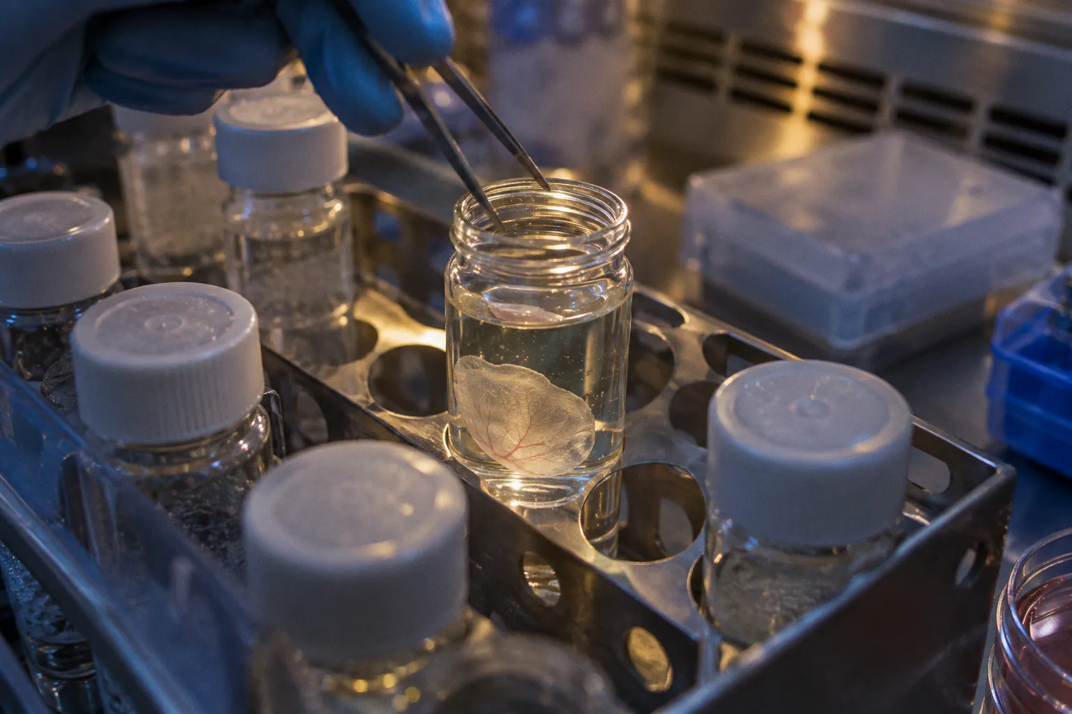
But the graft must also preserve its own architecture during harvest, transport, surgical delivery, and placement in the subretinal space. A retinal sheet that arrives intact but folds, rotates, or disperses at implantation may not deliver the same biological benefit as the tissue that left the culture dish.
That is the practical challenge behind current scaffold-free retinal organoid transplantation techniques: not simply keeping cells alive, but carrying organized retinal tissue through the entire chain from differentiation to functional integration. Preclinical studies provide a useful framework. Human embryonic stem cell-derived retinal organoid sheets have survived in immunodeficient retinal degeneration models for seven to ten months, with mature photoreceptors and evidence of synaptic and visual functional recovery. At the same time, the grafts can develop rosettes and disorganized cellular clusters when they lack the continuous basal lamina and Müller glia support found in native tissue.
The useful question, then, is not whether scaffold-free grafts work in principle. It is where the tissue loses structural control—and which parts of the workflow can be adjusted before that failure becomes visible.
The differentiation window determines whether the graft behaves like tissue
For sheet-based transplantation, developmental timing is not a minor culture variable. It determines whether the graft is sufficiently organized to be handled as a sheet while still retaining the developmental plasticity needed for integration.
The strongest preclinical results have involved intact retinal organoid sheets collected between days 30 and 70 of in vitro differentiation. This interval corresponds broadly to human embryonic developmental stages between approximately eight and fourteen weeks. At this stage, the organoids can preserve tissue polarity and recognizable retinal architecture without being so mature that the tissue becomes difficult to manipulate or unable to adapt to the host environment.
That window should not be treated as a universal release date. It is better understood as a starting range for process development. Two organoids cultured for the same number of days may not have equivalent architecture. Differentiation protocols, aggregate size, local nutrient gradients, and handling history can all influence the appearance and mechanical behavior of the tissue.
For a translational workflow, the differentiation window needs to be tied to release characteristics rather than time alone. A practical assessment should ask:
- Is the organoid sufficiently organized to maintain a clear orientation during harvest?
- Can the sheet be separated without extensive fragmentation?
- Does the tissue retain a coherent surface after transfer?
- Are the photoreceptor and inner retinal regions developing in a pattern consistent with the intended graft?
- Can the organoid tolerate the planned transport and delivery sequence without losing its architecture?
This is where a retinal progenitor cell sheet differs from a suspension of dissociated cells. The sheet carries spatial information into surgery. That information is biologically valuable, but it also creates a physical constraint: the tissue must remain intact long enough to be positioned correctly.
In scaffold-free transplantation, differentiation age is part of the surgical protocol. The graft is not ready when the calendar says so; it is ready when its architecture can survive the route to the retina.
Why early-stage tissue can integrate more effectively
An organoid sheet collected during an appropriate developmental stage offers two advantages. First, the tissue retains polarity. Second, the cells remain capable of maturing within the host environment. In preclinical models, this combination has supported the development of photoreceptors with inner and outer segments after subretinal implantation.
The host retina is not an empty landing site. It provides local signals, physical boundaries, glial responses, and remaining neuronal circuits. A graft that arrives with some internal organization can use those conditions more effectively than a randomly distributed population of dissociated cells. This does not mean that every layer will align perfectly. It means the graft begins with a structural advantage.
The risk is that the same developing tissue may respond poorly to mechanical disruption. Repeated pipetting, excessive shear, compression during transfer, or prolonged time outside controlled culture conditions can convert a potentially organized graft into a collection of damaged fragments. The final product may still contain viable cells, but viability alone does not confirm that the graft can form a functional retinal unit.
Graft survival depends on more than cell viability
Retinal graft survival in vivo is often discussed as though it were a single endpoint. It is not. A graft can remain present while showing limited organization, incomplete maturation, or weak communication with the host retina. Conversely, the appearance of mature photoreceptors is encouraging but does not by itself establish useful visual function.
The preclinical evidence is valuable because it connects several layers of outcome. In immunodeficient RCS rat models, scaffold-free human embryonic stem cell-derived retinal organoid sheets survived in the subretinal space for more than seven to ten months. The grafts developed mature photoreceptors with inner and outer segments, and the studies did not report tumorigenesis in those models. Functional signals were also observed: electroretinogram recovery reached a peak around two months after surgery, while superior colliculus electrophysiology recorded visual responses for six to eight months after grafting.
These findings suggest a sequence rather than a single event:
1. The tissue remains viable after implantation.
2. The graft maintains or reconstructs internal retinal organization.
3. Photoreceptors mature sufficiently to participate in retinal signaling.
4. Synaptic connections form between graft and host tissue.
5. The resulting activity is detectable through functional testing.
Each step can fail independently. A graft may survive without forming effective synapses. It may form synaptic contacts without producing a measurable improvement in the chosen functional assay. It may produce a signal in a rodent model without providing evidence that the same organization can be achieved in a human surgical setting.
For that reason, scaffold-free ocular tissue engineering should use a layered outcome strategy. Histology and immunostaining establish location and cellular identity. Structural assessment shows whether polarity and photoreceptor morphology are preserved. Electrophysiology and behavioral or circuit-level assays address function. None of these measurements replaces the others.
Immune privilege helps, but it does not remove the delivery problem
The subretinal space offers a relatively protected environment, and the eye has important immune-privileged characteristics. That protection is clinically relevant, but it should not be confused with a guarantee of graft acceptance. Surgical trauma, retinal degeneration, blood-retinal barrier disruption, graft composition, and the immune status of the host model all influence the result.
The published survival data come from immunodeficient retinal degeneration models. Those models are useful for studying tissue behavior, but they do not reproduce the full immune and surgical complexity of an immunocompetent human recipient. A long-surviving graft in an immunodeficient rat therefore supports feasibility; it does not establish long-term clinical safety or efficacy.
This distinction matters particularly for cell-replacement therapy ocular models. When the graft contains developing tissue rather than a narrowly purified cell population, the manufacturing and safety questions become broader. Researchers need to follow not only photoreceptor maturation but also the behavior of residual undifferentiated cells, proliferative activity, abnormal tissue organization, and the possibility of unwanted cell types. The absence of tumorigenesis in a defined preclinical study is reassuring, but it is not a universal safety claim.
Rosette formation is the central structural weakness
The most persistent limitation of scaffold-free retinal organoid grafts is rosette formation. These are circular, disorganized cellular arrangements that can appear after implantation in the subretinal space. They are not simply a cosmetic irregularity. They indicate that the graft has lost some of the planar organization required for a functional retinal layer.
Several forces may contribute. In native retina, the basal lamina and Müller glia help define physical orientation and cellular relationships. A scaffold-free organoid sheet enters the subretinal space without that continuous external support. Once the tissue is detached from its culture environment, it must preserve its own geometry while adapting to the host surface. Folding, local compression, uneven contact, and incomplete apposition can all create conditions in which cells reorganize into rosettes.
The practical response is not to assume that a synthetic scaffold is automatically superior. Biomaterials may improve handling or provide a supportive surface, but they introduce their own questions: degradation, inflammatory response, thickness, delivery behavior, and compatibility with photoreceptor maturation. Scaffold-free grafting avoids those risks, but it places more responsibility on tissue preparation and surgical placement.
Rosette prevention therefore has to be approached as a workflow problem:
- Select tissue with consistent polarity and limited spontaneous disorganization before harvest.
- Minimize mechanical stress during transfer and loading.
- Avoid allowing the sheet to fold or roll during delivery.
- Position the graft so that it maintains broad contact with the intended subretinal surface.
- Track whether rosettes appear immediately after implantation or during later remodeling.
That last distinction is important. If the tissue is already disorganized before implantation, the problem is likely rooted in differentiation or handling. If the sheet appears structurally coherent at delivery but develops rosettes later, the relevant variables may include host contact, local glial behavior, mechanical forces, or the absence of matrix support.
The precise molecular cues required to eliminate rosette formation without artificial matrix support remain unresolved. It would be premature to claim that current scaffold-free methods have solved the problem. The more realistic goal is to reduce the frequency and severity of disorganization while preserving the advantages of an intact, developmentally patterned graft.
Surgical delivery is where a good graft can become a poor implant
A retinal organoid may be biologically promising and still fail at the point of delivery. In the surgical field, the constraints are physical: the graft must pass through the delivery system, enter the subretinal space, and remain positioned without excessive manipulation. A sheet that is too fragile may fragment. A sheet that is too large or poorly oriented may fold. A suspension that is easy to inject may disperse before establishing useful contact with the host retina.
This is why retinal organoid surgical delivery should be treated as part of product development rather than as a separate final step. The culture protocol and the delivery method shape one another.
Sheet delivery
Intact sheets preserve polarity and architecture. That is their primary advantage. The cells do not need to reconstruct every spatial relationship from the beginning, and the developing photoreceptor layer can mature within a more organized tissue context.
The disadvantages are equally direct. Sheets are mechanically vulnerable, difficult to orient, and sensitive to folding. Their dimensions and thickness may influence how easily they can be delivered and how broadly they contact the host surface. A technically successful injection is not enough if the tissue arrives curled, fragmented, or only partially apposed.
A robust sheet-delivery workflow should record more than whether the graft entered the eye. It should document the condition of the tissue before loading, after loading, and after placement. Even simple categories—intact, partially folded, fragmented, or dispersed—can help connect surgical handling with later histological outcomes.
Suspension delivery
Single-cell suspensions made from digested retinal organoids allow more targeted purification and may offer greater flexibility in dosing or cell composition. They are also easier to distribute through a small delivery pathway. But digestion removes the original architecture and polarity. Cell survival may be lower than with sheet transplantation, and the implanted cells must establish their organization within the host environment.
Suspensions may be appropriate when the therapeutic aim is defined cell replacement rather than transfer of a developing retinal unit. They are less attractive when the intended benefit depends on preserving coordinated tissue structure. The choice should follow the biological objective, not simply the convenience of injection.
| Parameter | Intact organoid sheet | Dissociated cell suspension |
|---|---|---|
| Preserved architecture | Retains polarity and tissue organization | Architecture is lost during digestion |
| Handling risk | Vulnerable to folding, tearing, and fragmentation | Easier to distribute but vulnerable to cell loss |
| Cell selection | Less flexible once the sheet is formed | Allows more targeted purification |
| Integration requirement | Must maintain physical apposition and orientation | Cells must reorganize within the host |
| Main biological concern | Rosette formation and structural disorganization | Lower survival and weaker tissue-level organization |
| Best fit | Tissue replacement where retinal architecture matters | Defined cell delivery where composition is the priority |
The table does not identify a universal winner. It clarifies the trade-off. Sheets transfer more structure but demand more from the surgeon and the delivery system. Suspensions offer greater compositional control but ask the host retina to do more of the organizational work.
Transport is part of the surgical chain
One of the more practically important findings is that a specialized overnight transport protocol maintained survival, subretinal integration, and visual function improvement in retinally degenerate rats. That result expands the relevance of scaffold-free grafting beyond a single laboratory bench. It suggests that point-to-point tissue movement can be compatible with the biology of the graft when transport conditions are deliberately designed and validated.
Transport should therefore be evaluated as a controlled stage between manufacturing and surgery. The relevant question is not whether a dish can be moved from one room to another. It is whether the graft arrives with the same structural and functional potential it had at release.
A transport protocol needs to protect against changes that may not be visible immediately:
- loss of tissue cohesion;
- changes in orientation;
- mechanical damage during movement;
- prolonged deviation from the intended culture environment;
- delayed loss of viability that appears only after implantation.
The overnight transport finding is encouraging, but it remains preclinical. It supports the feasibility of coordinated workflows; it does not remove the need to validate transport for each tissue format, container, formulation, and surgical pathway.
The cold chain does not end when the organoid leaves the incubator. For a sheet graft, transport is another stage of tissue engineering—one that happens outside the culture system.
Functional integration has to be measured on its own timeline
A common mistake in regenerative therapy development is to expect structure and function to mature simultaneously. Retinal grafts do not necessarily follow that schedule. The tissue may survive first, develop photoreceptor morphology later, and establish measurable circuit activity after additional maturation.
In RCS rat models, electroretinogram recovery peaked around two months after surgery. Superior colliculus electrophysiology detected visual responses over a longer period of approximately six to eight months. These findings illustrate why a single early readout can be misleading. An initially modest signal does not necessarily mean the graft will remain inactive, while early structural presence does not prove functional connectivity.
A useful assessment plan separates three questions:
Is the graft present?
This requires anatomical localization and confirmation that the implanted tissue remains in the intended subretinal position. A graft that has migrated or dispersed may still be detectable, but it should not be interpreted as equivalent to a stable, apposed sheet.
Has the graft matured?
Researchers should look for the development of photoreceptor features, including inner and outer segments, while also assessing whether the tissue has retained an organized retinal pattern. Maturation without organization may not produce useful circuit integration.
Is the graft communicating with the host?
Synaptic integration and circuit-level responses are the critical translational endpoints. The 2024 transport study reported point-to-point response heatmaps in the superior colliculus, supporting the interpretation that grafted tissue contributed to visual pathway activity in the model. Such findings are stronger than simple cell survival, but they still need to be interpreted within the limits of the animal system.
The timing of these assessments should be planned before surgery. If tissue is collected only at one endpoint, the study may miss when the graft began to integrate, when rosettes developed, or whether functional recovery preceded later structural maturation.
A practical development path for scaffold-free grafts
The most reliable way to improve scaffold-free retinal organoid transplantation techniques is to treat the process as a sequence of linked decisions rather than a single implantation experiment.
1. Define the intended biological product.
Decide whether the goal is transfer of an organized retinal sheet or delivery of a selected cell population. This choice determines whether architecture or cellular composition is the primary manufacturing priority.
2. Map differentiation age to tissue behavior.
Use the day 30–70 range as a development window, then correlate culture age with polarity, mechanical integrity, cell identity, and susceptibility to fragmentation.
3. Characterize the graft before it reaches surgery.
Record whether the tissue is intact, folded, partially disorganized, or already showing rosette-like structures. Pre-implantation appearance provides an essential baseline.
4. Validate the delivery pathway with the actual tissue format.
A system that works for a suspension cannot be assumed to work for a sheet. Loading, transit, release, and placement must be assessed using the same type of graft intended for the animal study.
5. Treat transport as a validated process step.
If the graft must move between sites, evaluate the complete overnight or point-to-point workflow rather than validating only the storage container.
6. Use structural and functional endpoints together.
Survival, photoreceptor maturation, synaptic integration, electroretinography, and circuit-level responses answer different questions. The study should not allow one favorable measurement to stand in for all of them.
7. Separate feasibility from clinical readiness.
Long-term survival and functional responses in rodent models are meaningful preclinical milestones. They are not evidence that scaffold-free organoid sheets have reached routine clinical use or phase III validation in humans.
This sequence also helps identify where a failed experiment belongs. Poor graft survival may reflect tissue damage during delivery rather than an intrinsic limitation of the organoid. Weak function may reflect incorrect positioning, immature tissue, or incomplete synaptic connectivity rather than simple cell death. Without process-level records, these possibilities collapse into an unhelpful conclusion that the therapy did not work.
The translational value is in preserving organization without adding unnecessary materials
Scaffold-free retinal organoid grafts occupy an important middle ground in regenerative therapeutics. They are more organized than a simple cell suspension, but less mechanically supported than a scaffold-assisted construct. Their promise comes from carrying developmental architecture into the diseased retina. Their vulnerability comes from asking that architecture to survive every transition from culture dish to subretinal space.
The current evidence supports a measured position. Intact sheets collected during an appropriate differentiation window can survive for many months in retinal degeneration models, develop mature photoreceptors, and contribute to measurable visual function. Specialized transport can be compatible with this workflow. At the same time, rosette formation remains a real structural limitation, and the long-term human safety and efficacy of scaffold-free sheets have not been established.
The next advances are unlikely to come from one dramatic adjustment. They will come from better alignment between differentiation, tissue release criteria, transport, surgical delivery, and longitudinal assessment. A graft that preserves its polarity before surgery has a better starting point. A delivery system that respects the physical behavior of the sheet gives it a better chance of maintaining that advantage. And a study that measures function on the graft’s actual maturation timeline can distinguish temporary delay from genuine failure.
For clinicians and researchers, that is the practical lesson. Scaffold-free transplantation is not a shortcut around tissue engineering. It is tissue engineering concentrated into the organoid itself—and into the careful handling required to bring that tissue, still organized and viable, to the retina where it is meant to work.