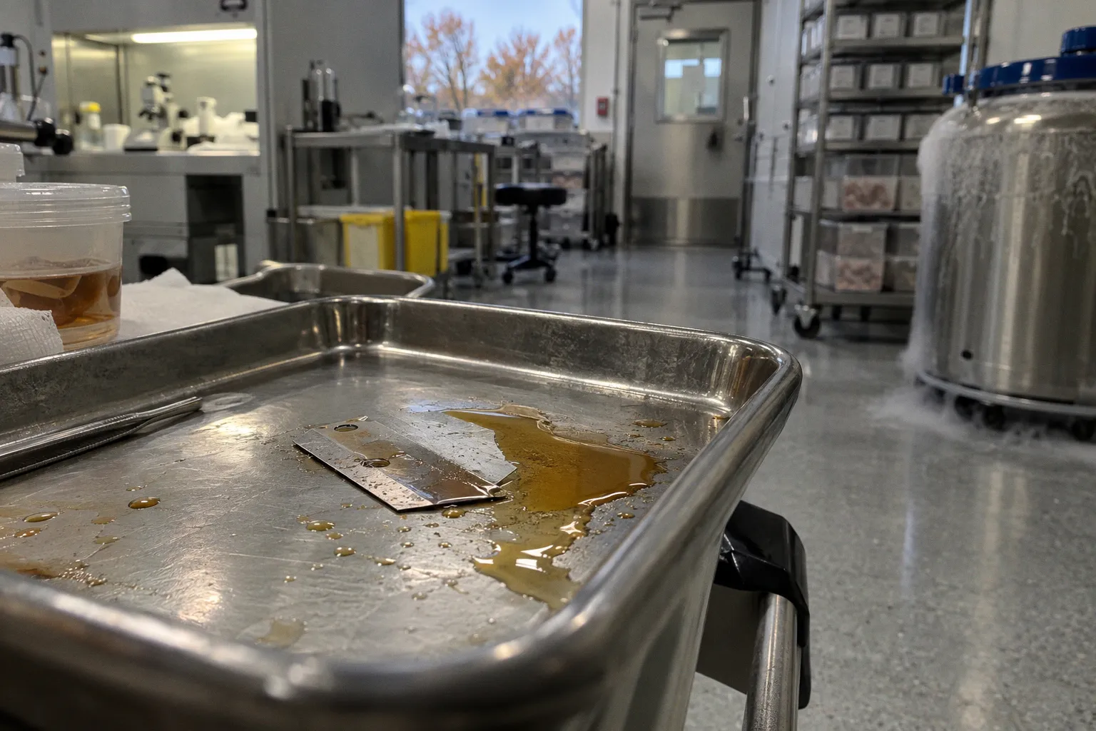
After death, circulation stops, tissue barriers deteriorate, and processing can alter the spaces a researcher later examines. The slide is therefore not a transparent record of disease. It is a record of disease and handling, often difficult to separate.
That distinction matters wherever donor tissue is used to study retinal vascular disease. A gap between the neurosensory retina and retinal pigment epithelium (RPE), or a fluid-filled space within the retina, may be compatible with pathology. It may also reflect post-mortem change or preparation. Without reliable case and handling records, the right interpretation is not to choose the more persuasive story. It is to mark the finding as ambiguous.
The Challenge of Post-Mortem Retinal Integrity
Once circulation stops, the retina’s living barriers and fluid regulation begin to fail. Endothelial cells no longer maintain their usual energy-dependent functions; the inner blood-retinal barrier becomes vulnerable. The outer barrier, formed by tight junctions between RPE cells, also loses the conditions that sustain it. Fluid distribution and tissue structure can change before fixation has stabilized the sample.
The result is a specimen shaped by more than one process. A donor retina may contain genuine antemortem disease, post-mortem change, and processing-related distortion at the same time. The longer and less completely documented the interval before fixation, the harder it becomes to assign a specific feature to one of those causes.
This does not make donor tissue unusable. Human retinal microvasculature can be examined directly in donor material, at a level of structural detail that clinical imaging cannot provide. But the value of that detail depends on knowing what the specimen can support. A beautiful section is not necessarily a faithful one: fixation may preserve some structures while displacing, extracting, or obscuring others.
Separation of the neurosensory retina from the RPE is a familiar concern in donor material, as are vacuolated spaces and disruptions in delicate retinal layers. Such findings need to be described before they are interpreted. Calling every separation a detachment, every cavity edema, or every pool exudate turns a processing uncertainty into a biological conclusion.
In donor tissue, an apparent leak is a finding to investigate—not a diagnosis the slide can make by itself.
The practical difficulty is that the same appearance can have more than one explanation. A space beneath the retina may be associated with antemortem disease, but the section alone may not reveal whether it formed before death, during the post-mortem interval, or during processing. The slide supplies morphology; case history and handling records supply context. When the latter are missing, confidence should narrow accordingly.
Histological Markers of Exudative vs. Transudative Fluid
Histological appearance can help frame the differential, but it should not be treated as a stand-alone clock. Fluid associated with vascular injury may contain plasma proteins and other material from blood. Erythrocytes, leukocytes, or fibrin can support an interpretation involving antemortem vascular or inflammatory events, depending on the tissue context. Their presence is not a universal requirement, and their absence does not prove that fluid accumulated after death.
Likewise, pale or granular material is not a dependable signature of post-mortem transudation. Autolysis and fixation can change cellular detail and the appearance of proteinaceous material. A smooth, eosinophilic pool may look persuasive, but staining intensity and texture alone cannot establish its origin. The safest reading combines morphology with the lesion’s distribution, adjacent tissue changes, clinical information, and the documented post-mortem and fixation intervals.
| Feature to assess | What it may contribute | What it cannot establish alone |
|---|---|---|
| Erythrocytes, leukocytes, or fibrin | May support a vascular or inflammatory process in the appropriate context | The timing or cause of every fluid space |
| Distribution around vessels or within retinal layers | Helps describe whether a finding follows a plausible anatomical pattern | That the pattern is necessarily antemortem |
| Tissue separation or vacuolation | Documents structural disruption relevant to interpretation | Whether it arose from disease, autolysis, or processing |
| Fixation and post-mortem records | Constrains which explanations are plausible | A definitive origin when records are incomplete |
| Protein staining or immunohistochemistry | Can characterize material in a space and complement routine stains | A complete causal history of the specimen |
The distinction between exudative and transudative fluid is therefore a working question, not a visual sorting exercise. A researcher can describe the material, note the presence or absence of associated features, and state how those observations affect interpretation. The claim should stop short of certainty when the evidence does.
This is particularly important in diabetic retinopathy ex vivo models. A fluid space that resembles edema may be relevant to the disease being studied, but it is not enough to demonstrate disease-related leakage. Comparisons with controls processed in the same way, together with donor history and handling information, are more informative than a single striking field. Even then, differences in tissue quality can complicate the comparison.
Quantifying Fixation-Induced Structural Disruptions
A study of vascular integrity needs to account for structural disruption as part of its measurement, not leave it as an informal caveat. The relevant question is not only whether vessels or fluid spaces are present, but whether the tissue preserves enough architecture to interpret them and whether preservation differs between groups.
The Henle fiber layer illustrates the problem. Its oblique arrangement makes it vulnerable to separation and distortion. Cavities in this region may resemble cystoid spaces, but resemblance is not identity: the appearance has to be assessed alongside the layer’s organization, neighboring retinal structure, and the specimen’s history. A single H&E section may not resolve the distinction.
Several classes of findings deserve explicit description in retinal microvascular histology analysis:
- Retina–RPE separation: Record its location, extent, and relation to other abnormalities. Do not assume that a detached-looking area represents a clinical serous detachment.
- Disruption of the Henle fiber layer: Note whether spaces follow the oblique fiber arrangement or occur alongside other signs of tissue breakdown.
- Vacuolation in the inner retina: Describe which layers are affected and whether the changes are focal or widespread.
- Vascular fragmentation or loss of recognizable vessel structure: Distinguish an apparent absence of vessels from evidence that the local tissue is poorly preserved.
- Fluid pools: Record their location and contents where these can be assessed, while keeping origin separate from appearance.
Quantification should make these distinctions visible. A study can report how many sections or fields show separation, vacuolation, or disrupted vessels, and explain how those features were defined. It should also state whether the same criteria were applied to every donor and control. This is not administrative detail: if one group has more compromised tissue, an apparent disease effect may partly reflect differential preservation.
A useful discipline is to keep three statements separate: what is seen, what it may mean, and how certain that interpretation is. For example, a report can describe subretinal fluid and retinal–RPE separation, note that both are compatible with several processes, and qualify the inference because the interval to fixation is unknown. That is more informative than converting an equivocal feature into a firm label.
Bridging the Gap Between Clinical FFA and Ex Vivo Microvascular Density
Fundus fluorescein angiography (FFA) and histology do not measure the same thing. FFA follows dye passage through the living circulation and can show dynamic leakage. Histology examines fixed tissue and can reveal vessel structure at high resolution. Their results may complement one another, but neither serves as a simple substitute for the other.
Clinical imaging has limits in resolving the smallest vessels, and leakage can make vascular boundaries harder to interpret. Histology, meanwhile, may display vessels that were below the imaging modality’s practical resolution, but only where the relevant structures survived procurement and processing. It cannot reproduce blood flow, and a visible vessel in a section does not show whether it was perfused or leaking before death.
That makes cross-modality comparisons easy to overstate. A higher vessel count in histological material does not, by itself, mean that FFA missed a specific set of capillaries. Differences may reflect resolution, staining, sampling, tissue preservation, or how vessels were counted. Similarly, a lower count in a donor specimen could indicate disease-related change, post-mortem fragmentation, or a combination of both.
Paired clinical and tissue data can help connect the methods when the same donor’s imaging and specimen history are available. Such comparisons should be read as evidence about the particular samples and methods used, not as a universal conversion between FFA and ex vivo microvascular density. The interpretation depends on image quality, anatomical matching, tissue orientation, fixation, and the criteria used to identify a vessel in each modality.
For a study comparing diabetic and control donor retinas, the most defensible approach is to state separately what each method shows. FFA may support a claim about leakage during life; histology may support a claim about vascular structure in the sampled tissue. If the data do not link those observations in the same donor, the article should not imply that one directly verifies the other.
Optimizing Tissue Processing to Minimize Diagnostic Bias
Processing cannot restore information lost before fixation, but a careful protocol can make the losses more visible and reduce avoidable variation. The first requirement is a usable record: when death occurred, when the eye was recovered, how it was stored, when fixation began, and how the tissue was prepared. If any interval is unknown, that uncertainty belongs in the analysis.
Formalin is widely used, but fixation is not instantaneous. Neural tissue may continue to change while fixative penetrates, and the condition of the globe before immersion affects what is preserved. Any comparison of donor tissue should therefore treat fixation method and timing as part of the experimental design, not as background logistics.
The choice of embedding and sectioning should follow the question. Routine paraffin sections are useful for many histological assessments. When the endpoint depends on fine detail in a fluid space or vessel wall, other preparation methods may preserve features differently and can be considered alongside routine stains. No method removes the need to interpret morphology in context.
A practical workflow can keep that context attached to each sample:
1. Record the timeline and its limits. Preserve the available post-mortem, recovery, storage, and fixation information. Report missing intervals rather than filling them with assumptions.
2. Standardize handling across comparison groups. Process diabetic and control specimens as consistently as possible, and document any unavoidable differences.
3. Define structural artifacts before scoring. Specify how retinal–RPE separation, vacuolation, and vessel disruption are recognized, then apply those definitions consistently.
4. Match methods to the endpoint. Use a preparation and staining strategy suited to the feature under study, and avoid treating a single stain as a complete test of fluid origin.
5. Report tissue quality with the biological findings. If preservation differs across samples, readers need to see that alongside the vascular measurements.
The goal is not to exclude every imperfect specimen. In human tissue research, that may be unrealistic and can introduce its own bias. The goal is to make specimen quality legible enough that a reader can judge whether a vascular interpretation is supported, qualified, or unresolved.
The Question That Still Has No Number
A central uncertainty remains: how often are post-mortem or processing-related changes interpreted as antemortem vascular leakage in donor retina research? Without evidence that establishes the frequency, it should not be presented as a known rate or as a settled direction of error. The answer may vary with procurement, fixation, disease context, and the criteria used to classify fluid.
What can be said is narrower and more useful. Fluid pooling in donor tissue is ambiguous unless case records, tissue context, or other evidence support a particular origin. Structural disruption can compromise vascular measurements. Clinical imaging and ex vivo histology provide different kinds of evidence, and comparisons between them require care.
For vascular leakage in donor retina models, restraint is not a retreat from interpretation. It is part of the measurement. The stronger study is not the one that gives every space a confident name; it is the one that distinguishes observation from inference and makes the specimen’s history visible enough for others to judge the difference.